


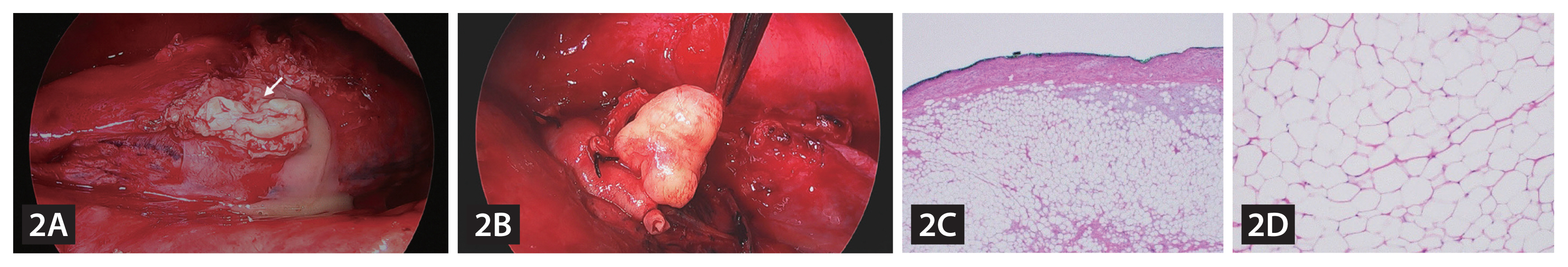
A 70-year-old woman was admitted to the hospital with left pleuritic chest pain for 3 days. She complained of a cough, sputum, and chilling sensation but no dyspnea. She had no history of smoking or trauma. Pneumonia with a parapneumonic effusion and endobronchial nodule were evident on the chest computed tomography and flexible bronchoscopy (Fig. 1A, B). Despite receiving intravenous antibiotics and undergoing toilet bronchoscopy, the patient’s fever persisted. A pneumothorax was also detected, which recurred despite a closed thoracostomy (Fig. 1C). To address the obstruction of the left main bronchus, which was causing prolonged obstructive pneumonia and barotrauma, the patient underwent surgery. Thoracoscopically, the left lower lung was totally consolidated and purulent pus with a thickened pleura was noted (Fig. 2A). An endobronchial mass was removed through the bronchus (Fig. 2B). We performed a lobectomy of the left lower lung and the final histological diagnosis was endobronchial lipoma (Fig. 2C, D).
Endobronchial lipomas are extremely rare and benign (0.1–0.5% of all lung tumors). Bronchoscopic intervention can be effective in preserving the lung function. However, despite the lipoma being benign, our patient required a lobectomy due to post-obstructive pneumonia intractable to medical treatment and irreversible lung damage. It is important to tailor the treatment according to the patient’s clinical condition.

 PDF Links
 PubReader
 ePub Link
 Full text via DOI
 Download Citation
 Print
Print


