


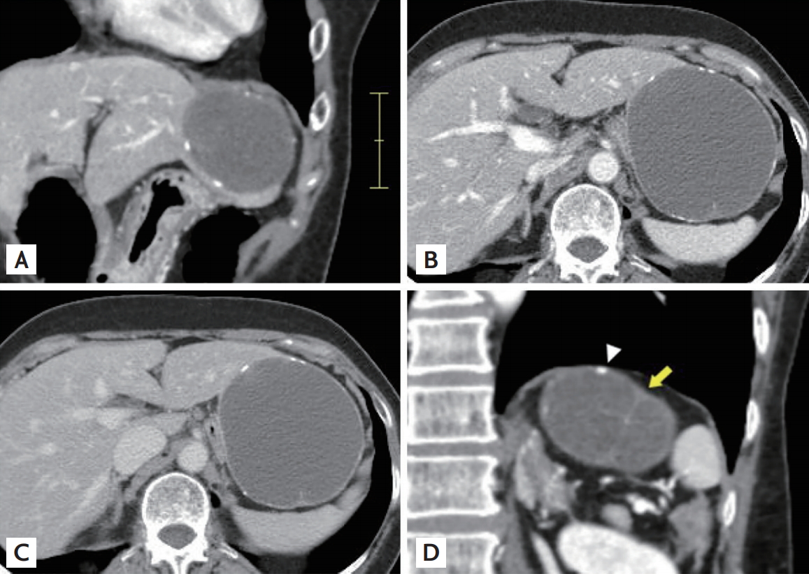
A 62-year-old female chronic hepatitis B patient was referred to our clinic after an incidental cystic tumor in her liver was found on abdominal ultrasound. Her serum alanine aminotransferase level was 31 U/L and the hepatitis B virus DNA titer was 1,900 IU/mL. An abdominal ultrasound revealed a hypoechoic cystic tumor measuring 8.2 ├Ś 7.2 cm in the lateral lobe of the liver. A computed tomography (CT) scan showed a large exophytic cyst in the lateral margin in the left lobe (Fig. 1A-1C). A unilocular cyst with multifocal rim calcifications and a thin intracystic septum was observed (Fig. 1D).
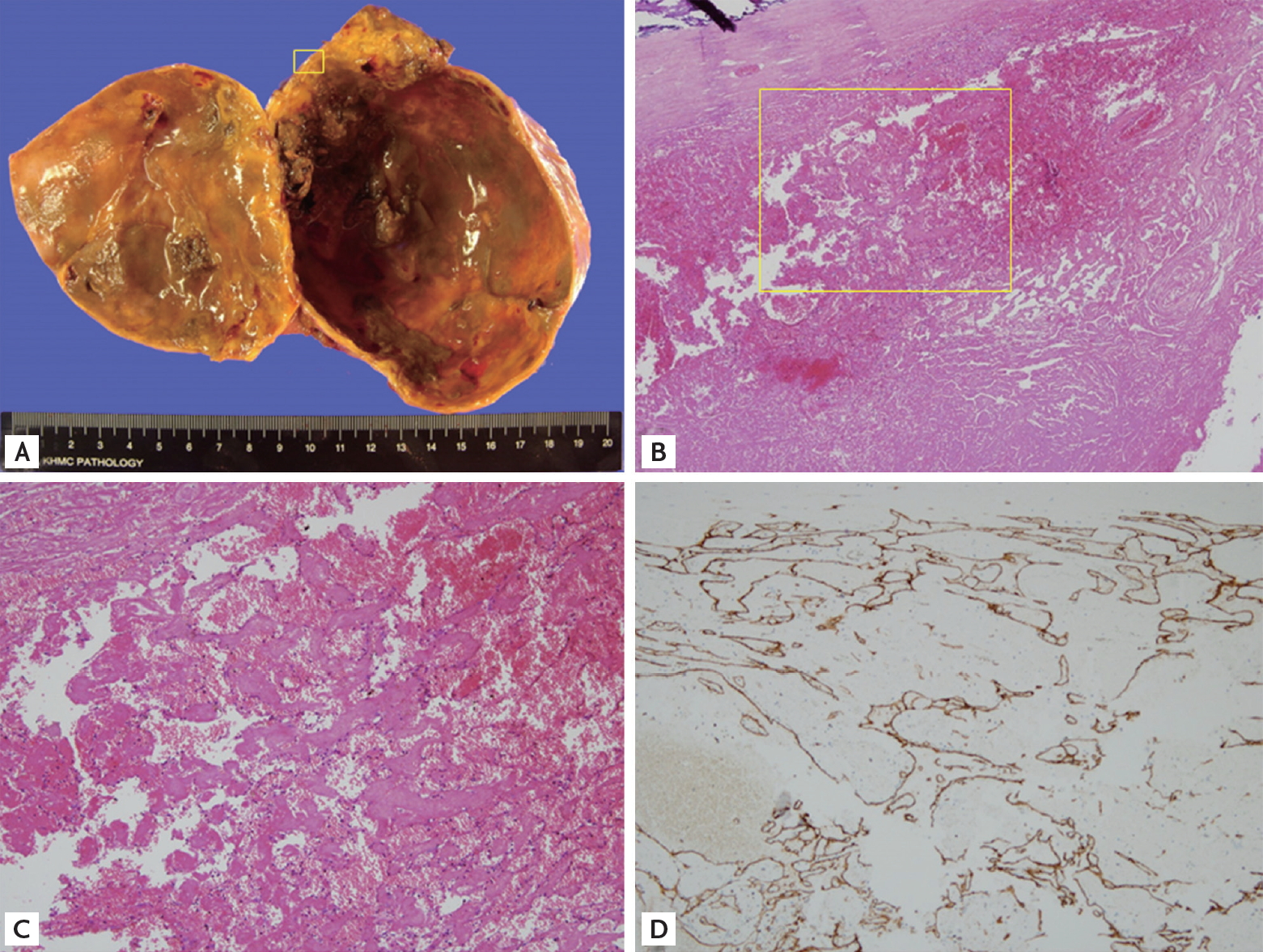
A biliary cystadenoma was initially suspected and a laparoscopic approach was employed for tumor resection. The macroscopic cut section showed a brownish round unilocular cyst measuring 9.0 ├Ś 8.0 ├Ś 8.0 cm with a dense fibrous wall having small multifocal calcifications (Fig. 2A). The microscopic view of the cystic wall and small papillary remnants in that showed a dense fibrous wall without lining epithelium. The majority of the solid portion was almost completely necrotized and the remaining lesion had mono-layered endothelial cells and fibrous stroma mixed with blood cells, which indicated hepatic hemangioma (Fig. 2B and 2C). Immunohistochemical staining revealed strong positivity for cluster of differentiation 34 (CD34) and CD31 and negative for cytokeratin 19 (CK19), suggesting an endothelial origin (Fig. 2D). The cystic degeneration following the near-complete necrosis may have altered the presentation of this exophytic hemangioma into that of a cystic tumor.
Hepatic hemangiomas are the most common benign solid tumors in the liver. With ultrasound and CT scans becoming more accessible than ever, the detection of atypical hemangiomas mimicking malignant tumors has increased as well. As this case showed that a hepatic hemangioma can be misdiagnosed as a biliary cystic neoplasm. Thus, hepatic hemangiomas should be included in differential diagnoses of cystic tumors.
Written informed consent was obtained from the patient.
 PDF Links
 PubReader
 ePub Link
 Full text via DOI
 Download Citation
 Print
Print


