


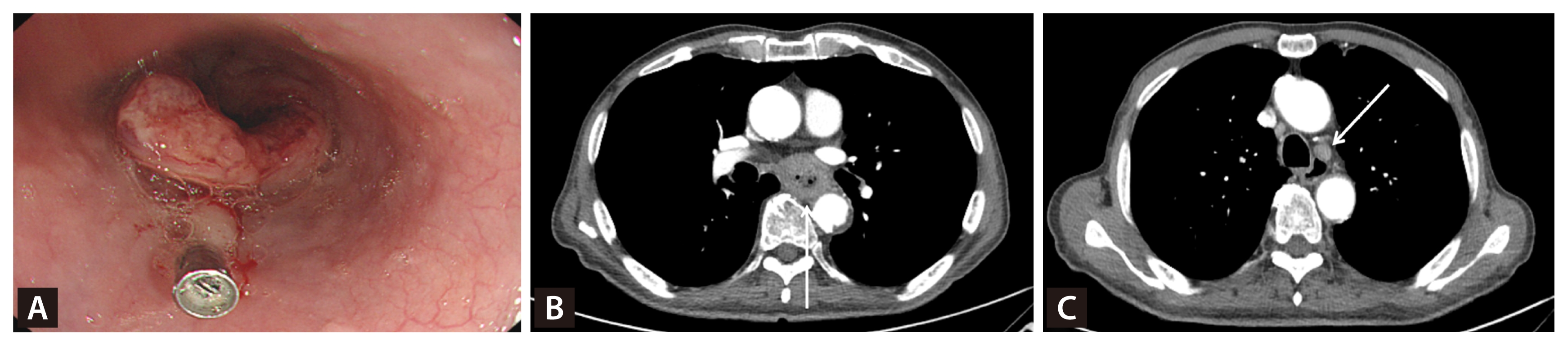
A 72-year-old male presented with progressive dysphagia. Esophagogastroduodenoscopy (EGD) revealed an ulcerofungating mass at the mid-esophagus, located 38 cm from the incisors (Fig. 1A). Biopsy confirmed moderately differentiated squamous cell carcinoma (SCC). Chest and abdominal contrast-enhanced computed tomography (CT) demonstrated circumferential esophageal wall thickening extending into the adventitia (Fig. 1B) and enlarged left paratracheal lymph node (Fig. 1C), consistent with a clinical stage of cT3N1M0. Positron emission tomography (PET)/CT performed for initial staging revealed hypermetabolic activity in the primary esophageal lesion and regional lymph nodes, with no evidence of distant metastases.
The patient subsequently underwent neoadjuvant concurrent chemoradiotherapy, consisting of paclitaxel and carboplatin, along with radiation at a total dose of 50 Gy delivered in 25 fractions. Follow-up EGD demonstrated complete disappearance of the previously noted esophageal mass (Fig. 2A). During the preoperative workup, a screening colonoscopy revealed an incidental 5 mm slightly elevated lesion in the sigmoid colon (Fig. 2B). Narrow band image showed irregular microvascular and surface patterns (Fig. 2C). Endoscopic polypectomy was subsequently performed. Unexpectedly, histopathological examination of the resected specimen revealed nests of moderately differentiated SCC infiltrating the submucosal layer (Fig. 2D). Immunohistochemistry demonstrated strong positivity for CK5/6 (Fig. 2E) and p63 (Fig. 2F), supporting the diagnosis of metastatic SCC originating from the esophageal primary rather than primary colorectal SCC.
Colonic metastasis from esophageal SCC is extremely rare, with fewer than 20 cases reported in the English literature [1–4]. Common metastatic sites of esophageal cancer include liver, lung, and bone, while gastrointestinal tract metastases are exceptional [5]. Most previously reported cases presented with symptomatic metachronous lesions manifesting as intestinal obstruction or hematochezia several months to years after initial diagnosis [1,2]. Synchronous asymptomatic colonic metastasis discovered incidentally during preoperative evaluation, as seen in our case, is exceptionally rare [3]. The mechanism of colonic metastasis remains unclear but may involve hematogenous dissemination through circulating tumor cells or retrograde lymphatic spread via the complex esophageal lymphatic drainage system [6].
Distinguishing metastatic SCC from primary colorectal SCC is essential for appropriate staging and management. Primary colorectal SCC is exceedingly rare, accounting for only 0.1–0.3% of all colorectal malignancies. In the present case, the colonic lesion demonstrated submucosal infiltration without evidence of intraepithelial dysplasia or squamous metaplasia in the overlying mucosa, arguing against a primary colorectal origin. Furthermore, the histological features were concordant with those of the previously diagnosed esophageal SCC. The strong immunoreactivity for CK5/6 and p63, markers of squamous differentiation commonly expressed in esophageal SCC, further supported a metastatic origin. In the absence of another primary squamous malignancy, these findings collectively support the diagnosis of metastatic spread from the esophageal primary.
Detection of colonic metastasis led to upstaging of the disease to stage IV, and the planned curative esophagectomy was deferred. The treatment strategy was subsequently modified to systemic therapy. The prognosis of patients with colonic metastasis from esophageal cancer is poor, with most reported survival ranging from 2.5 to 12 months despite multimodal therapy [3,4]. This case therefore illustrates how identification of unexpected distant metastasis during preoperative evaluation including colonoscopy may directly alter therapeutic planning and prognostic assessment.


 PDF Links
 PubReader
 ePub Link
 XML Download
 Full text via DOI
 Download Citation
 Print
Print