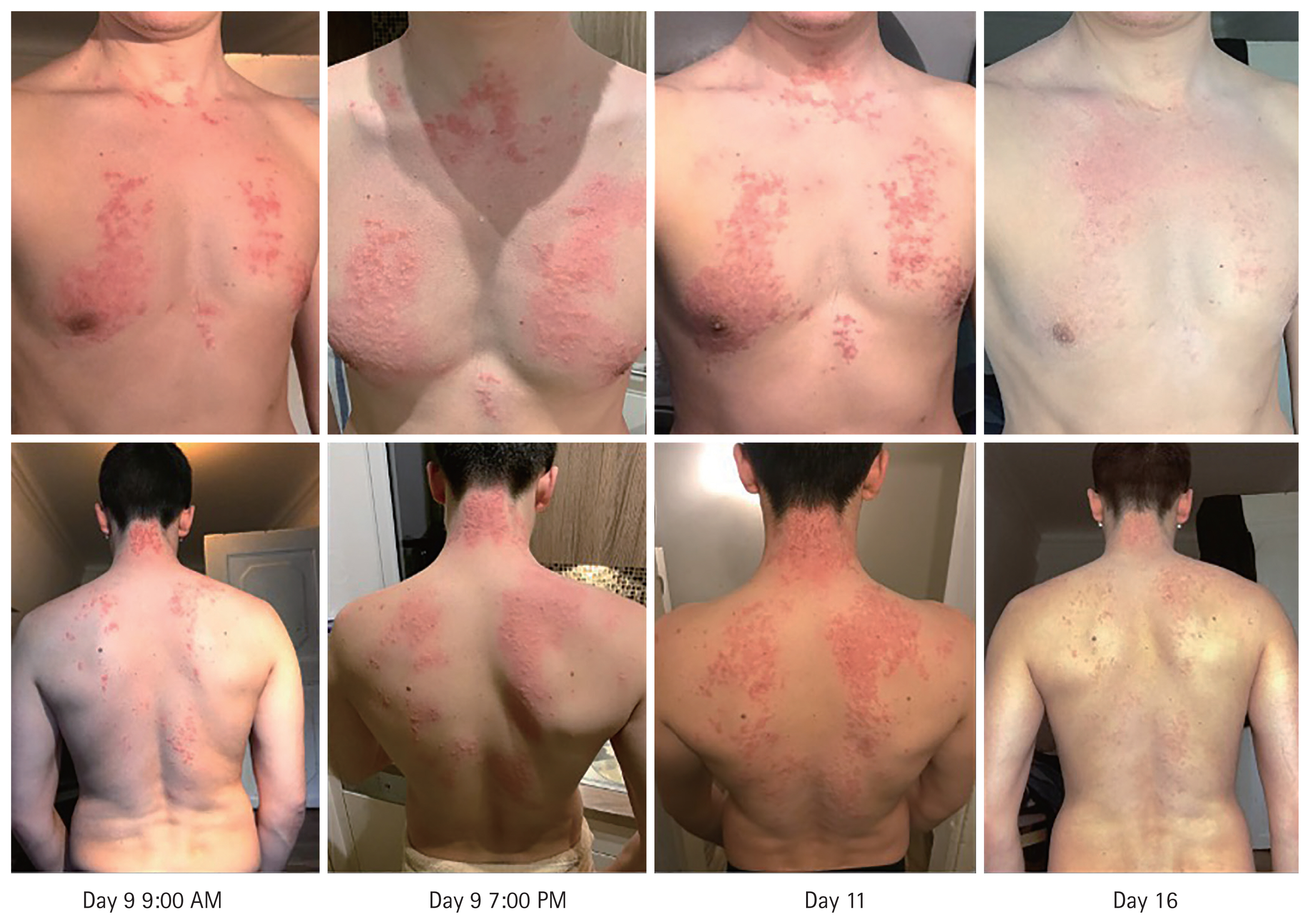
A 24-year-old healthy, Caucasian, male non-diabetic athlete, free of any allergies or intolerance reactions, followed a ketogenic diet in the course of a nutritional intervention trial. After 9 days of nearly complete carbohydrate restriction, he developed a rapidly advancing symmetrical trunk- and neck-accentuated skin rash of brownish-red, itchy papules in a reticular pattern, with no further systemic complaints. Laboratory investigations revealed no signs of infection (leukocyte count 3.9 g/L [reference range, 3.7 to 9.9]; C-reactive protein < 0.6 mg/L), glycemic disturbances (blood glucose 90 mg/dL [reference range, 70 to 100]), impairment of liver or kidney function, and a normal blood cell count. Betahydroxybutyrate in the urine was significantly increased (2.7 mmol/L [reference range, 0.4 to 0.5]) indicating ketotic state.
After returning to a balanced diet and treatment with mild topical corticosteroids, the skin lesions vanished spontaneously within a few days, leaving a mild reticular hyperpigmentation resembling prurigo pigmentosa. Allergy testing did not reveal any suspicious cause, and histology was not justified due to the benign and promptly self-limiting course of the condition. The progress of the skin lesions are illustrated in Fig. 1.
In view of the patient’s history of a ketogenic diet and spontaneous recovery after resumption of a regular diet, a diet-related keto rash in a previously healthy person was the most plausible diagnosis. Ketogenic diets, characterized by a maximum of 50 g of oral carbohydrate intake daily, have become popular among communities due to the benefits of the diets in terms of weight loss and improved physical performance. A keto rash is a very rare side effect, believed to occur twice as often in women than in men, and appears to be more prevalent in the Asian population. In most of the cases it appears in attribution to diabetic ketoacidosis which was not present in our normoglycemic patient. The skin lesions are usually characterized by erythematous pruritic coalescing of papules distributed in a reticulate pattern, which resolve later (usually after discontinuation of the diet) and recover with persistent post-inflammatory hyperpigmentation. Histologic assessment typically reveals perivascular neutrophilic infiltrates during the early stages, which subsequently evolve into necrotic keratinocytes and spongiosis of the epidermis, leaving melanophages and lymphocytic infiltrates of the superficial dermis. In practice, diagnostic procedures should take allergic contact dermatitis, drug eruption, as well as viral co-infection affecting the skin into account. The registration of a detailed medical history, including dietary behavior, is the cornerstone of diagnosis in such ambiguous cases.
A written informed consent has been obtained in which the presented person agreed to publish his images.


 PDF Links
 PubReader
 ePub Link
 Full text via DOI
 Download Citation
 Print
Print


