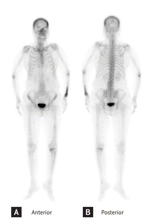
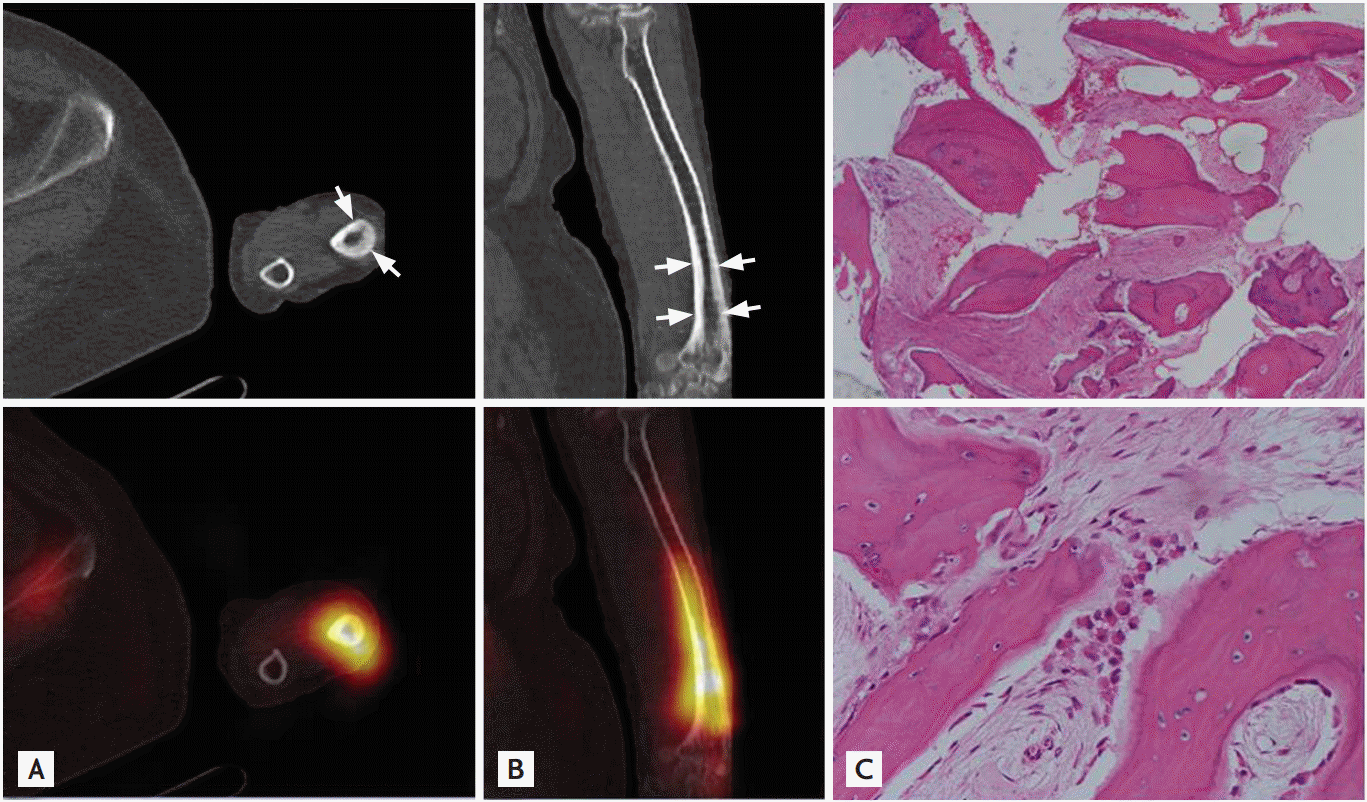
A 59-year-old female patient with a 2-year history of lung cancer complained of pain throughout her whole body, especially in the left forearm, that had lasted 1 month. No evidence of fever or trauma was found, and no significant abnormalities were observed in laboratory examinations that investigated the tumor marker, serum alkaline phosphatase, calcium, and parathyroid hormone levels. She was referred for an evaluation of metastatic disease through a whole-body bone scan using 99mTc-methylene diphosphonate (MDP), which revealed a marked striped tracer accumulation in the left distal forearm (Fig. 1). Single photon emission computed tomography/computed tomography (CT) hybrid imaging of the left forearm was performed and only revealed cortical thickness in the radius on CT corresponding to the high tracer uptake region (Fig. 2A and 2B). The C-terminal cross-linking telopeptide of type I collagen value was 1.04 ng/mL (normal range, 0.12 to 0.75). Using the radiologic, biochemical, and biopsy (Fig. 2C) findings a final diagnosis of Paget disease (PD) was established.
PD is a benign, focal metabolic bone disease that is initiated by aggressive osteoclast-mediated bone resorption followed by compensatory and imperfect increased bone formation. It is usually polyostotic and generally involves the pelvis, femur, spine, skull, and tibia. The characteristic radiographic features are an increase in bone density with cortical thickening and coarsening of the bony trabeculae; however, the majority of cases are asymptomatic. Bone scintigraphy represents a useful imaging technique that can evaluate the scope and extent of the damage. In PD, the upper extremities are rarely affected; to date, only several case reports have described presentations of this disease in the thumb, metacarpal, carpal, and humerus. In this case, the involvement of the radius in PD was incidentally detected through a conventional 99mTc-MDP bone scan. The bone pain was relieved following treatment with bisphosphonate, and the 6-month follow-up was uneventful.


 PDF Links
 PubReader
 ePub Link
 Full text via DOI
 Download Citation
 Print
Print


