 |
 |
| Korean J Intern Med > Volume 30(2); 2015 > Article |
|
Abstract
Most gastric cancers are caused by infection with the common human bacterial pathogen, Helicobacter pylori. It is now accepted that gastric cancer can be prevented and virtually eliminated by H. pylori eradication and this knowledge was responsible for country-wide H. pylori eradication combined with secondary cancer prevention for those with residual risk that was introduced in Japan in 2013. Korea is a high H. pylori prevalence and high gastric cancer incidence country and a good candidate for a gastric cancer elimination program. The presence of an H. pylori infection is now considered as an indication for treatment of the infection. However, antimicrobial drug resistance is common among H. pylori in Korea making effective therapy problematic. Country-wide studies of the local and regional antimicrobial resistance patterns are needed to choose the most appropriate therapies. H. pylori and gastric cancer eradication can be both efficient and cost effective making it possible and practical to make Korea H. pylori and gastric cancer free. There is no reason to delay.
Korea is a country with a high incidence of gastric cancer. One hundred years ago, most western countries also experienced a high incidence of gastric cancer but mysteriously the incidence began to decline to where in many countries it has become an uncommon disease [1]. In the early 20th century gastric cancer was the number one cause of cancer in many countries (e.g., the United States, England, and Germany) leading to considerable interest in finding the cause. In the late 1800's it was recognized that there was a relationship between gastric cancer and achlorhydria (atrophic gastritis) and in the first half of the 20th century research focused on and confirmed the strong association between gastric cancer and atrophic gastritis [2,3]. Numerous epidemiology studies on gastritis were done [4] and hypotheses were developed including how gastric cancer developed as part of the progression of the histologic damage [5,6].
The major breakthrough occurred in the early 1980's when Warren and Marshall [7] cultured Helicobacter pylori and proved that it caused gastritis. They also speculated that, if H. pylori caused gastritis, it was also likely etiologically involved in the gastritis-associated diseases, peptic ulcer and gastric adenocarcinoma. Rapid progress followed. In 1994 the World Health Organization declared H. pylori to be a class I (Definite) human carcinogen [8,9]. In 2014 the World Health Organization published a new monograph entitled "Helicobacter pylori eradication as a strategy for preventing gastric cancer" describing the recent and ongoing epidemiologic studies regarding H. pylori eradication and gastric cancer [10]. Probably the most important recent change in thinking and management of H. pylori occurred in 2013 when the Japanese government approved a test and treat strategy to find and eliminate H. pylori in order to eliminate gastric cancer in Japan [11,12]. We now understand that cure of the infection will stop the progression of gastric damage but it will not completely reverse the accumulated risk of developing gastric cancer [6,13]. Thus, the new Japanese gastric cancer program also includes a secondary cancer prevention program utilizing endoscopic surveillance to find and remove early gastric cancers that appear despite H. pylori eradication.
H. pylori infection is classified as a "necessary but insufficient cause" of gastric cancer [14]. That is the same classification used for other infectious agents that are etiologically involved in carcinogenesis such as human papilloma virus (cervical cancer), hepatitis B and C (liver cancer) and the Epstein-Barr virus (also known as human herpesvirus 4) with causes Burkitt's lymphoma and nasopharyngeal carcinoma. The "not sufficient" means that presence of one of these infections does not guarantee that the infected person will develop cancer but the specific type of associated cancer will not develop without the infection. There are other less common causes of gastric cancer besides H. pylori (e.g., Epstein-Barr virus, primary genetic abnormalities) such that eradication of H. pylori will not completely eliminate gastric cancer. However, because H. pylori infection is responsible for more than 95% of gastric cancers, its eradication will make gastric cancer a truly rare disease. Thus, the decision of the Japanese government to eliminate H. pylori was based on the premise that doing so would virtually eliminate the problems associated with the present high burden of gastric cancer.
The natural history of H. pylori gastritis is one of progressive histologic damage ultimately leading to atrophic gastritis and gastric atrophy. Gastric cancer is an inflammation-associated cancer [15,16] and the link to gastric cancer is through atrophic gastritis [6]. An individual's risk can be stratified based on the extent and severity of the gastric damage. A population's risk can be estimated based on the average rate of development of atrophic changes. The progression to atrophic gastritis is influenced by the virulence of the infecting H. pylori strain, environmental factors particularly diet, and host genetics. Dietary factors play an important role such that the gastric cancer environment is typically one where diets are seasonal (e.g., lack of fresh fruits and vegetables in winter) and salt is commonly used as a food preservative. For example, a change of diet to include one where refrigeration largely replaces salt in food preservation and fresh fruits and vegetables are available year around can result in rapid decline in the incidence of gastric cancer (e.g., by 60% in Japan between 1965 and 1995) [1,17]. In Western countries where the incidence of gastric cancer has fallen from the most common malignancy to an uncommon cancer has been associated both with a change in diet and improvements in sanitation resulting in a progressive decline in the prevalence of H. pylori infection.
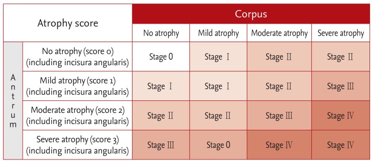
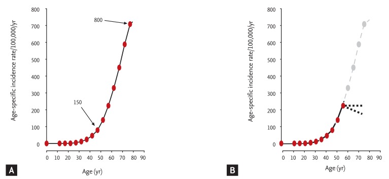
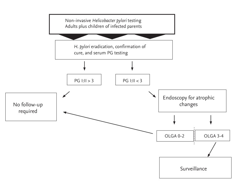
H. pylori infections are typically acquired in childhood and remain active throughout an individual's life. Improvements in standards of living and sanitation as has occurred in Korea following World War II resulted in a decline in transmission in families and thus a lower prevalence in each new birth cohort [18]. This natural decline in prevalence of the infection requires many generations to reach sufficiently low levels to affect the incidence of gastric cancer. The rate of disappearance can be accelerated by programs designed to identify and treat active infections as is being done in Japan. However, if all H. pylori infections were eliminated today it would not immediately eliminate gastric cancer because those with irreversible damage would still have some remaining risk of developing cancer. This is most easily visualized using a gastric cancer risk assessment tool such as the OLGA histologic staging system (Fig. 1) [19]. This system scores gastric cancer risk on a 5 point scale (from 0 to 4) with 0 being no risk to 4 representing very high risk. The scoring system evaluates both the severity and extent of damage (Fig. 1). The natural history of H. pylori infection is for the cancer risk stage to increase over time (i.e., this is responsible for the age-related increase in gastric cancer) (Fig. 2A). H. pylori eradication will stop the progression of damage and prevent further increases in risk which then stabilizes or declines (Fig. 2B). Cancer prevention in high risk countries such as Korea requires that a program of H. pylori detection and eradication be combined with an estimation of cancer risk at the time of H. pylori eradication (e.g., histology, serum pepsinogen testing, or a combination of both) (Fig. 3) [6].
Korea is a high H. pylori incidence country. The most common H. pylori strain infecting Koreans is highly virulent and the Korean diet tends to be of high salt content such that H. pylori infection frequently results in atrophic gastritis reflecting in the high rate of gastric One solution would be to follow Japan's lead and search out and eradicate H. pylori infections. Because H. pylori-induced atrophic gastritis is common, a program to identify and perform surveillance on those with a significant gastric cancer risk should also be considered [20,21]. The current secondary cancer prevention program of annual or every 2 year endoscopy or radiology to detect cancer can be modified to include from for include primary prevention. Those in the current programs with no H. pylori infection or with non-atrophic gastritis receive little or no benefit (Fig. 4) [22,23] and those at higher risk of cancer continue to experience the expected age-related increase in cancer risk despite surveillance. Probably more than one-half of those currently enrolled in secondary cancer prevention programs fall into the "no possible benefit" category (Fig. 4). The resources currently utilized for secondary gastric cancer prevention could possibly be better utilized by converting current secondary prevention programs into surveillance of the high risk group who have continuing risk despite H. pylori eradication (Fig. 2) [3,6].
H. pylori is a bacterial infection and thus require antibiotics to cure. Treatment is a problem in Korea because antibiotic resistance is common. The currently approved regimen (a triple therapy consisting of a proton pump inhibitor, amoxicillin, and clarithromycin) has proven to be relatively ineffective because of the high rate of clarithromycin resistance [24,25,26,27,28]. This therapy is considered obsolete in most countries but as it is often the only one approved by the government insurance, doctors have a dilemma. Choice of an effective regimen for most infections is based on antibiotic susceptibility testing. While this is potentially available in Korea it is rarely done and are there are few data about country-wide or even regional antibiotic resistance patterns. In the United States we have moved toward four drugs combinations (e.g., 14 day therapy with a proton pump inhibitor, clarithromycin, metronidazole, and amoxicillin or concomitant therapy which is effective except in the presence of clarithromycin-metronidazole dual resistance) or the combination of a bismuth, tetracycline, metronidazole and a proton pump inhibitor which is generally effective despite metronidazole resistance provided it is given a full dose and for 14 days [29,30]. In Asia the combination of a high dose proton pump inhibitor and amoxicillin such as 20 mg of rabeprazole and 500 to 750 mg of amoxicillin every 6 hours for 14 days appears effective [31]. An organized program to identify the resistance patterns in Korea and define regimens that are highly effective is clearly needed. Human experimentation with empiric therapies without susceptibility testing is an extremely inefficient method of discovery of effective therapies and should be strongly discouraged.
The guidelines for management of H. pylori have varied somewhat over time and between regions [32]. It is important to recognize that in most instances these "indications" were not designed as being indications to treat the infection but rather were indications to test for H. pylori. This was based on the premise that H. pylori was a very common disease (i.e., at least one-half of the world's population is infected) and as most would not develop a clinical outcome, testing should be focused on those in whom a definite benefit was likely. Nonetheless, it was recommended that whenever the infection was diagnosed, the patient be treated unless there were compelling reasons not to. Recently, the focus has turned to the problem of gastric cancer. The recognition that H. pylori eradication would also essentially eliminate gastric cancer caused a rethinking of whom to test. It is important to recognize that the natural history of the infection is to cause progressive damage such that the presence of non-atrophic changes is likely only a stage in the infection but one in which H. pylori eradication is almost certain to prevent gastric cancer. In populations where atrophic gastritis and gastric cancer is common, population wide screening and treatment is indicated. Korea, China, Japan have such populations, High risk populations exist even in low gastric cancer incidence countries and are also potential targets for a test and treat strategy along with targeted secondary cancer prevention.
Most gastric cancers result from an infectious disease caused by the common human bacterial pathogen, H. pylori. Gastric cancer can be prevented and virtually eliminated by elimination of the cause (i.e., H. pylori eradication). The current focus is on how to implement a test and treat program for high gastric cancer risk populations. In Korea there are probably 30 million H. pylori infected individuals a proportion of whom already are at high risk and would likely benefit most if H. pylori eradication was followed by a secondary cancer prevention program utilizing endoscopic surveillance. The majority of Korean's have not yet progressed beyond non-atrophic gastritis and only require H. pylori eradication to eliminate or greatly reduce their gastric cancer risk. The advantage of H. pylori therapy is that cure is a one-off such that the benefits are permanent. What needs to be done is clear. However it is yet unclear how to accomplish this goal in the most efficiently and cost-effective manner. However, there is no reason to delay in approaching the goal of making Korea an H. pylori and gastric cancer free country.
Acknowledgments
The author thanks Dr. Sun-Young Lee for reading the draft manuscript and making important suggestions. Dr. Graham is supported in part by the Office of Research and Development Medical Research Service Department of Veterans Affairs, Public Health Service grants DK067366 and DK56338 which funds the Texas Medical Center Digestive Diseases Center. The contents are solely the responsibility of the authors and do not necessarily represent the official views of the VA or NIH.
Conflict of Interest
Dr. Graham is a unpaid consultant for Novartis in relation to vaccine development for treatment or prevention of H. pylori infection. Dr. Graham is a paid consultant for RedHill Biopharma regarding novel H. pylori therapies and has received research support for culture of Helicobacter pylori. He is a consultant for Otsuka Pharmaceuticals regarding diagnostic breath testing. Dr. Graham has received royalties from Baylor College of Medicine patents covering materials related to 13C-urea breath test.
References
1. Graham DY. History of Helicobacter pylori, duodenal ulcer, gastric ulcer and gastric cancer. World J Gastroenterol 2014;20:5191ŌĆō5204PMID : 24833849.


2. Comfort MW. Gastric acidity before and after development of gastric cancer: its etiologic, diagnostic and prognostic significance. Ann Intern Med 1951;34:1331ŌĆō1348PMID : 14838496.


3. Graham DY, Asaka M. Eradication of gastric cancer and more efficient gastric cancer surveillance in Japan: two peas in a pod. J Gastroenterol 2010;45:1ŌĆō8PMID : 19714291.


4. Cheli R, Perasso A, Giacosa A. Gastritis: A Clinical Review. Berlin: Springer-Verlag, 1987.
6. Shiotani A, Cen P, Graham DY. Eradication of gastric cancer is now both possible and practical. Semin Cancer Biol 2013;23(6 Pt B):492ŌĆō501PMID : 23876852.


7. Warren JR, Marshall B. Unidentified curved bacilli on gastric epithelium in active chronic gastritis. Lancet 1983;1:1273ŌĆō1275PMID : 6134060.

8. International Agency for Research on Cancer. IARC Monographs on the Evaluation of Carcinogenic Risks to Humans. Vol. 100B, Biologic Agents: A Review of Human Carcinogens. Lyon: International Agency for Research on Cancer, 2012;385ŌĆō435.
9. International Agency for Research on Cancer. IARC Monographs on the Evaluation of Carcinogenic Risks to Humans. Vol. 61, Schistosomes, Liver Flukes and Helicobacter pylori. . Lyon: International Agency for Research on Cancer, 1994.
10. International Agency for Research on Cancer. IARC Working Group Reports. Vol. 8, Helicobacter pylori Eradication as a Strategy for Preventing Gastric Cancer. Lyon: International Agency for Research on Cancer, 2014.
11. Asaka M, Kato M, Sakamoto N. Roadmap to eliminate gastric cancer with Helicobacter pylori eradication and consecutive surveillance in Japan. J Gastroenterol 2014;49:1ŌĆō8PMID : 24162382.


12. Asaka M. A new approach for elimination of gastric cancer deaths in Japan. Int J Cancer 2013;132:1272ŌĆō1276PMID : 23180638.


13. Graham DY, Shiotani A. The time to eradicate gastric cancer is now. Gut 2005;54:735ŌĆō738PMID : 15888771.


14. Bracken MB. Risk, Chance, and Causation: Investigating the Origins and Treatment of Disease. New Haven: Yale University Press, 2013.
15. Hanada K, Uchida T, Tsukamoto Y, et al. Helicobacter pylori infection introduces DNA double-strand breaks in host cells. Infect Immun 2014;82:4182ŌĆō4189PMID : 25069978.


16. Hanada K, Graham DY. Helicobacter pylori and the molecular pathogenesis of intestinal-type gastric carcinoma. Expert Rev Anticancer Ther 2014;14:947ŌĆō954PMID : 24802804.


17. Wang C, Weber A, Graham DY. Age, period, and cohort effects on gastric cancer mortality. Dig Dis Sci 2015;60:514ŌĆō523PMID : 25274157.


18. Lim SH, Kwon JW, Kim N, et al. Prevalence and risk factors of Helicobacter pylori infection in Korea: nationwide multicenter study over 13 years. BMC Gastroenterol 2013;13:104. PMID : 23800201.


19. Rugge M, Correa P, Di Mario F, et al. OLGA staging for gastritis: a tutorial. Dig Liver Dis 2008;40:650ŌĆō658PMID : 18424244.


20. Shin DW, Cho J, Kim SH, et al. Preferences for the "screen and treat" strategy of Helicobacter pylori to prevent gastric cancer in healthy Korean populations. Helicobacter 2013;18:262ŌĆō269PMID : 23384480.


21. Park JM, Hahm KB. The Korean perspective of Helicobacter pylori infection: lessons from the Japanese government's policy to prevent gastric cancer. Dig Dis 2014;32:290ŌĆō294PMID : 24732195.


22. Ohata H, Kitauchi S, Yoshimura N, et al. Progression of chronic atrophic gastritis associated with Helicobacter pylori infection increases risk of gastric cancer. Int J Cancer 2004;109:138ŌĆō143PMID : 14735480.


23. The Research Group for Population-based Cancer Registration in Japan. Cancer incidence and incidence rates in Japan in 1986: estimates based on data from nine population-based cancer registries. Jpn J Clin Oncol 1991;21:318ŌĆō323PMID : 1942562.


24. Chung JW, Lee GH, Han JH, et al. The trends of one-week first-line and second-line eradication therapy for Helicobacter pylori infection in Korea. Hepatogastroenterology 2011;58:246ŌĆō250PMID : 21510323.

25. Yoon JH, Baik GH, Sohn KM, et al. Trends in the eradication rates of Helicobacter pylori infection for eleven years. World J Gastroenterol 2012;18:6628ŌĆō6634PMID : 23236238.


26. Jung YS, Lee SH, Park CS, et al. Trends in the eradication rates of Helicobacter pylori infection in Daegu and Gyeongsangbuk-do, Korea: multicenter study over 13 years. Korean J Gastroenterol 2014;63:82ŌĆō89PMID : 24561694.


27. Jeon HK, Kim GH. Does standard triple therapy still have a role in first-line Helicobacter pylori eradication in Korea? J Korean Med Sci 2014;29:619ŌĆō620PMID : 24851014.


28. Gong EJ, Yun SC, Jung HY, et al. Meta-analysis of first-line triple therapy for helicobacter pylori eradication in Korea: is it time to change? J Korean Med Sci 2014;29:704ŌĆō713PMID : 24851029.


29. Graham DY, Lee YC, Wu MS. Rational Helicobacter pylori therapy: evidence-based medicine rather than medicine-based evidence. Clin Gastroenterol Hepatol 2014;12:177.e3ŌĆō186.e3PMID : 23751282.


30. Wu JY, Liou JM, Graham DY. Evidence-based recommendations for successful Helicobacter pylori treatment. Expert Rev Gastroenterol Hepatol 2014;8:21ŌĆō28PMID : 24410470.


Figure┬Ā1
Gastritis staging: the OLGA (Operative Link on Gastritis Assessment) system. Atrophy is defined as loss of appropriate glands (with or without metaplasia). In each compartment (i.e., mucous-secreting antral and oxyntic/corpus mucosa), atrophy is scored in a five-tiered scale (0 to 4) according to the visual analogue scale of the Houston-updated Sydney system. The stage result from the combination of atrophic changes was assessed in the two mucosal compartments considered. Adapted from Rugge et al. [19] with permission of Elsevier.

Figure┬Ā2
(A) Illustration of the natural history of subjects entering a secondary prevention program after Helicobacter pylori eradication at age 50. At age 50 the average risk of gastric cancer is 150/100,000 per year and increases to 800/100,000 per year at age 80. Thus, despite annual surveillance the gastric cancer risk would increase 533%. (B) Illustrates the postulated effect if H. pylori eradication were done at age 60. After that point their risk stop increasing and would likely remain stable or decrease as healing occurred. Importantly, the risk does not return to zero. Adapted from Shiotani et al. [6] with permission from Elsevier.

Figure┬Ā3
Possible scenario of population-wide detection and eradication program to eliminate gastric cancer. The proposal is based on initially identifying those with Helicobacter pylori infections and assessing the health of the gastric mucosa. This considers using non-invasive testing with a locally or regionally validated immunoglobulin G H. pylori serology and serum pepsinogen (PG) testing. Those without H. pylori infection or atrophic gastritis would require no further evaluation or follow-up. All those with H. pylori infections would undergo eradication therapy with confirmation of cure, preferably using non-invasive testing with a urea breath or stool antigen testing. After H. pylori eradication, those with non-atrophic gastritis would require no further follow-up. Those with suspected atrophic gastritis (based on pepsinogen testing) would undergo endoscopy for proper risk stratification (e.g., using a validated histologic staging system). Those with cured H. pylori and healed non-atrophic gastritis would require no further follow-up. Those with confirmed atrophic gastritis (e.g., OLGA [Operative Link on Gastritis Assessment] stage III or IV) would be entered in to a long term endoscopic surveillance program. Because the cancer risk if likely to decline over time, they are also candidates for research regarding surveillance intervals and whether adjuvant therapy such as anti-inflammatory or anti-oxidant therapy would further reduce the risk current data does not allow firm recommendations for those after H. pylori eradication with mild atrophy (e.g., OLGA I and II) and they are considered candidates for research regarding the best strategy. Adapted from Shiotani et al. [6] with permission from Elsevier.

Figure┬Ā4
Proportion of an asymptomatic Japanese cohort in each risk group for gastric cancer. The illustration shows the risk groups identified by a surveillance program of a cohort of 4,655 asymptomatic Japanese average age 50 years followed by Ohata et al. [22] Helicobacter pylori status was determined by ELISA and chronic atrophic gastritis (CAG) by pepsinogen values. The pie chart is superimposed on the age-specific gastric cancer incidence among Japanese men from 1986 [23]. If H. pylori eradication had been done at the outset, only a minority of subjects would have been candidates for annual surveillance (i.e., those with mild [28.3%] and severe CAG [0.7%]). Adapted from Graham et al. [3] with permission of Springer.


 PDF Links
 PubReader
 ePub Link
 Full text via DOI
 Download Citation
 Print
Print


