To the Editor,
Systemic sclerosis (SSc) is a chronic multi-systemic disorder of unknown etiology characterized by immune activation, tissue fibrosis due to excessive synthesis and deposition of extracellular matrix, and vasculopathy due to vascular endothelial cell activation and injury. Peripheral vascular involvement manifesting as Raynaud's phenomenon (RP) is one of the common clinical features of SSc [1]. However, neurological complications and cerebral vasculopathies representative of macrovascular complications occur uncommonly [2]. Here we report a patient with cerebral vasculopathy associated with limited SSc. Cerebral angiography showed an occlusive lesion of the middle cerebral arteries (MCAs) and abnormal collateral vessels similar to those observed in patients with moyamoya disease.
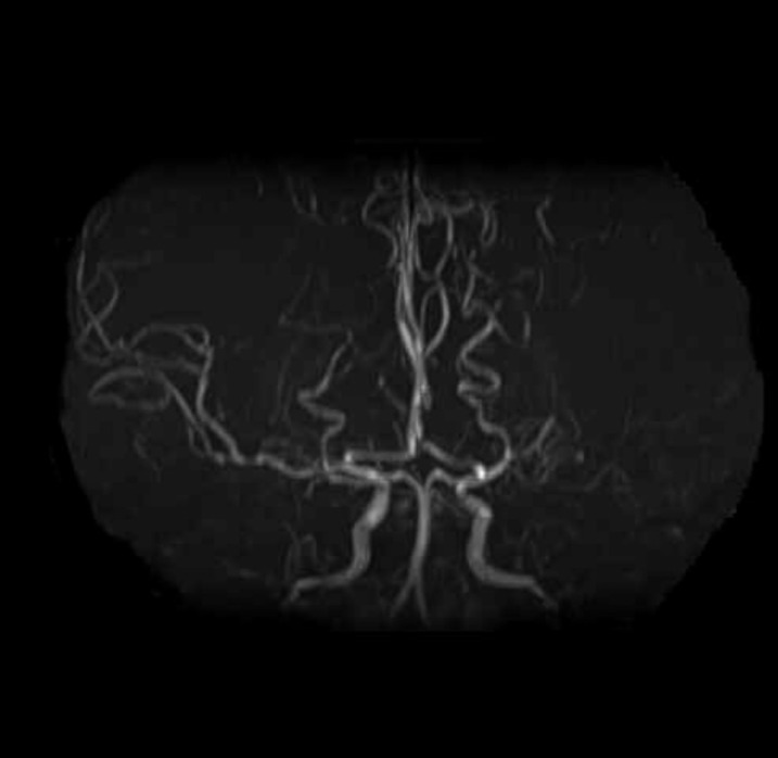
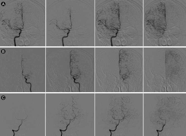
A 48-year-old woman presented with RP, xerostomia, arthritic pain, general swelling, and thickening of the skin below the forearm level. She was diagnosed with limited scleroderma and had been prescribed oral D-penicillamine and low-dose methylprednisolone. Laboratory examination revealed positive reactions for serum antinuclear (1:640, speckled pattern) and anti-Scl 70 antibodies, and negative results for anti-neutrophil cytoplasmic, anti-centromere, and anti-SSA and -SSB antibodies. One year later, she complained of a headache and severe dizziness that lasted for 2 months, and was admitted to a general ward via the rheumatology outpatient department. Her lipid profile was within normal limits. She was normotensive and other vital signs were within the normal range. She had no other past medical history except SSc, and in terms of familial history, her mother had diabetes mellitus. She did not complain of otolaryngologic symptoms. There were no other focal neurologic findings such as blurred vision, ataxia, or pathologic nystagmus. She underwent brain magnetic resonance imaging and magnetic resonance angiography (MRA); these revealed an occlusive lesion in the left MCA, and collateral vascular supply from the left external cerebral artery, which are similar to cerebral artery-mimicking moyamoya vessels (Fig. 1). For further evaluation of this abnormal vasculature, digital subtract angiography (DSA) of the internal carotid artery (ICA), external carotid artery and vertebral artery was performed. The DSA result was similar to that of the MRA, and several collateral circulations from both posterior cerebral arteries to both MCAs were observed (Suzuki grade II [3]) (Fig. 2). Blood chemistry tests (total cholesterol and low-density lipoprotein) and cardiovascular evaluation (trans-thoracic echocardiogram and 3D cardiac computed tomography) revealed no specific findings or risk factors for atherosclerotic disease. A single-photon emission computed tomography study showed no definitive abnormality in cerebral blood flow either at baseline or after acetazolamide injection (Fig. 3). After symptomatic treatment, she was discharged and has since been without neurological deficit.
Moyamoya disease is a macrovascular disease that involves the ICA and development of collateral vessels. In a typical case, vascular imaging reveals the appearance of a "puff of smoke" due to obliteration of the cerebral microvasculature [3,4]. Although the exact pathophysiologic mechanism has not yet been elucidated, vessel occlusion in moyamoya disease results from a combination of hyperplasia of smooth muscle cells and luminal thrombosis. The media are often attenuated with irregular elastic lamina. Caspase-dependent apoptosis has been proposed as a contributory mechanism in the associated degradation of the arterial wall [4].
In contrast, limited scleroderma usually involves small vessels. Comorbid vasculopathy in SSc involves all layers of the vessel wall and is characterized by fibrotic intimal hyperplasia. As a result, vessels lose their elasticity and become narrower. In time, the arterial intima may thicken and occlusion of the small arteries can facilitate the formation of in situ thrombosis. These vasculopathies can occur in the form of pulmonary arterial hypertension, scleroderma renal crisis, digital ulceration, or infarct.
A unique finding in our patient was the combination of limited scleroderma and cerebral angiographic findings showing abnormal vasculature. Although microvascular disease is a hallmark of SSc, there is an ongoing debate regarding the presence and extent of macrovascular diseases and the presence of accelerated atherosclerosis in limited SSc. It is usually limited to the ulnar or cerebral arteries, but its causal relationship with vascular endothelial damage has not yet been elucidated. A previous study suggested that there is a progressive and highly significant reduction in the elastic properties of the carotid artery of SSc patients compared to non-SSc subjects [5]. Arterial elastic properties are good markers of early atherosclerosis, and augmented carotid intima-media thickness may be more prevalent in scleroderma patients and related to antibodies against human heat shock protein (HSP)-60 and mycobacterial HSP-65 [6]. However, these moyamoya-like changes, which represent MCA occlusion and collateral circulation for the stenotic cerebral arteries, occur not only in patients with moyamoya disease but also in those with other disorders, such as atherosclerosis, Down's syndrome, neurofibromatosis type 1, and systemic lupus erythematosus. Terajima et al. [7] reported a typical case of moyamoya disease with transient ischemic attack showing bilateral obliterative cerebral vasculopathy. However, our case showed non-specific neurologic symptoms and unilateral occlusion of cerebral vessels. In this case, because the patient had no risk factors for atherosclerosis and showed no signs of ischemic stroke, we considered moyamoya-like vascular changes a possible cause of MCA occlusion. Because up to two-thirds of moyamoya patients may exhibit symptomatic progression over a 5-year period, the clinical symptoms and signs of ischemic cerebral occlusion in this SSc case will be followed carefully.


 PDF Links
 PubReader
 ePub Link
 Full text via DOI
 Download Citation
 Print
Print


