 |
 |
| Korean J Intern Med > Volume 27(1); 2012 > Article |
|
Abstract
Brugada syndrome can be unmasked by several conditions including a febrile state, marked leukocytosis, and electrolyte disturbances. Herein, we describe a 62-year-old man with cholangiocarcinoma in the first reported case of Brugada syndrome onset following photodynamic therapy.
Brugada syndrome is typified by an electrocardiographic (ECG) pattern of elevated ST-segments in the right precordial leads (V1-V3), a morphology similar to that seen in right bundle branch block, an absence of structural heart disease, and a high risk of ventricular fibrillation and sudden death [1]. Sodium channel blockers, fever, α-adrenergic agonists, β-adrenergic blockers, vagotonic agents, hyperkalemia, hypokalemia, hypercalcemia, acidosis, tricyclic antidepressants, alcohol, and cocaine toxicity all reportedly unmask the characteristic ST-segment elevation in right precordial leads [2,3]. In this article, we report a case of Brugada syndrome developing after photodynamic therapy (PDT) in a patient diagnosed with cholangiocarcinoma.
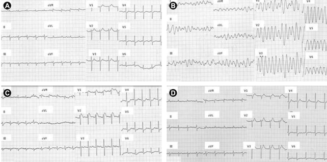
A previously healthy 62-year-old man was admitted to the hospital for PDT following a diagnosis of cholangiocarcinoma (Klatskin tumor, type IV) made 1 month prior to admission. His only complaint upon admission was mild abdominal pain. An endoscopic retrograde biliary catheter for draining bile was already in place. The patient had a cigarette smoking history of 40 packs per year and was a heavy drinker. His vital signs were stable, and no significant cardiopulmonary signs were evident. The electrocardiogram was normal (Fig. 1A). There was no prior family history of ventricular arrhythmias or sudden cardiac death.
For scheduled PDT, the patient was injected intravenously for over 5 minutes with a hematoporphyrin derivative-type photosensitizer (Photogem, 2 mg/kg; Photogem LLC. Co., Moscow, Russia). After 40 to 50 hours of PDT, light at a release power of 150 J/cm2 was applied.
The patient was stable until 7 hours following the light application, at which time he began to complain of feeling febrile and having chills. His body temperature was 38.4℃. Emergency laboratory tests revealed hepatic biochemical abnormalities: an aspartate transaminase (AST) level of 247 U/L, alanine transaminase (ALT) of 140 U/L, and direct bilirubin of 0.8 mg/dL.
One hour following the injection of antipyretics, the patient's condition stabilized. His only complaint was general weakness, but he went into cardiac arrest 10 hours later. The ECG performed during the cardiopulmonary resuscitation revealed polymorphic ventricular tachycardia (Fig. 1B). After electrical cardioversions (300 J × 3), the cardiac rhythm recovered to a sinus rhythm. The 12-lead ECG showed a right bundle branch block and a pronounced ST-segment elevation in the precordial leads (V1, V2) consistent with Brugada syndrome (Fig. 1C). Echocardiography revealed global left ventricular systolic dysfunction (ejection fraction 45%) with aneurysmal dilation of the mid to apical wall, suggestive of stress-induced cardiomyopathy.
Recurrent ventricular tachyarrhythmia was absent. The 12-lead ECG was normal when assessed 2 days post-attack (Fig. 1D), but the patient was comatose and showed irreversible hypoxic brain damage upon neurologic examination. One day following the attack, an intractable fever persisted despite antipyretics and antibiotics. Laboratory evaluation revealed increases in the white blood cell count (14,200/uL, 80% polynuclear neutrophils), C-reactive protein (19.9 mg/dL), and myocardial enzymes. The hepatic biochemistry deteriorated steadily with an AST level of 615 U/L, ALT of 174 U/L, and total bilirubin of 8.5 mg/dL. The patient's condition worsened with oliguria and azotemia. On the sixth day post-attack, the patient died of fulminant hepatic failure and sepsis due to obstruction of the biliary tract. Additional electrophysiologic examinations could not be performed.
Several studies have demonstrated that fever unmasks the characteristic ECG pattern of Brudaga syndrome and precipitates ventricular arrhythmias [4]. Although one episode of fever had occurred prior to the attack in this patient, no fever arose during the ventricular arrhythmia. The fever was transient, returning 1 day post-attack, and then persisted, but the ECG had normalized, which is why the Brugada syndrome was not associated with a fever in this patient.
PDT is an ablative treatment for rapidly proliferating tissues, including dysplastic and malignant lesions. It employs the administration of a photosensitizing drug followed by the application of a specific wavelength of light, leading to intracellular photoexcitation and injury. Three groups of photosensitizers are generally available, the most common of which comprises hematoporphyrin derivatives, which we used to treat this patient. The complications and toxicities occurring from PDT for gastrointestinal disease are related to the associated endoscopic procedure, acute effects of the photosensitizing agent, the local inflammatory and scarring effects in the region of therapy, and systemic phototoxicity [5]. Although some complications such as atrial fibrillation and congestive heart failure following PDT administration are reported rarely [6,7], the occurrence of ventricular arrhythmia such as Brugada syndrome post-PDT in cholangiocarcinoma has not been documented. The exact mechanism by which PDT causes cardiac arrhythmia is incompletely understood, and because this patient was not febrile and lacked any abnormal laboratory findings except hepatic biochemical abnormalities before the PDT, we speculate that the PDT itself may have caused Brugada syndrome in this patient.
In conclusion, we report the first case of Brugada syndrome following PDT administration in a patient with cholangiocarcinoma. When treating patients with PDT, physicians should be aware of the potential risk of arrhythmic complications.
References
1. Brugada P, Brugada J. Right bundle branch block, persistent ST segment elevation and sudden cardiac death: a distinct clinical and electrocardiographic syndrome: a multicenter report. J Am Coll Cardiol 1992;20:1391–1396PMID : 1309182.


2. Gavrielatos G, Letsas KP, Pappas LK, Efremidis M, Sideris A, Kardaras F. Brugada electrocardiographic pattern induced during febrile state with marked leukocytosis. Pacing Clin Electrophysiol 2007;30:135–136PMID : 17241329.


3. Araki T, Konno T, Itoh H, Ino H, Shimizu M. Brugada syndrome with ventricular tachycardia and fibrillation related to hypokalemia. Circ J 2003;67:93–95PMID : 12520160.


4. Antzelevitch C, Brugada P, Borggrefe M, et al. Brugada syndrome: report of the second consensus conference: endorsed by the Heart Rhythm Society and the European Heart Rhythm Association. Circulation 2005;111:659–670PMID : 15655131.


5. Petersen BT, Chuttani R, Croffie J, et al. Photodynamic therapy for gastrointestinal disease. Gastrointest Endosc 2006;63:927–932PMID : 16733105.


Figure 1
Development of Brugada syndrome following photodynamic therapy in a patient with cholangiocarcinoma. (A) Electrocardiogram upon admission. (B) Polymorphic ventricular tachycardia during arrest. (C) Brugada pattern electrocardiogram following cardioversion. (D) Repeat electrocardiogram 2 days following ventricular tachycardia.

-
METRICS

- Related articles
-
The Role of Photodynamic Therapy for Hilar Cholangiocarcinoma2010 December;25(4)


 PDF Links
 PubReader
 ePub Link
 Full text via DOI
 Download Citation
 Print
Print


