INTRODUCTION
Eosinophilic gastroenteritis is an uncommon disease that's pathologically characterized by marked eosinophilic infiltration in any area of the gastrointestinal tract without the presence of parasitic infections, drug reactions or malignancy. Since it was first described in 1937 by Kaijer1), approximately 300 cases have been reported worldwide by 19962) and there have been Korean 31 cases3). The clinical manifestations are related to the site of gastrointestinal (GI) involvement and the layer of the involved bowel wall4). To the best of our knowledge, eosinophilic enteritis presenting as intussusception in adult has not been reported in the literature. We report here on such a rare, interesting case.
CASE REPORT
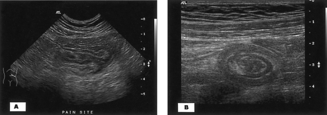
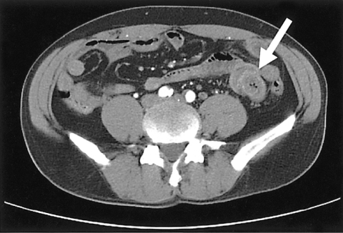
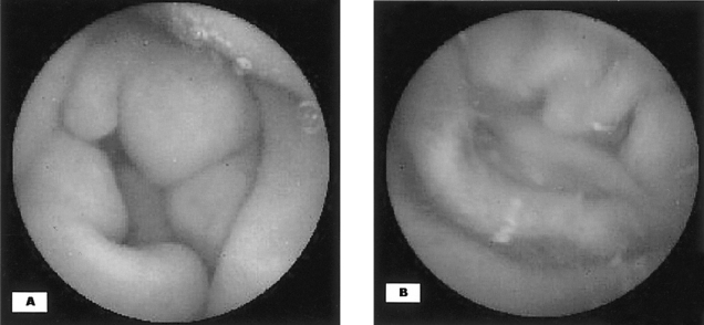
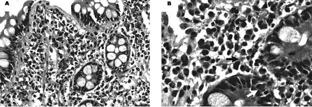
A previously healthy 55-year-old Korean male was admitted to our hospital with a 7 day history of intermittent left abdominal pain without nausea, vomiting, diarrhea or hematochezia. The man's pain had intensified and loose stool had occurred over the previous 24 hours. He had no history of medications, atopy or food allergy. The physical examination on admission revealed an acute ill appearance, but the patient was alert and mentality orientated. His body temperature was 36.8Ōäā, the blood pressure was 130/90 mmHg, the pulse 80/min and the respiratory rate 18/min. He was tender upon palpation in the left mid abdomen; he had no rebound tenderness. The cardiovascular, respiratory and neurological examinations were normal. Laboratory investigation revealed a white blood cell count of 9,100/mm3 with 16.8% eosinophils, an eosinophil count of 1,824/mm3, a hemoglobin level of 14.1 g/dL, a prothrombin percentage of 51% and an international normalized ratio (INR) of 1.72. The serum total protein (= 7.3 g/dL) and albumin (= 4.2 g/dL) were within the reference ranges. The ECG showed normal sinus rhythm. The chest radiograph was within normal limits, and the abdominal film revealed a nonspecific bowel gas pattern. Abdominal ultrasound demonstrated a central echogenic core surrounded by a hypoechoic rim in the left midabdomen (Figure 1A. transverse section: note the target sign). The longitudinal section demonstrated jejunojejunal intussusception (Figure 1B). Computed tomography scanning demonstrated diffuse circumferential wall thickening at the duodenal bulb and jejunum, and this was combined with short segmental enteroenteric intussusception (arrow) without a definite mass or any enlarged lymph nodes (Figure 2). However, we could not rule out a small bowel mass as a leading point of intussusception in this adult. Capsule endoscopy was performed to investigate the small bowel mucosal lesion. It showed only diffuse mucosal edema and erythematous change at the jejunum without a mass lesion (Figure 3). The peripheral blood smear showed eosinophilia. A stool exam, a serologic test for parasitic infection via micro-ELISA and the serum IgE level (42.8 IU/mL, normal range: 0~170) were all within the normal ranges. Esophagogastroduodenoscopy and colonoscopy showed normal mucosa except for mucosal edema and the erythematous change at the duodenum. We performed multiple endoscopic biopsies. Histologically, normal mucosa was observed at the terminal ileum, cecum, ascending colon, transverse colon, descending colon, sigmoid colon, rectum, gastric antrum, body and fundus. At the duodenum, a diffuse infiltration by inflammatory cells and mainly eosinophils (more than 20/High Power Field) were observed. In addition, there were extracellular eosinophilic granules in the lamina propria, and eosinophils in the epithelium were also observed (Figure 4). The final diagnosis was eosinophilic enteritis presenting as intussusception in an adult. The patient was treated with 30 mg/day of oral prednisolone. His abdominal pain was improved and the follow up eosinophil count after 7 days medication was within the normal range. The patient has been asymptomatic for about 1 year after discharge.
DISCUSSION
Eosinophilic gastroenteritis is defined as a disorder that selectively affects the gastrointestinal tract with eosinophil-rich inflammation in the absence of any known causes for this eosinophilia such as drug reactions, parasitic infections or malignancy. The etiology and prevalence of this disorder are still unknown. The clinical manifestations vary according to the site of the eosinophilic infiltrated layer of the bowel wall. Klein et al.4) classified it into three forms based on the dominantly involved layer of the bowel wall (e.g. the mucosal, muscular and subserosal types). In the mucosal type, symptoms such as diarrhea, hematochezia and cramping abdominal pain usually occur, which are similar to those of inflammatory bowel diseases. In the other hand, thickening of the muscular layer in the muscular type narrows the intestinal lumen and leads to intestinal obstruction or perforation. Ascites may develop if the subserosal layer is involved. Therefore, eosinophilic gastroenteritis should be considered in the differential diagnosis for the above mentioned, unexplained gastrointestinal signs and symptoms. Most patients with this disorder can be treated successfully without surgical therapy if the correct diagnosis is made. Although in our case the dominant affected layer of the bowel wall was not confirmed histologically, it was thought to be the muscular type based on the clinical feature of the muscular type (intussusception), and there was no diarrhea of the mucosal type or ascites of the subserosal one.
There are currently no diagnostic standards for eosinophilic gastroenteritis5). However, some GI symptoms and eosinophilic infiltration of the GI tract without involvement of other organs such as heart or CNS may support the diagnosis. The presence of food allergy and peripheral eosinophilia are not required for the diagnosis6). Although a normal esophagus is devoid of eosinophils, the rest of the gastrointestinal tract contains readily detectable eosinophils. Thus, differentiation of eosinophilic gastroenteritis from the normal condition relies on several factors, including eosinophil quantification and comparisons with the normal values at each medical center, the location of the eosinophils (e.g. their presence in abnormal positions such as he intraepithelial, superficial mucosal and intestinal crypt regions), the presence of extracellular eosinophilic staining constituents (often free granules), and the absence of pathologic features suggestive of other primary disorders (e.g. neutrophilia associated with IBD or vasculitis associated with Churg-Strauss syndrome)6). Our patient, who had no history of medications, atopy or food allergy, presented with intussusception together with peripheral eosinophilia and a typical eosinophilic infiltration of the GI tract without involvement of the other organs (Figure 4).
Intussusception is an extremely rare complication of eosinophilic gastroenteritis and only 4 childhood cases of this disease have been reported so far7-10). However, eosinophilic enteritis presenting as intussusception in adult has not been reported in the literature. Intussusception remains a rare condition in adults, and it represents 1% to 3% of all bowel obstructions11). In adults, a diagnosis of intussusception is often difficult due to the variable clinical findings. Acute intestinal obstruction is not common and most patients present with subacute, chronic or intermittent symptoms. The classic clinical triad of conventional intussusception consists of abdominal pain, a palpable sausage-shaped mass and occult blood positive stool, but this complete spectrum of signs is rarely present12). Our patient only has an intermittent cramping abdominal pain for about a week, yet based on ultrasonography, the correct diagnosis of intussusception was easy to make. Because a demonstrable etiology can be found in 70% to 90% of the adult cases of intussusceptions, and about 40% of them are caused by a primary or secondary malignant neoplasm11-13), we performed CT and capsule endoscopy. The CT scan revealed diffuse circumferential wall thickening at the duodenal bulb and jejunum, and this was combined with short segmental jejunojejunal intussusception without a definite mass or any enlarged lymph nodes. Capsule endoscopy also showed only diffuse mucosal edema and erythematous mucosal change at the jejunum without a mass lesion.
The patient was diagnosed as suffering with intussusception due to eosinophilic gastroenteritis, and he was treated with oral prednisolone at 30 mg/day. From the therapeutic point of view, an appropriate therapeutic approach includes a trial of food elimination if sensitization is found on the basis of food skin testing, radioallergosorbent testing (RAST) or both. However, because no sensitization was found, we choose the anti-inflammatory drug prednisolone. Drugs such as ketotifen, montelukast, sodium cromoglycate or alternative Chinese medicines have been advocated, but controlled trials are lacking for any therapies14-17). Our patient's abdominal pain and tenderness were dramatically improved and this disappeared after 7 days of medication; the eosinophil count was normalized. The natural history of eosinophilic gastroenteritis has not been well documented, but regular evaluation with endoscopy and abdominal ultrasonography, and obtaining the eosinophil count are necessary due to the chronic waxing and waning nature of this disorder. In addition, routine surveillance of the cardiopulmonary systems is recommended6) because these diseases can often be a manifestation of another primary disease process. For our patient, the dose of oral prednisolone was tapered for 2 months. The patient has been asymptomatic for about 1 year after discharge without any recurrence.
We report here on a first case of eosinophilic enteritis that presented as intussusception in a 55-year-old man. The man was successfully treated with steroid, and surgery wasn't
necessary.


 PDF Links
 PubReader
 ePub Link
 Full text via DOI
 Download Citation
 Print
Print


