INTRODUCTION
Urinary tract obstruction usually causes dilatation of the collecting system. Although ultrasonography is regarded as the gold standard for the diagnosis of obstructive nephropathy, in a situation of minimal or no dilatation of the renal pelvis, ultrasonography cannot detect obstructive nephropathy despite severe acute renal failure1, 2). This may be due to normal hydrostatic pressure in the collecting system or a modification in physical property of the urinary tract3). Malignant diseases encasing the distal ureters and idiopathic retroperitoneal fibrosis are major causes in such cases.
The degree of dilatation in obstructive nephropathy partly depends upon the patient’s state of hydration3). Dehydrated patients with a mild degree of obstruction may show no signs of hydronephrosis despite obstructive nephropathy. In patients with a solitary kidney, delayed management of obstructive nephropathy may cause the permanent loss of renal function. Here we report upon a case of minimally dilated obstructive nephropathy initially suspected as pre-renal azotemia in a kidney donor with volume depletion.
CASE REPORT
A 54-year-old priest was admitted because of anuria and a diffuse abdominal pain of 2 days duration. Ten years previously, he had donated his right kidney to a non-related recipient for transplantation. He had been doing well with the solitary kidney after the unilateral nephrectomy. He had no medical history of diabetes or hypertension. One month before admission, he abstained from all food except water and salt for 30 days and denied taking any medicine during this period, as a result his weight had fallen from 60 kg to 52 kg.
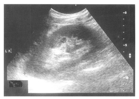
On admission, his blood pressure was 110/70 mmHg, his pulse rate 72/min and his body temperature was within the normal range. His tongue was slightly dehydrated. No neck vein engorgement was evident and heart sound and breathing sounds were normal. The abdomen was not distended and no abdominal mass was detected. Skin turgor was slightly reduced., the prostate was not enlarged and bladder catheterization did not make urine. The chest X-ray was normal and the abdominal X-ray (KUB) was unremarkable with no urinary tract opacities. By ultrasonography, the right kidney was absent and the left kidney enlarged (13.0 cm in length), with prominent corticomedullary differentiation and minimally dilated lower calices (Figure 1). The ureter was not traced and the urinary bladder was empty. On complete blood counts, WBC was 6,300/mm3, hemoglobin 11.8 g/dL, hematocrit 33.7%, and platelet count 126,000/mm3. Blood chemistry showed the following: fasting sugar, 99 mg/dL; urea nitrogen, 107.3 mg/dL; serum creatinine, 16.5 mg/dL; sodium, 129 mEq/IL potassium, 5.6 mEq/IL chloride, 95 mEq/IL calcium, 8.4 mg/dL; phosphorous, 4.2 mg/dL; total protein, 5.1 g/dL; albumin, 3.0 g/dL; creatine phosphokinase, 135 IU/IL and lactate dehydrogenase, 374 IU/L.
Because he had fasting history for 1 month and had lost 8 kg of weight loss, and because he showed clinical signs of volume depletion, we initially suspected pre-renal azotemia due to volume depletion as a cause of acute renal failure. Thus, intravenous hydration and furosemide were prescribed. But, diuresis did not occur. We considered that pre-renal azotemia had progressed to acute tubular necrosis, and decided to start hemodialysis in order to manage the severe renal failure. Anuria continued for 5 days in spite of adequate hydration during hemodialysis. Doppler ultrasonography was performed to rule out diseases of the renal artery and vein, but it revealed no abnormality of the renal vessels, though it showed slight progression of the dilatation of the renal collecting system, compared with our previous study (Figure 2). Cystoscopy was done for retrograde pyelography and a 1 cm-sized impacted stone was found at the uretero-vesical junction. The stone was removed with foreign body forcep and the retrograde pyelography was not pursued. After the stone removal, a total of 4,800 mL of urine was drained within 24 hours. His renal function improved dramatically, with serum creatinine dropping to 2.7 mg/dL within 24 hours, and then to 1.0 mg/dL over the next 24 hours. Follow-up ultrasonography showed improved renal collecting system dilatation, though minimal dilatation persisted at a level similar to that observed in the initial study (Figure 3).
DISCUSSION
This case was one of minimally dilated obstructive nephropathy due to a ureter stone in a kidney donor. To our knowledge, this is the first report of minimal or no dilated obstructive nephropathy in a kidney donor. The renal failure was so severe (serum creatinine 16.9 mg/dL) that hemodialysis was started. KUB showed no urinary renal tract opacities. On admission, ultrasonography revealed only minimal dilatation of the collecting system. Therefore, we did not suspect obstructive nephropathy. On the 5th hospital day, follow-up ultrasonography showed a slight progression of this dilatation and we considered obstructive nephropathy to be a cause of the acute renal failure. Cystoscopy demonstrated an impacted stone at the uretero-vesical junction, and removal of the stone initiated dramatic diuresis with a rapid return of renal functions to normal by the third day.
The degree of dilatation in obstructive nephropathy partly depends upon the patient’s state of hydration3). Dehydrated patients with a mild degree of obstruction may show no signs of hydronephrosis. In contrast, well-hydrated patients may have signs of minimal hydronephrosis in the absence of obstruction. In this case, the patient was in a dehydrated state because he had lost 8 kg of body weight after fasting for 1 month. We believe that this dehydrated state may in part have contributed to the minimal dilatation of the collecting system.
We initially considered pre-renal azotemia as a cause of the acute renal failure. The patient had abstained from all food, except water and salt, for 30 days, and had clinical signs of dehydration and had lost 8 kg of body weight. Generally, the ratio of blood urea nitrogen to serum creatinine is usually over 20 in pre-renal azotemia4), but in our case the ratio was 6.5. We believed that this low ratio was related to the 1month long fast. Ultrasonography showed kidney enlargement with minimal dilatation of the collecting system. The ureter was not traced. We believed that the kidney enlargement could have resulted from compensatory growth due to unilateral nephrectomy, and that the minimal dilatation could have been a change secondary to kidney enlargement. Given the situation described aboves, the clinical significance of the minimal dilatation was overlooked and consequently diagnosis was delayed.
In patients with a solitary kidney, prompt diagnosis and management of obstructive nephropathy is very important, because obstructive nephropathy is a reversible condition and adequate treatment improves renal function; however, treatment delay may cause a permanent loss of renal function. Although ultrasonography is regarded as the gold standard for the diagnosis of obstructive nephropathy, ultrasonography cannot demonstrate obstructive nephropathy in cases showing minimal or no dilatation of the collecting system5–7). Therefore, in patients at risk of obstructive nephropathy, more extensive and invasive evaluation such as retrograde or antegrade pyelography are necessary, despite the absence of a sonographically demonstrable dilatation. In this case, cystoscopy was undertaken for retrograde pyelography, and demonstrated a small impacted stone at the uretero-pelvic junction, which was removed with a foreign body forcep. Thus the retrograde pyelography was not performed.
In conclusion, obstructive nephropathy can occur with minimal or no dilatation of the collecting system in patients with a solitary kidney, especially in situations of dehydration, which might reduce dilatation. If obstructive nephropathy is highly suggested, extensive evaluation such as, retrograde or antegrade pyelography will be necessary despite the absence of a sonographically demonstrable dilatation. These diagnostic procedures enabled us to find and eliminate the cause of this patient’s renal failure.


 PDF Links
 PubReader
 ePub Link
 Full text via DOI
 Download Citation
 Print
Print


