INTRODUCTION
Thyroid nodule is a common disorder whose prevalence on clinical evaluation varies from 1ŌĆō7% according to iodine intake. However, thyroid cancer represents just 1ŌĆō2% of all malignancies, and only 5ŌĆō24% of thyroid nodules treated surgically are malignant. Fine-needle aspiration biopsy (FNAB) is the first line tool for the evaluation of thyroid nodules. However, misdiagnoses may occur, due to either an insufficiency or the unsuitability of the aspirated material for cytologic evaluation or to a sampling mistake. Furthermore, the main limitation of FNAB is its lack of sensitivity in the evaluation of follicular neoplasms, due to its inability to differentiate follicular adenoma from follicular carcinoma. If a more reliable marker for the presence of thyroid cancer were available for preoperative evaluation, unnecessary thyroid surgeries could be avoided.
Cyclooxygenase (COX) catalyzes the formation of prostaglandins from arachidonic acid1). Recent animal studies suggest that prostaglandins play a key role in tumor progression via their influence on angiogenesis, tumor growth, and metastasis2ŌĆō4). At least two COX enzymes are present in humans: COX-1 and COX-2. COX-1 is known as a housekeeping gene and is constitutively expressed in most tissues. In contrast, COX-2 is an early response gene and is induced by various factors, such as growth factors, oncogenes, cytokines, and carcinogens. Moreover, COX-2 is up-regulated in numerous neoplasms, including colorectal5), pancreatic6), prostate7), esophageal8), lung9), gastric10), and skin carcinoma11).
Recently, Specht et al.12) reported that the COX-2 mRNA level is higher in malignant thyroid nodules than in adjacent normal tissue or in benign thyroid nodules. Thus, we investigated COX-2 expression in various thyroid specimens in order to elucidate its physiological role in pathologic conditions, and to evaluate the efficiency of COX-2 protein expression as a molecular marker of malignancy in the thyroid gland, and thus its potential usefulness in the differentiation of benign and malignant tumors.
MATERIALS AND METHODS
1) Specimens
Sixty-four paraffin-embedded thyroid specimens were obtained from surgical resections performed at Cheonan Hospital, Soon chun hyang University, Cheonan, Korea. These consisted of 19 papillary carcinomas, 8 follicular carcinomas, 14 follicular adenomas, 2 H├╝rthle cell carcinomas, 4 H├╝rthle cell adenomas, 8 nodular hyperplasias, 3 GravesŌĆÖ diseases, 3 HashimotoŌĆÖs thyroiditis, 2 medullary carcinomas, 1 anaplastic carcinoma, and 20 normal thyroid tissues. Normal thyroid tissue samples were taken from histologically normal areas adjacent to neoplastic lesions.
2) Immunohistochemistry
Immunohistochemical analysis of COX-2 was performed using established protocols. Briefly, paraffin-embedded tissue was cut into 5 m section and dried for 1h at 57┬░C in an oven. After routine deparaffination and rehydration, tissue sections were microwaved for 20 min in 0.01 M sodium citrate buffer (pH 6.0). Endogenous peroxidase activity was blocked with 3% H2O2 in methanol for 30 min, and sections were then incubated with rabbit polyclonal antibody against human COX-2 (Cayman Chemical, Ann Arbor, MI) at a dilution of 1:10 for 60 min at room temperature. All slides were then washed 3 times for 3 min each with phosphate-buffered saline (PBS). Samples were incubated with PicTure-plus bulk kit (Zymed Lab, San Francisco, CA), i.e., ZymedŌĆÖs HRP polymer detection system, for 20 min at room temperature, washed and incubated with Liquid DAB substrate kit (Zymed Lab, San Francisco, CA) for 5 min, and then counterstained with MeyerŌĆÖs hematoxylin for 5 min and mounted. For negative controls, incubation with the primary antibody was omitted. Staining was scored as follows: (ŌłÆ) = absent, (+/ŌłÆ) = weak or focal staining, and (+) = moderate to strong staining in all or most epithelial cells.
RESULTS
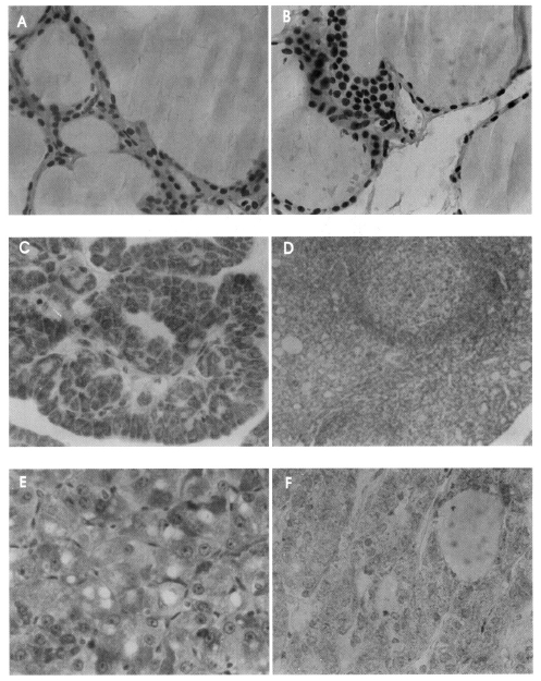
Table 1 summarizes the results obtained for COX-2 immunostaining in the thyroid tissues of the 64 patients. COX-2, when expressed, was found in the cytoplasm of follicular cells. COX-2 staining was not seen in any of the 20 normal thyroid (Figure 1A), 3 GravesŌĆÖ disease, or 8 nodular hyperplasia (Figure 1B) specimens. In contrast, COX-2 staining was observed in all of 19 papillary carcinomas (Figure 1C), 3 HashimotoŌĆÖs thyroiditis (Figure 1D), 2 H├╝rthle cell carcinomas, and 4 H├╝rthle cell adenomas, and in 7 of 8 follicular carcinomas (Figure 1E) and 11 of 14 follicular adenomas (Figure 1F). One of 2 medullary carcinomas showed diffuse COX-2 staining, but the another showed focal staining. One anaplastic carcinoma showed weak COX-2 positivity.
DISCUSSION
In contrast to the findings of Smith et al13), we did not observe COX-2 expression in the normal thyroid epithelium. This result was in accordance with reports by Cornetta et al.14) and Specht et al.12) Moreover, in this study, we found increased COX-2 expression in HashimotoŌĆÖs thyroiditis, follicular adenoma and in well-differentiated thyroid cancer. Chronic inflammation may enhance carcinogenesis and aid neoplastic growth and progression by promoting genomic instability15). For example, ulcerative colitis has been shown to carry an increased risk of colon cancer development16). In addition, patients with HashimotoŌĆÖs thyroiditis have a 10 to 40% increased risk of developing papillary thyroid carcinoma17,18). The association of lymphocytic infiltrates with papillary thyroid carcinomas and HashimotoŌĆÖs thyroiditis suggest that inflammatory conditions within the thyroid may promote the development of thyroid cancer.
The incidence of COX-2 over-expression in follicular carcinoma did not significantly differ from that of follicular adenoma, which indicates that COX-2 is not useful as a marker of malignancy. Since COX-2 expression was evident in follicular adenomas, and papillary and follicular carcinomas, this enzyme may be involved in the early processes of thyroid tumorigenesis.
The cyclooxygenase pathway has been implicated as a mediator of inflammation and cellular growth in the thyroid. Berg et al.19) demonstrated that IL-1 and TNF, which are two well-known proinflammatory mediators of thyroiditis, induced COX-2 expression in thyroid epithelial cells. Based on immunohistochemistry, immunoblot, and Northern analysis results, Smith et al.13) reported that COX-2 is constitutively expressed in multinodular goiter, GravesŌĆÖ disease and in papillary carcinoma. In contrast, Specht et al.12) reported that the levels of COX-2 mRNA and protein are elevated in human thyroid cancer as compared with adjacent normal tissue. In particular, they found no expression of COX-2 by immunohistochemistry, in contrast to SmithŌĆÖs result. Nose et al.20) reported that stepwise increments in the over-expression of COX-2 were shown by epithelial cells of HashimotoŌĆÖs thyroiditis, follicular adenoma, papillary carcinoma, and follicular carcinoma. Cornetta et al.14) also reported that COX-2 expression was observed in HashimotoŌĆÖs thyroiditis, and in papillary and follicular carcinoma, but not in normal thyroid tissue, multinodular goiter, or in anaplastic carcinoma. Recently Ito et al.21) reported that positive COX-2 staining was only observed occasionally in normal follicles or stromal cells, and that COX-2 over-expression was found in only 20% of follicular adenomas, 41% of follicular carcinomas, and in 81% of papillary carcinomas.
In our study, COX-2 was expressed in all 19 papillary carcinomas, 7 of 8 follicular carcinomas, and 11 of 14 follicular adenomas, which is a higher rate of positivity expression than found by Ito et al21). But, Cornetta et al.14) reported that 4 of 5 papillary carcinomas and both 2 follicular carcinomas were COX-2 positive, which is similar to our result.
Prostaglandins, the products of COX-2 activity, have been implicated in carcinogenesis via multiple mechanisms, such as by promoting angiogenesis22), inhibiting apoptosis23), increasing malignant cell invasion24), stimulating cell proliferation25), and inhibiting immune surveillance2).
Moreover, there is evidence that nonsteroidal anti-inflammatory (NSAIDs) can lower the risk of developing certain epithelial cancers and inhibit carcinogenesis. Epidemiological studies have demonstrated a 50% reduction in the rate of mortality from colorectal cancer in patients taking NSAIDs26), and regular NSAIDs use can significantly reduce the risk of developing breast cancer by approximately 50%27). In addition, the risk of certain esophageal and gastric cancers is significantly reduced by the regular administration of NSAIDs28).
No epidemiologic study has found that NSAIDs or selective COX-2 inhibitors can reduce thyroid tumor development or are effective in the treatment of thyroid tumor. But, based on our findings of an increased expression of COX-2 in thyroid tumor, we expect that use of COX-2 may be effective in the prevention or treatment of thyroid nodules. Thus further study should be conducted to determine whether NSAIDs or selective COX-2 inhibitors lower the risk of developing thyroid nodule and/or the development of thyroid cancer.


 PDF Links
 PubReader
 ePub Link
 Full text via DOI
 Download Citation
 Print
Print


