INTRODUCTION
Hypersensitivity pneumonitis (HP), such as farmer’s lung, pigeon breeders disease and bagassosis, has been generally associated with inhalation of specific organic dusts. Sensitive individuals intermittently exposed to these materials develop acute symptoms of fever, malaise, cough, dyspnea and chest tightness within four to eight hours after exposure. The organic dusts are usually contaminated with thermophilic actinomyces or fungi1,2). A few cases of HP by a cool mist vaporizer (HPCMV) have previously been reported, but they failed to identify the causative organism3,4).
Recently we had an opportunity to evaluate a patient with HPCMV. A detailed immunologic and bacteriologic study was done to identify the causative agent.
CASE REPORT
A 25-year-old housewife was admitted to Yeungnam University Hospital in February of 1986 with cough and anterior chest tightness.
She was well until ten days prior to admission, when she developed symptoms of gastroenteritis. She was treated with unknown medicines by a local physician and stayed home. The gastrointestinal symptoms subsided in two days, but she noticed tightness in her chest, dyspnea, nonproductive cough, fever, chills and headaches. She was treated again for “acute bronchitis” with different medicines by the same physician. But the symptoms progressed over the next eight days and she was referred for pulmonary evaluation.
She was a garments salesperson, nonsmoker, and had no significant past medical history. Her family history was unremarkable and she had no known allergies. She denied recent travels, changes of home environment, and exposure to any pets, trees, or bird extracts.
On admission, she was afebrile, and her respiratory rate was 34/min. On auscultation of the chest, there were inspiratory rales on all lung fields, but no wheezing or friction rub. The peripheral blood hemoglobin was 14.5gm/dl, hematocrit 44, WBC 14,800 (poly 74%, lymphocyte 14%, eosinophil 7%), and ESR was 43mm/hr. The chest x-ray showed bilateral patchy infiltrates (Fig. 1), No organisms were found on sputum smears and cultures. After treatment with erythromycin, she had a prompt recovery and was discharged. At the time of discharge, she was in an asymptomatic condition and had normal vital signs. The chest was clear on auscultation. The peripheral leukocyte count and ESR were normal.
Twelve hours after discharge, she was brought back to the emergency room with the same symptoms of severe chest tightness, cough and dyspnea. She was apprehensive and tachypneic. Body temperature was 39°C, pulse rate 142/min and respiratory rate 36/min. Inspiratory rales were heard on all lung fields again, but no wheezing. The peripheral leukocyte count rose to 31,600 (poly 61%, band 35, lymph 4%). The arterial blood gases on room air were pH 7.50, PCO2 33.2 mmHg, and PO2 50.0 mmHg.
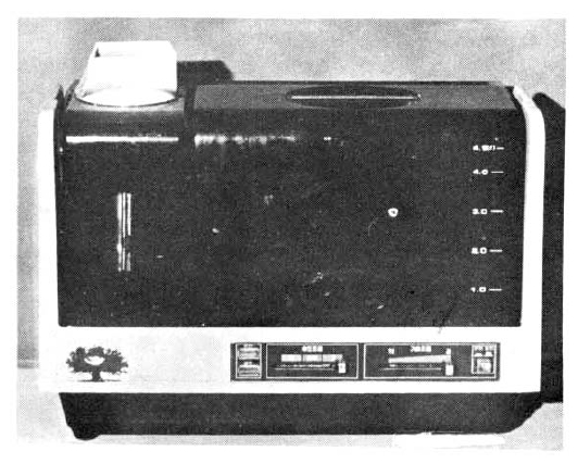
HP or other inhalational diseases were suspected. On careful interview, it was found that she had used a small cool-mist vaporizer (Fig. 2) every night since she suffered from the gastroenteritis and had used it again on the night of discharge from hospital. HPCMV was suspected. In the reservoir of the vaporizer, the water was slightly cloudy and some floating material in the water and sediment on the bottom were seen. She claimed that “fresh tap water” was used for the vaporizer and it was occasionally rinsed out with tap water, but no further efforts to clean the container were made.
She recovered without any specific therapy. Three days later when she became asymptomatic, spirometry was done and showed FVC 1.82L (56.2% of predicted), FEV1 1.64L (54.7% of predicted), and FEV1/FVC 90%, consistent with restrictive lung disease. On the fourth day, an inhalational challenge test was tried by keeping the vaporizer on to confirm the possibility of HPCMV, and nine hours after the vaporizer was on, she suddenly developed symptoms of HP. Body temperature rose to 39.6°C, pulse rate to 120/min and peak expiratory flow dropped from 260 to 230 L/min. The diagnosis of HP due to a contaminated cool-mist vaporizer was made. She was discharged from hospital and remained in a stable condition. A follow-up spirometric study was done three weeks later and showed normal FVC, FEV1 and FEV1/FVC ratio. The peak flow was 360L/min.
MYCOLOGIC AND BACTERIOLOGIC STUDIES
The smear of the floating materials and sediment in the reservoir showed molds, yeasts and gram negative bacilli which were also identified by cultures. Culture specimens were taken 1) from water in the reservoir, 2) from debris on mechanical parts and the mist-outlet and 3) by direct exposure of media to mist. All specimens were placed in 1) blood agar plate, 2) Sabouraud’s media and 3) brain heart infusion broth, and were incubated at room temperature, 37°C and 56°C.
SEROLOGIC STUDIES
Seventy ml of blood was obtained on the day of the second admission and the serum was stored below −70°C. Six harvested organisms were tested serologically with the stored serum by the 1) hemagglutination test, 2) double immuno-diffusion test and 3) counter immune electrophoresis test. Precipitation studies with additional antigen were not done because the antigens were not available.
SKIN TESTS
Skin tests with the extracts of the individual organisms were done. The extract was prepared from the cultured fungi and bacteria cultured and diluted on a 1:1000 wt/vol basis in saline.
INHALATIONAL CHALLENGE TESTS
An inhalational challenge test with the untouched contaminated vaporizer produced all the symptoms and signs of HP as above (Fig. 3). To identify the causative agent, inhalational challenges were done with 2ml of the following materials: 1) untreated fresh tap water at her house, 2) millipore-filtered vaporizer water and 3) phenol-killed suspension. Challenges with phenol-killed suspensions of other harvested organisms were planned but the patient refused. Each test was done on different days after confirmation of stable vital signs and an asymptomatic condition. Inhalation was done by nebulizer (Inhaleboy made in West Germany). Vital signs and peak flow were measured every 30 minutes up to 10 hours or until she developed symptoms of HP.
RESULTS
On microbiologic studies, the following six organisms were identified: a Pseudomonas species, a Candida sp., a Foncelea sp., a Fusarium sp., a Rodotorala sp. and an Aureobasidium sp., No thermophilic organisms or amoebas were identified. On serologic studies, her serum only reacted weakly to the harvested Pseudomonas species by hemagglutination test, but did not react to the other harvested organisms on all methods. All skin tests were negative.
Inhalational challenges with untreated fresh tap water and millipore-filtered vaporizer water failed to produce any symptoms and signs of HP. On inhalational challenge with a phenol-killed suspension of the Pseudomonas species, she developed coughing and mild irritation of the throat immediately after inhalation. Peak flow dropped from the 360L/min to 340L/min in 10 minutes and returned to 360L/min over the next 30 minute. Eight hours later, it dropped again to 240L/min (Fig. 3) and she became febrile, developed a cough and complained of chest tightness.
DISCUSSION
Hypersensitivity pneumonitis is a granulomatous lung disease caused by repeated exposure to various inhaled organic dusts or occupational agents. Numerous conditions causing HP have been reported since 1713 when the first case of HP was reported by Ramazzini, and many causative agents have been identified1,2,3,6). Thermophilic organisms are the main causative organisms of HP and they grow in warm and humid conditions and reach the human lung by inhalation.
Since humidifiers and air conditioners have been developed and widely used, they have become one of the common causes of HP. In HP caused by humidifiers (“humidifier fever”), the thermophilic organisms are also the main causative organisms. They grow in warm water and moisture in the filter or the air passway of the humidifiers, and are distributed by fans2). Different organisms such as amoebas and gram negative bacilli have been added to the list of the etiologic agents of HP caused by humidifiers as well as other types of HP2,5).
In our case, a gram negative bacilli, Pseudomonas species, was identified as the etiologic agent on the basis of a positive serologic test and inhalational challenge. It was unfortunate that inhalational challenges with other harvested organisms were refused. Even though it was not done, they are unlikely to be the cause of her illness because of negative serologic and skin tests. On inhalational challenge with the phenol-killed suspension of the isolated Pseudomonas species, there was an early drop of peak flow which was not seen on the initial challenge with the untouched vaporizer (Fig. 3), and it made this positive result uncertain as the definite cause of her HPCMV. The reason for this difference is still unclear, but might be a nonspecific reaction of bronchial irritation with the strong-smelling phenol added for sterilization. No thermophilic organisms were isolated.
In English literature, only a few cases of HP by cool-mist vaporizer (HPCMV) have been reported3,4,6). They failed to identify the causative agent and no thermophilic organisms were found. It could be expected because their vaporizer was not a heated system. Dr. Hodges did a careful study on a patient of HPCMV to identify the causative organism, and Gram-negative bacilli was clearly eliminated3). Therefore, the identified organism in our case, Pseudomonas species, seems not to be the sole cause of HPCMV. Many different organisms and materials could be expected to cause HPCMV, as with HP.
These small, inexpensive, cool-mist vaporizers have been distributed widely to many families, offices and even hospital rooms in the last few years. All users should be aware that these can cause HP and should try to keep them clean. Physicians should give HPCMV a high index of suspicion where these vaporizers are being used and in any patient presenting with symptoms of HP to ensure early diagnosis and prevent its sequelae.
In conclusion, we are presenting the first case of HP in Korea, and on the basis of the above results, we concluded that the cause of HP in this patient was due to a cool-mist vaporizer and Gram-negative organisms of Pseudomonas species were the etiologic organism.


 PDF Links
 PubReader
 ePub Link
 Full text via DOI
 Download Citation
 Print
Print


