INTRODUCTION
Swallowed foreign objects are a common problem1). Many objects spontaneously pass through the gastrointestinal tract without causing symptoms. The potential dangers of impaction, obstruction, and perforation may requrie a physicion to remove the foreign body2). Through technical improvements in flexible fiberoptic endoscopes and their accessories, it has become apparent that the fiberendoscopic management of gastrointestinal foreign bodies is the procedure of choice when removal of the objects is indicated3ŌĆō5).
We now report on the endoscopic retrieval of a wire perforating the stomch wall.
REPORT OF CASE
A 50 year old female was admitted to the Wonkwang University Hospital. The patient complained of an epigastic pain of one month duration.
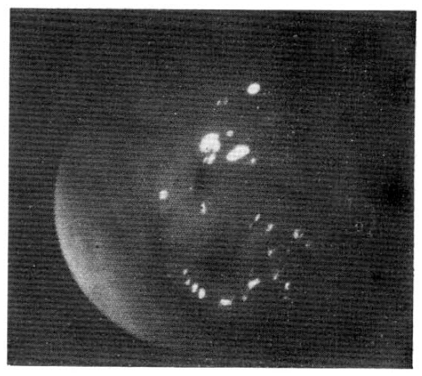
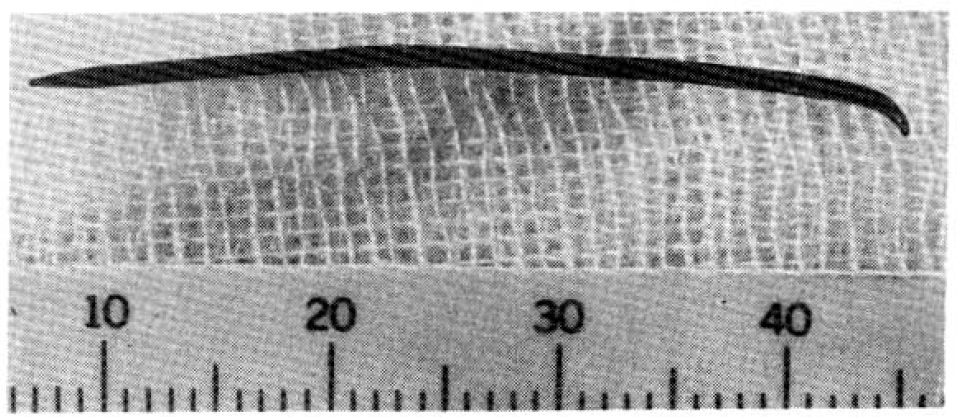
She had no other significant symptoms, and her past history was negative. Physical examination revealed only mild tenderness of the epigastrium but was otherwise normal. Routine laboratory data was also within normal limits. Gastroscopy showed a thin wire in the prepyloric region sticking into the gastric mucosa (Fig. 1), The visible part of the foreign body was caught with biopsy forceps and was extractd together with the endoscope. The extracted wire was pointed at both ends (Fig. 2). There remained superficial ulcer of 5 ├Ś 7 mm diameter. Neverthless, no clinical symptoms of perforaton or peritoneal irritation were prsent.
The patient had an unevenful recovery for 2 weeks and is now free of gastrointestinal symptoms.
DISCUSSION
To our Knowedge, the perforation of the stomach wall by a wire has rarely been reported. Sartory and Trabant6) escribed a patient where a perforating paperclip was endoscopically removed without complications. Its length was 2.8 cm.
The Ingestion of foreign bodies can produce difficult diagnostic problems when the patient is unaware of the ingestion. Complications such as asphixation or perforation occur in about 1% of the cases, with perforation sometimes causing infection, hemorrhage, or fistulization7).
Most probably the perforation made by the wire in our patient had occurred one month previously, when the epigastric pain started. The patient did not remember that she had swallowed any foreign bodies. The Endoscopic extraction of a perforating foreign body apparently can be done without any danger of peritonitis either because the foregin body may have been sterilized by contact with hydrochloric acid or because the perforation occurs in adjacent organs like the pancreas6).
Foreign bodies with sharp edges or points present a special problem to the endoscopist. Rogers and his coworkers8) reported using the endoscopic overtube so that it was advanced from its initial position on the distal end of the endoscope to envelope a guitar pick lodged in the esophagus. The whole assembly was then withdrawn with the foreign body safely inside the overtube. One disadvantage of the overtube is that it covers the markings on the endocope so that the total length of the insertion is not readily apparent.
Also, the overtube interferes with the torquing of the endoscope, which causes scanning more difficult. These disadvantages are more noticeable when used in the stomach. Therefore, diagnostic gastroscopy should be performed prior to the utilization of the overtube when a foreign body is in the stomach. For esophageal foreign bodies it is advantageous to use the overtube with the endoscope together9).
Removal of hard, smooth, foreign bodies with the biopsy forceps, as accomplished by Gelzay and Jetly10), or with an intragastric magnet as by Kleckner11) is inherently hazardous. Hard surfaced metallic objects can not be firmly gripped with forceps and magnets, in addition to them being limited to the extraction of metallic objects they also restrict vision through the endoscope. Direct visualization of the objects permits the careful timing of the withdrawal procedure to coincide with the interperistaltic periods so as to limit pin-mucosa contact, time, and pressure.
With the development of the snare cautery technique for the removal of polyps through an endoscope, there has been a concurrent application of this instrument to the removed foreign bodies from the upper gastrointestinal tract. The first reported success was by DeGerome12) who removed a hair pin by this technique. Subsequently, a paper given by Schiller and Salmon13) to the British Endoscopy Society indicated multiple foreign body removals. In our experience, this method appears to be a safe, rapid, and successful way to remove foreign bodies. We do not consider the snare to be the ultimate foreign body extractor but its success should be an impetus to design further instrumentation for the removal of all objects of varing sizes and shapes through our fiberotic instruments.
In the case of penetrating or perforating items the use of endoscopic removal seems justified when easy access if possible and the foreign body can be tightly grasped. A close follow-up of the patientŌĆÖs condition for the following 24 hours is advisable.


 PDF Links
 PubReader
 ePub Link
 Full text via DOI
 Download Citation
 Print
Print


