Korean J Intern Med > Volume 2(2); 1987 > Article
Kim, Moon, Lee, Moon, Kim, and Kim: Clonogenic Assay of Gastric Adenocarcinoma Stem Cells

Abstract

Clonogenic assay is the culturing of tumor stem cells, which are responsible for tumor growth by self renewal and differentiation to tumor end cells. Elimination of the malignant stem cells or their self renewal will lead cure of the tumor. Thus, the behavior of the stem cells by clonogenic assay has been correlated with prognosis and outcome of therapy.
We studied clonogenic assay by means of double agar system for 21 patients with advanced stomach cancer. The colony formation evaluted on 14th day of the culture was grown from 14 of 19 malignant effusions plated and 1 of 2 tumor nodules plated. The number of the colonies ranged from 5 to 96 per petri dish, and the median number was 20. The plating efficiency ranged from 0.001 to 0.036%, and the median was 0.003%. In the morphologic studies, the colonies made of tightly packed cells were grown from the poorly differentiated adenocarcinoma, whereas the colonies made of loosely packed cells with mucin formation were grown from the mucin secreting adenocarcinoma of stomach. The chemosensitivity in vitro tests to cisplatin were performed in 5 patients, and the results showed 4 sensitive and 1 resistant patient. The result of in vivo study with instillation of intraperitoneal cisplatin revealed that 3 patients “in vitro sensitive” were “responsive in vivo”, and 1 patient “in vitro resistant” was “non-responsive in vivo”.
We conclude that clonogenic assay of stomach cancer is useful method to understand the biology of the stem cell pool and select proper chemotherapy according to the chemosensitivity test.

INTRODUCTION

Adenocarcinoma of the stomach is the most common malignancy in Korea1). Although patients with early localized disease are often curable by surgery, the majority of patients present advanced disease with metastasis which is not curable and the prognosis is very poor. Until now no form of cytotoxic chemotherapy for these advanced diseases has made a significant improvement of response rate and survival2,3). The poor prognosis and the low response rate to chemotherapy provide considerable impetus to search for more effective forms of chemotherapy. Advances in the chemotherapy of stomach cancer appear likely to come either from the new discovery of drugs and combinations of drugs more active or from developing methods for the selection of specific forms of chemotherapy for individual patients based on the drug sensitivity of their tumors.
The human tumor stem cell clonogenic assay developed by Salmon and Hamburger4) provides good methods not only for in vitro chemosensitivity tests but for studying tumor biology. Several investigators have reported results suggesting a good correlation between in vitro drug sensitivity measured by clonogenic assay and the in vitro responsiveness5).
Since 1984, the authors have studied clonogenic assay of human tumor stem cell using a double agar method of Salmon and Hamburger in an attempt to applicate this assay to a variety of clinical situations6). In this paper, the results of clonogenic assay including morphologic study of colonies and in vitro chemosensitivity tests in stomach cancer are presented. This information should provide a basis for future clinical application of the clonogenic assay, especially for clinical trial designs.

MATERIALS AND METHODS

1. Collection of Tumor Specimens

In a 22-month period from October 1984 to July 1986, the authors tried in vitro clonogenic assay on 21 specimens from advanced stomach cancer patients. Nineteen were from malignant effusions (ascites 17, pleural effusions 2) and 2 from tumor nodules.

2. Preparation of Tumor Single Cell Suspension

Malignant effusions were collected in heparinized bottles (50 μ/ml). After centrifugation at 1,500 rpm for 10 min, the cells were harvested and washed twice in McCoy’s wash (McCoy’s 5A Medium containing 10% heat-inactivated fetal calf serum and penicillin/streptomycin/fungizone solution). Tumor cells were collected by centrifugation over a Ficoll-Conray (specific gravity 1,080) gradient at 1,850 rpm for 45 min, then washed twice with McCoy’s wash.
Tumor nodules obtained immediately after surgery were mechanically minced. They were minced with a iris scissor in McCoy’s wash and then washed by centrifugation at 1,000 rpm for 5 min. Cells thus freed were separated from the tissue fragments by pouring them twice over a 120 stainless steel mesh screen. Remaining tissues were gently returned to the petri dish and the mincing procedure was repeated until cell release was no longer significant.
The viability of single cell suspension determined by a trypan blue dye exclusion method ranged from 45 to 95% (mean 83±15%).

3. Culture Assay for Tumor Colony-Forming Cells

The cells were cultured according to the technique of Salmon and Hamburger6,7). Briefly, the tumor cells collected were incorporated into 0.3% agar with double enriched CMRL 1066 medium to yield a final viable cell number of 5×10⁵/ml. One ml aliquots of the agar-cell mixture (top layer) were pipetted onto 35 mm plastic petri dishes previously prepared with an under layer which contained 0.5% agar with double enriched McCoy’s 5A medium.
After preparation of both the under and top layers, the dishes were examined with inverted microscope to assure the presence of a good single cell suspension and the even distribution. The dishes were incubated at 37°C in a fully humidified chamber of 7.5% CO2 for 2 weeks.
After incubation for 14 days, the number of colonies on the triplicate dishes was counted with an inverted microscope.
Clusters were considered aggragates of less than 30 cells. Aggregates of 30 or more cells were considered colonies. We defined cultures as positive growth when at least 5 tumor colonies grew in a dish. The plating efficiency (PE) was calculated as follows:
PE(%)=NumberofcolonyperdishNumberofviabletumorcellsplated×100

4. In Vitro Chemosensitivity and in Vivo Correlation

Stock solutions of intravenous formulations of 5-fluorouracil, adriamycin and cisplatin were prepared in sterile buffered saline and stored at −70°C in aliquots sufficient for individual assays. Subsequent dilutions were made in medium for cell incubation. Tumor cell suspensions were transferred to tubes and adjusted to a final number of 5×10⁵ cells/ml in the presence of the appropriate drug dilution or a control medium. Each drug was tested at a minimum of three dose levels, including pharmacologically achievable concentrations. Cells were incubated with drugs in McCoy’s medium for one hour at 37°C.
The cells were then centrifuged at 1,500 rpm for 5 min, washed twice in medium, and prepared for culture. The number of colonies on control plates and drug-treated plates was counted. At least 30 tumor colonies per plate were required in the control plates to assure an adequate range for measurement of drug effect. We defined drug sensitivity of a given tumor as a more than 50% reduction of colony formation (less than 50% survival) in the presence of the drug compared to colony formation in the absence of the drug. Four patients with malignant ascites were treated with cisplatin intraperitoneally8). In vivo response was defined as clearing of the effusion without reaccumulation of fluid accompanied with disappearance of tumor cells in ascitic fluid.

5. Morphologic Studies of Tumor Colonies

Serial examination with an inverted microscope was performed to observe the process of cell division and colony formation. After 14 days, we fixed wet agar cultures with a 3% solution of glutaraldehyde in Hank’s balanced salt solution and placed them in a humidified plastic box and stored in the cold room for the morphologic study.
Permanent dry slides were prepared from top plating layer separated9). The plating layer then was poured gently onto a microscope slide and a prewetted cellulose acetate membrane was carefully placed on top of the wet agar. Once the slide dried, the cellulose acetate strip was removed. The dried slide was stained with Papanicolaou method.

RESULTS

  1. The overall growth rate was 71.4% (15 of 21). The growth rates of tumor cells obtained from ascites, pleural effusions and tumor nodules were 70.5% (12 of 17), 100% (2) and 50% (1 of 2) respectively (Table 1, 2).

  2. The number of the colonies ranged from 5 to 96 per petri dish, and the median was 20. The PE ranged from 0.001 to 0.036%, and the median was 0.003% (Table 1, 2).

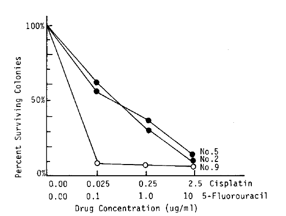
  3. The in vitro chemosensitivity tests were performed in 5 patients, and the result showed 4 sensitive and 1 resistant to cisplatin (Table 3). In 3 cases sensitive to cisplatin, dose-related responses were seen (Fig. 1). The result of trial with intraperitoneal instillation of cisplatin revealed that 3 patients in vitro sensitive were responsive in vivo, and 1 patient in vitro resistant was non-responsive in vivo.

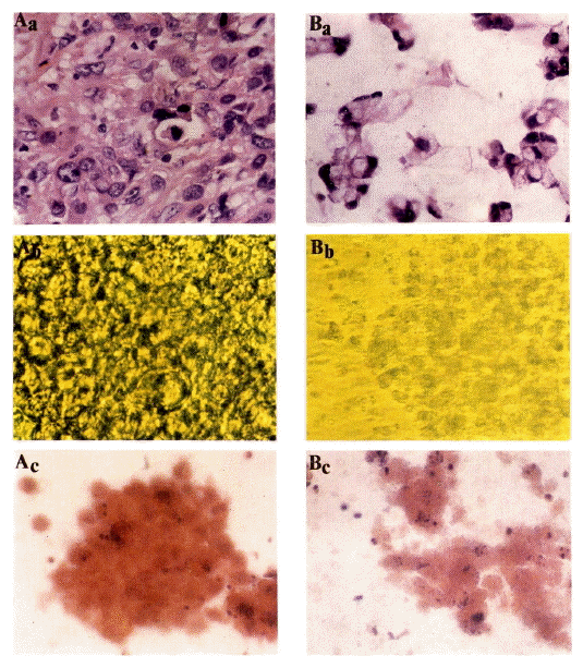
  4. In morphologic studies, the colonies made of tightly packed cells were grown from the pooly differentiated adenocarcinoma whereas the colonies made of loosely packed cells with mucin formation were grown from the mucin secreting adenocarcinoma (Fig. 2).

DISCUSSION

This study has shown that gastric adenocarcinoma can be grown successfully in a human tumor clonogenic assay using a double agar method. The overall growth rate was 71.4% including 14 of 19 (73.7%) malignant effusions and 1 of 2 tumor specimens. This data is similar to the previously published results by several investigators. Von Hoff et al.5,10) reported the growth rate as 70% in a rather large variety of tumor types and noted that the solid tumor and lymph node specimens were more difficult to grow than malignant effusions because of technical problems in maintaining viability while making single cell suspensions.
The median number of colonies formed from stomach cancer specimens was 20 (ranged from 5 to 96) and the PE was 0.001 ∼ 0.036% (median 0.003%). Von Hoff et al.10) showed the PE in neuroblastoma, ovarian cancer, breast cancer, melanoma and colorectal cancer as 0.01%, 0.01%, 0.01%, 0.006% and 0.005% respectively. Laboisse et al.11) reported the PE in stomach cancer as ranged from 0.006 to 0.82%. Therefore, the PE varied considerably both in terms of median PE from different tumor type and in terms of range of PE within a same tumor type. Also, approximately 70% of all tumors plated in the stem cell assay exhibit colony formation, but in over one half of the cases, cloning efficiency is too low to permit further detailed studies12). The low PE may reflect not only a small cell population in a tumor and a large number of non-tumor cells in the inoculum but also an inherent feature of spontaneous human tumor. It is believed that less than 0.1% of tumor cells are usually clonogenic. The variation within a type of tumor could reflect differences in stage of tumor, time since prior treatment, site of tumor sampled, type of specimens sampled and method preparing tumor specimens. The PE will likely be improved somewhat with better techniques for cell disaggregation, adjustment in medium components, and/or addition of appropriate growth factors and hormones. Increasing the number of nucleated cells per plate did not usually increase the number of colonies10).
The authors experienced that it is not easy to obtain sterile single cell suspensions from stomach cancer tissue enough to study clonogenic assay. Because the tumor specimens were usually contaminated and frequently too necrotic to get enough cells. The viability of cells from tumor specimens was lower than from malignant effusions. Also in the case of mucinous adenocarcinoma, it is very diffcult to disaggregate mechanically due to the presence of mucin (data not shown). The method of clonogenic assay in stomach cancer will be improved with enzymatic disaggregation and careful decontamination.
Patterns of chemosensitivity obtained for 5 cases are shown in Table 3. All but one patient (case No. 8) had prior chemotherapy (5-fluorouracil, adriamycin and mitomycin). Using the authors’ criterion, 4 of 5 tumors were sensitive in vivo to cisplatin and one was resistant. For the in vivo study, 4 patients received cisplatin intraperitoneally to control malignant ascites. The results of in vivo and in vitro study showed that 3 patients responsive to intraperitoneal cisplatin were sensitive to cisplatin in vitro. Whereas, 1 patient non-responsive to intraperitoneal cisplatin was resistant to cisplatin in vitro. Generally speaking, the in vitro chemosensitivity tests using clonogenic assay is more valuable in predicting which drugs would not be useful clinically (in vitro resistant and in vivo non-responsive) than in predicting which drugs would be useful clinically (in vitro sensitive and in vivo responsive)13,14). Overall, the true negative rate is about 90% while true positive rate is ranged from 60 to 70%.
Cultures were examined with an inverted microscope to observe the process of cell division and colony formation. In general, 2 cells, 4 cells and 8 cells stage were observed on the 3rd, 6th and 7th day after plating respectively. Clusters appeared on the 8th day and colonies began to appear from the 10th day after plating. Colonies consisted of 30 to several hundred large round cells (data not shown). We prepared permanent slide stained with Papanicolaou method from a wet agar for the morphologic study. The morphology of human stomach cancer colonies was similar to stomach cancer as identified in the original specimens (Fig. 2).
In this study, the authors could grow colonies from stomach cancer tissues and observed good correlation between in vitro chemosensitivity tests and in vivo results. However, the PE was low and the number of colonies was not enough to determine the chemosensitivity in all patients. By improving the assay techniques, we could applicate this clonogenic assay to a variety of clinical research including further chemosensitivity test, morphologic study with electron microscope and chromosomal analysis.

Notes

This Work was supported by Clinical Research Funds of Catholic Medical Center in 1987.

Fig. 1.
In vitro chemosensitivity of case No. 2 and No. 5 to cisplatin (●) and case No. 9 to 5-fluorouracil (○).
kjim-2-2-163-6f1.gif
Fig. 2.
A. Poorly differentiated adenocarcinoma.
a. Original histology, HE stain ×400.
b. Colony characterized by numerous tightly packed cells ×400.
c. Papanicolaou stain of colony after permanent fixation ×400.
B. Mucin secreting adenocarcinoma.
a. Original histology, HE stain ×400.
b. Colony made of loosly packed cells with mucin formation ×200.
c. Papanicolaou stain of the colony ×400.
kjim-2-2-163-6f2.gif
Table 1.
Characteristics of Patients with Stomach Cancer and Colony Formation in a Double Agar System
Case No. Age/Sex Histology* Specimen Viability (%) No. of colony Plating efficiency (%)
1 43/M AWD Ascites 95 5 0.001
2 66/M AWD Ascites 95 45 ± 9 0.010
3 70/M AMUCIN Ascites 53 96 ± 20 0.036
4 50/M AMD Ascites 93 5 0.001
5 54/M APD Ascites 95 59 ± 20 0.012
6 63/M APD Ascites 83 16 + 4 0.003
7 40/F APD Ascites 80 5 0.001
8 35/F APD Ascites 79 47 ± 12 0.012
9 56/F APD Ascites 87 38 ± 9 0.009
10 62/F APD Ascites 95 10 0.002
11 49/M AWD Ascites 95 23 ± 22 0.005
12 59/F APD Ascites 80 7 0.001
13 29/F AMD Ascites 70 0 0.
14 74/M APD Ascites 95 0 0.
15 59/F APD Ascites 75 0 0.
16 70/M APD Ascites 89 0 0.
17 43/M APD Ascites 88 0 0.
18 40/F APD Pleural effusion 80 5 0.001
19 23/M APD Pleural effusion 90 6 ± 3 0.001
20 66/M APD Mass 45 33 ± 7 0.007
21 70/F APD Mass 83 0 0.

Mean viability : 83.0 ± 15.3%
Median No. of colony/dish (range) : 20 (5 – 96)
Median plating efficiency (range) : 0.003% (0.001 – 0.036)

* AWD  : Adenocarcinoma, well differentiated

AMD  : Adenocarcinoma, moderately differentiated

APD  : Adenocarcinoma, poorly differentiated

AMUCIN  : Mucin secreting adenocarcinoma

Table 2.
Growth of Tumor Cells by Source of Specimen
Specimen Growth rate
Ascites 12/17 (70.5%)
Pleural effusion 2/2 (100.0%)
Tumor nodule 1/2 (50.0%)

Total 15/21 (71.4%)
Table 3.
Correlation of in vitro and in vivo Sensitivity of Stomach Cancer to Chemotherapy
Case No. Previous chemotherapy In vitro* In vivo*

5-FU Adriamycin Cisplatin Cisplatin
2 FAM S S S S
3 FAM ND ND R R
5 FAM ND ND S S
8 None ND ND S S
9 FAM S ND S ND

* R : Resistant,

S : Sensitive,

ND : Not done

REFERENCES

1. Ministry of Health and Social Affairs. Three years’ report for cancer register programme in Republic of Korea (July 1, 1980∼June 30, 1983). J Kor Cancer Res Assoc 16:73. 1984.

2. MacDonald JS, Woolley PV, Smythe T, Ueno SV, Hoth D, Shein PS. 5-Fluorouracil, adriamycin, and mitomycin-C (FAM) combination chemotherapy in the treatment of advanced gastric cancer. Cancer 44:42. 1979.
crossref pmid
3. Moon H, Kim HK, Kim CC, Yoo JY, Lee KS, Park DH, Kim BS, Kim DJ. The efficacy of High-FAM in advanced gastric cancer. J Kor Cancer Res Assoc 16:272. 1984.

4. Hamburger AW, Salmon SE. Primary bioassy of human myeloma stem cells. J Clin Invest 60:846. 1977.
crossref pmid pmc
5. Von Hoff DD, Clark GM, Stogdil BJ. Prospective clinical trial of a human tumor cloning system. Cancer Res 43:749. 1983.
pmid
6. Kim HK, Lee KS, Kim BS, Kim DJ. Clonogenic assay of human tumor stem cell. Kor J Intern Med 29:470. 1984.

7. Salmon SE, Hamburger AW. Primary bioassay of human tumor stem cells. Science 197:401. 1978.

8. Chung JM, Moon H, Hong YS, Park DJ, Kim HK, Lee KS, Kim DJ, Hong DS. Intraperitoneal chemotherapy in patients with malignant ascites. Kor J Intern Med 32:821. 1987.

9. Salmon SE. Morphologic studies of tumor colony. In: Salmon SE, ed. Cloning of human tumor stem cells. 135. New York: Alan R Liss Inc, 1980.

10. Von Hoff DD, Harris GJ, Hohnson G, Glaubiger D. Initial experience with the human tumor stem cell assay system: Potential and Problems. In: Salmon SE, ed. Cloning of human tumor stem cells. 113. New York: Alan R Liss Inc, 1980.

11. Laboisse CL, Augeron C, Potet F. Growth and differentiation of human gastrointestinal adenocarcinoma stem cells in soft agarose. Cancer Res 41:310. 1980.

12. Salmon SE. Background and overview. In: Salmon SE, ed. Cloning of human tumor stem cells. 3. New York: Alan R Liss Inc, 1980.

13. Careny DD, Winkler CF. In vitro assay of chemotherapeutic sensitivity. In: DeVita VT, Hellman S, Rosenberg S, eds. Important advances in oncology. 78. Philadelphia: JB Lippincott company, 1985.
pmid
14. Salmon SE, Alberts DS, Meyskens FL, Durie BGM Jr, Jones SE, Soehnlen B, Young L, George Chen HS, Moon TE. Clinical correlations of in vitro drug sensitivity. In: Salmon SE, ed. Cloning of human tumor stem cells. 223. New York: Alan R Liss Inc, 1980.


Editorial Office
101-2501, Lotte Castle President, 109 Mapo-daero, Mapo-gu, Seoul 04146, Korea
Tel: +82-2-2271-6792    Fax: +82-2-790-0993    E-mail: kaim@kams.or.kr                

Copyright © 2024 by Korean Association of Internal Medicine.

Close layer
prev next