


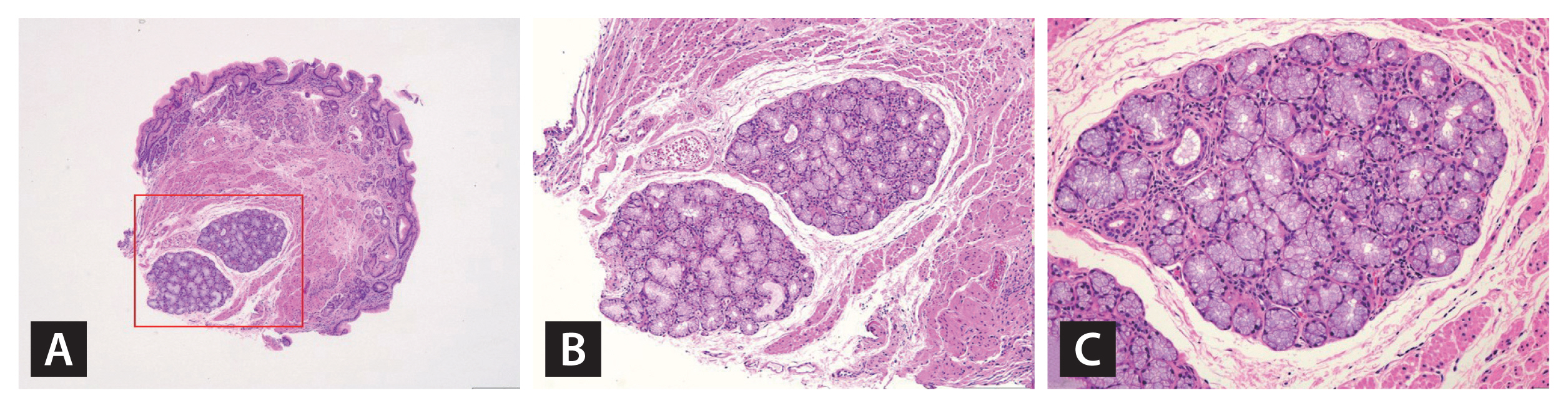
A chest computed tomography (CT) performed for detecting a solitary pulmonary nodule in a 31-year-old woman incidentally revealed distal esophageal wall thickening (Fig. 1A). The patient had no specific symptoms. Upper endoscopy was performed. Endoscopic examination revealed diffuse erythema and whitish nodules at the esophagogastric junction (EGJ), accompanied by mucosal breaks (Fig. 1B). Hiatal hernia was also observed (Fig. 1C). Biopsy of the lesion at the EGJ demonstrated salivary gland heterotopia (Fig. 2). Without any additional procedures, the patient is taking proton pump inhibitor and is being observed.
Esophageal wall thickening on CT can be observed not only in esophageal cancer but also in esophagitis, hiatal hernia, and esophageal motility disorders. Salivary gland heterotopia is most commonly reported in the head and neck. Salivary gland heterotopia of the gastrointestinal tract is rare, and the reported occurrence of salivary gland heterotopia in EGJ is extremely rare. It is unclear whether salivary gland heterotopia in EGJ is the result or cause of chronic reflux esophagitis. We report a case of endoscopic diagnosis of salivary gland heterotopia in the EGJ due to esophageal wall thickening discovered incidentally on chest CT. The patient provided informed consent for the publication of this report.

 PDF Links
 PubReader
 ePub Link
 Full text via DOI
 Download Citation
 Print
Print


