Korean J Intern Med > Volume 34(2); 2019 > Article
Sun, Jin, Jin, Lim, Chung, and Yang: The effects of addition of coenzyme Q10 to metformin on sirolimus-induced diabetes mellitus

Abstract

Background/Aims

This study was performed to determine whether adding coenzyme Q10 (CoQ10) to metformin (MET) has a beneficial effect as a treatment for sirolimus (SRL)-induced diabetes mellitus (DM).

Methods

DM was induced in rats by daily treatment with SRL (0.3 mg/kg, subcutaneous) for 28 days, and animals were treated with CoQ10 (20 mg/kg, oral) and MET (250 mg/kg, oral) alone or in combination for the latter 14 days of SRL treatment. The effects of CoQ10 and MET on SRL-induced DM were assessed with the intraperitoneal glucose tolerance test (IPGTT) and by determining plasma insulin concentration and the homeostatic model assessment of insulin resistance (HOMA-R) index. We also evaluated the effect of CoQ10 on pancreatic islet size, apoptosis, oxidative stress, and mitochondria morphology.

Results

IPGTT revealed overt DM in SRL-treated rats. The addition of CoQ10 to MET further improved hyperglycemia, decreased HOMA-R index, and increased plasma insulin concentration compared with the SRL group than MET alone therapy. While SRL treatment induced smaller islets with decreased insulin staining intensity, the combination of CoQ10 and MET significantly improved insulin staining intensity, which was accompanied by a reduction in oxidative stress and apoptosis. In addition, co-treatment of CoQ10 and MET significantly increased the levels of antiperoxidative enzymes in the pancreas islet cells compared with MET. At the subcellular level, addition of CoQ10 to MET improved the average mitochondrial area and insulin granule number.

Conclusions

Addition of CoQ10 to MET has a beneficial effect on SRL-induced DM compared to MET alone.

INTRODUCTION

Sirolimus (SRL) is a promising drug for replacing calcineurin inhibitors in solid organ transplantation; however, it has diabetogenic properties [1]. The mechanism of SRL-induced diabetes mellitus (DM) is still undetermined; however, impaired insulin signaling or direct pancreatic islet cell injury by SRL is associated with the pathogenesis of SRL-induced DM [2-5], and oxidative stress plays an important role in SRL-induced pancreatic islet injury [6].
According to the current guidelines for treating new onset diabetes after transplantation (NODAT), metformin (MET) is recommended as a first-line drug, and the addition of a second-line drug is suggested to achieve better control of hyperglycemia [7]. Coenzyme Q10 (CoQ10), which is a lipid-soluble molecule derived mainly from endogenous synthesis, has glycemic control properties [8,9] and acts as an antioxidant by functioning as an electron carrier in mitochondrial oxidative phosphorylation [10,11]. Therefore, CoQ10 may have beneficial effects on controlling hyperglycemia and decreasing oxidative stress in SRL-induced DM.
In the present study, we investigated whether the addition of CoQ10 to MET has a beneficial effect on SRL-induced DM in an experimental rat model. First, we evaluated whether the addition of CoQ10 to MET exerts better control of hyperglycemia in an experimental model of SRL-induced DM. Second, we investigated the antioxidant effects of CoQ10 on SRL-induced pancreatic islet injury. Third, we evaluated the effects of CoQ10 on SRL-induced mitochondrial injury using electron microscopy.

METHODS

Animals and drugs

All the animal experiments in this study were performed in accordance with the Guidelines for the Care and Use of Laboratory Animals, Catholic University of Korea (CUMC-2015-0028-02), and all procedures in this study were performed in accordance with ethical guidelines for animal studies. Eight-week-old male Sprague Dawley rats (Charles River Technology, Seoul, Korea) that initially weighed 220 to 230 g were housed in cages (Nalge Co., Rochester, NY, USA) in a temperature- and light-controlled environment at The Catholic University of Korea’s animal care facility. The rats received a low-salt diet (0.05% sodium, Teklad Premier, Madison, WI, USA). SRL (Wyeth-Ayerst Research, Princeton, NJ, USA) was diluted in Tween 80 (10%), N, N-dimethylacetamide (20%), and polyethylene glycol 400 (70%) to a final concentration of 0.3 mg/kg. CoQ10 was kindly supplied by LG Life Sciences (Seoul, Korea) and was diluted in drinking water to a final concentration of 5 mg/kg. MET (Dongwha Pharmaceutical Co., Seoul, Korea) was diluted in drinking water to a final concentration of 250 mg/kg.

Experiments and designs

The primary goals of this study were to determine whether CoQ10 is helpful to treat SRL-induced pancreatic injury and whether concomitant administration of CoQ10 and MET is superior to MET monotherapy for SRL-induced pancreatic injury.
  • Rats were randomized into five groups, nine animals per group, as follows:

  • (1) Vehicle (VH) group: rats received a daily subcutaneous injection of olive oil (1 mg/kg/day) and oral administration of sterile water for 4 weeks.

  • (2) SRL group: rats received a daily subcutaneous injection of SRL (0.3 mg/kg/day).

  • (3) SRL + CoQ10 group: rats received a daily subcutaneous injection of SRL (0.3 mg/kg) for 4 weeks and oral administration of CoQ10 (20 mg/kg) for the latter 2 weeks.

  • (4) SRL + MET group: rats received a daily subcutaneous injection of SRL (0.3 mg/kg) for 4 weeks and oral administration of MET (250 mg/kg) for the latter 2 weeks.

  • (5) SRL + CoQ10 + MET: rats received a daily subcutaneous injection of SRL (0.3 mg/kg) for 4 weeks and oral administration of CoQ10 (20 mg/kg) plus MET (250 mg/kg) for the latter 2 weeks.

In rats that received a daily subcutaneous injection of SRL, an intraperitoneal glucose tolerance test (IPGTT) were performed to check diabetic status before the administration of CoQ10 and MET. The doses of CoQ10 were chosen based on preliminary studies [12,13]. Before starting drug treatment, the rats were pair fed, and body weight was measured every day thereafter. Pancreatic islet cell function was measured with an IPGTT at 4 weeks. Before sacrifice, animals were housed individually in metabolic cages (Tecniplast, Buguggiate, Italy) for the measurement of water intake and urine volume for 24 hours. The following day, animals were anesthetized with Zoletil 50 (10 mg/kg, intraperitoneal, Virbac Laboratories, Carros, France) and Rompun (15 mg/kg, intraperitoneal, Bayer, Leverkusen, Germany), and blood samples and pancreas tissue were obtained for further analysis. Serum creatinine concentration was measured using an autoanalyzer (Coulter-STKS, Coulter Electronics, Hialeah, Finland). The whole-blood SRL concentration was measured using a microparticle enzyme immunoassay (Abbott Diagnostics, Abbott Park, IL, USA). Insulin immunohistochemistry was also examined. Oxidative stress and apoptosis were measured using immunohistochemistry.

Preservation of pancreatic tissues

Pancreases were preserved by in vivo perfusion through the abdominal aorta. The animals were perfused with 0.01 mol/L phosphate-buffered saline to flush blood from the tissues. Dissected pancreases were immersed in periodate-lysine-2% paraformaldehyde solution and embedded in paraffin for further histologic observation.

Pancreatic β-cell function

An IPGTT was performed at the end of the 4-week treatment period as previously described [14,15], and the area under the curve of glucose (AUCg) was calculated by trapezoidal estimation using the values obtained in the IPGTT. Plasma insulin level was measured in duplicate by an enzyme-linked immunosorbent assay (ELISA) kit (Dainabot Corp., Tokyo, Japan). Glycated hemoglobin (HbA1c) level was measured using a hemoCue B-Glucose Analyzer (HemoCue AB, Angelholm, Sweden) and DCA 2000+HbA1c kit (Bayer, Elkhart, IN, USA). The homeostatic model assessment of insulin resistance (HOMA-R) index was calculated using the following formula: HOMA-IR = fasting insulin (international units/mL) × fasting glucose (mmol/L) / 22.5.

Measurement of pancreatic β-cell area

A minimum of 20 fields per section was assessed using a color image analyzer (TDI Scope Eye version 3.0 for Window, Olympus, Tokyo, Japan). Briefly, captured images from insulin immunohistochemistry were quantified using the Polygon program by measuring the pancreas area that was positively stained for insulin, except vacuoles, when viewed under ×200 magnification. Histopathologic analysis was performed on randomly selected fields of pancreas sectioned by a pathologist blinded to the identity of the treatment groups.

Measurement of 8-hydroxy-2′-deoxyguanosine

Oxidative DNA damage was evaluated based on the level of the DNA adduct 8-hydroxy-2′-deoxyguanosine (8-OHdG) in serum and 24-hour urine using a competitive ELISA (Cell Biolabs, San Diego, CA, USA).

Immunohistochemistry of pancreatic tissue

Immunohistochemistry was performed to assess oxidative stress markers, antioxidative stress-related molecules, and apoptosis using the methods described previously [14]. The oxidative stress marker 8-OHdG was detected by incubating 4-μm tissue sections for 12 hours with specific antibodies against 8-OHdG (both from JaICA, Shizuoka, Japan) at 4°C. The antioxidative stressrelated molecules manganese superoxide dismutase (MnSOD) and catalase were also detected by incubating 4-μm tissue sections with primary anti-MnSOD antibody and anticatalase antibody, respectively (both from Abcam, Cambridge, MA, USA), at 4°C for 12 hours. Furthermore, the activity of catalase and MnSOD in serum samples from experimental animals were measured using a commercially available colorimetric ELISA kit, according to the manufacturer's recommendations (Cell Biolabs). The most representative apoptotic marker, caspase-3, was detected by incubating 4-μm tissue sections with specific antibodies against active caspase-3 (Millipore, Billerica, MA, USA) at 4°C for 12 hours, and apoptosis was identified in the tissue sections using the ApopTag In Situ Apoptosis Detection Kit (Millipore). The number of terminal deoxynucleotidyl transferasemediated dUTP-biotin nick end labeling (TUNEL)-positive cells was counted in 20 different fields in each section at 200× magnification.

Transmission electron microscopy

Processing for the electron microscopic observation was performed as previously described [16]. Using an image analyzer, the number and area of mitochondria per cell were measured from 20 random pancreatic β-cells (TDI Scope Eye version 3.0 for Windows).

Statistical analysis

The data are expressed as the mean and standard error of at least three independent experiments. Multiple comparisons between groups were performed using one-way analysis of variance with the Bonferroni post hoc test (SPSS software version 19.0, IBM, Armonk, NY, USA). Statistical significance was assumed as p < 0.05.

RESULTS

Addition of CoQ10 to MET effectively controls SRL-induced DM

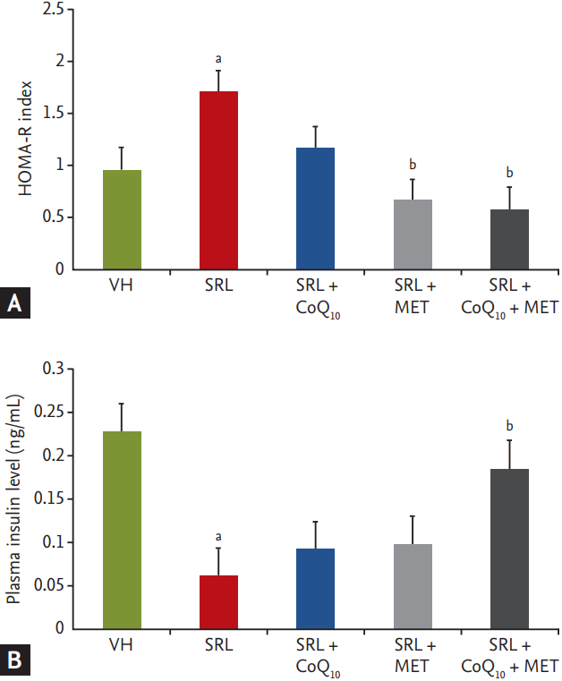
After 3 weeks of SRL treatment, 24-hour water intake (18 ± 4 vs. 30 ± 5, p < 0.05) and urine volume (14 ± 2 vs. 20 ± 3, p < 0.05) of the rats were significantly increased. However, MET and combined CoQ10 and MET suppressed the elevation of water intake and urine excretion. SRL treatment slowed the rate of body weight increase (85 ± 5 vs. 19 ± 4, p < 0.05), while both MET (41 ± 2 vs. 19 ± 4, p < 0.05) and the combined use of CoQ10 and MET (62 ± 3 vs. 19 ± 4, p < 0.05) significantly recovered these changes (Table 1). The IPGTT was used to assess the basal metabolism of plasma glucose concentration, and SRL treatment significantly increased the AUCg, whereas both MET and the combination of CoQ10 and MET attenuated the increase in AUCg induced by SRL (Table 2). Four weeks of SRL treatment increased the HOMA-R index and decreased the plasma insulin level. Consistent with our data thus far, the combination of CoQ10 and MET reverted these changes, resulting in a decreased HOMA-R index and increased plasma insulin level compared with the SRL group (Fig. 1).

Addition of CoQ10 to MET attenuates SRL-induced pancreatic islet dysfunction and reduces islet size

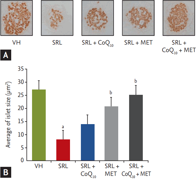
Pancreatic β-cell area was evaluated using immunohistochemistry. The SRL group exhibited smaller islets with decreased insulin staining intensity within islets compared with the VH (27.17 ± 2.32 μm2) group. However, addition of CoQ10 to MET resulted in a significant improvement in insulin staining intensity within islets (SRL: 8.19 ± 1.12 μm2 vs. SRL + MET + CoQ10: 25.18 ± 2.17 μm2, p < 0.05) (Fig. 2).

Addition of CoQ10 to MET suppresses SRL-induced oxidative injury in pancreatic islets

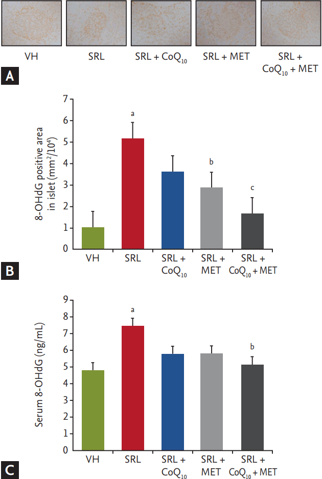
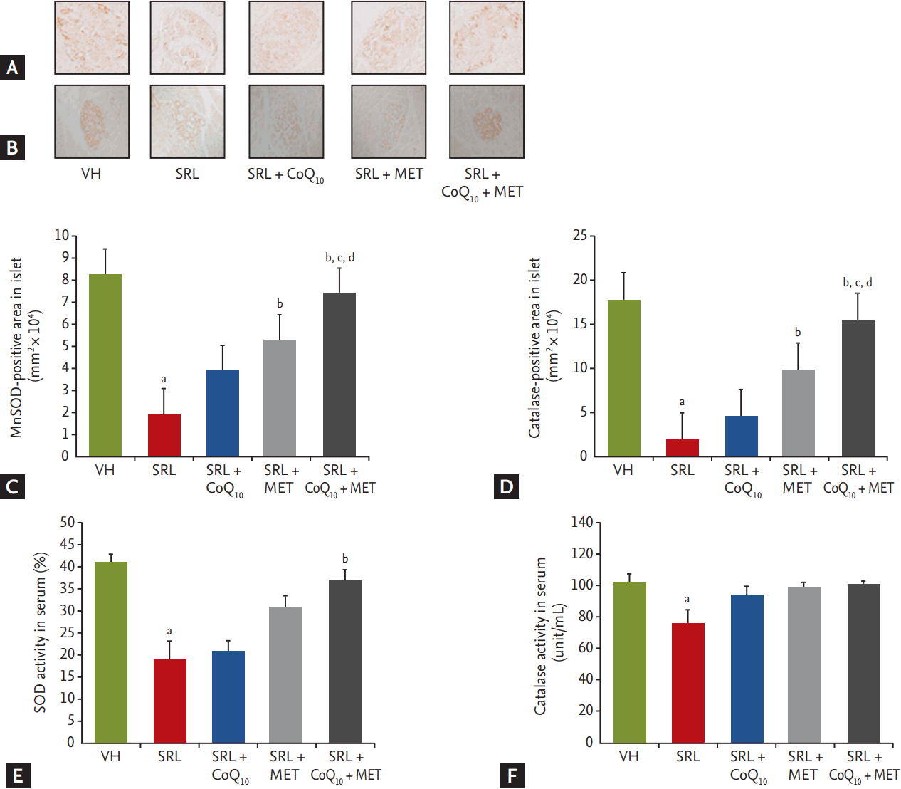
In the present study, 8-OHdG was used as a marker of oxidative DNA damage. Fig. 3 shows the results of immunohistochemistry for 8-OHdG and the levels of serum 8-OHdG in the experimental groups. The strong nuclear expression of 8-OHdG in islets and its area of positive staining were markedly increased in the SRL group (5.17 ± 0.53 mm2/104) compared with the control group (1.02 ± 0.13 mm2/104), and this effect was reversed by CoQ10 and MET combination treatment (1.66 ± 0.14 mm2/104) (Fig. 3A and 3B). A higher level of serum 8-OHdG was also detected in the SRL group; however, the combination of CoQ10 and MET treatment significantly decreased these changes (SRL: 7.47 ± 0.82 ng/mL vs. SRL + MET + CoQ10: 5.16 ± 0.67 ng/mL, p < 0.05) (Fig. 3C). Likewise, concomitant administration of CoQ10 and MET significantly reduced 8-OHdG levels in islets compared to treatment with MET alone. Rats in the SRL group exhibited a significant (p < 0.001) decrease in the levels of antiperoxidative enzymes (MnSOD and catalase) in pancreatic tissue compared to the normal control group; however, addition of CoQ10 to MET increased MnSOD (SRL: 1.96 ± 0.15 mm2 × 104 vs. SRL + MET + CoQ10: 7.39 ± 0.81 mm2 × 104, p < 0.05) (Fig. 4A and 4C) and catalase activities (SRL: 2.03 ± 0.19 mm2 × 104 vs. SRL + MET + CoQ10: 15.44 ± 1.67 mm2 × 104, p < 0.05) (Fig. 4B and 4D) in pancreatic tissue. Similar results were also shown in serum MnSOD (SRL: 19.22% ± 7.88% vs. SRL + MET + CoQ10: 37.98% ± 4.48%, p < 0.05) (Fig. 4E) and catalase activities (SRL: 76.09 ± 7.89 unit/mL vs. SRL + MET + CoQ10: 101.91 ± 0.48 unit/mL, p < 0.05) (Fig. 4F).

Addition of CoQ10 to MET suppresses SRL-induced apoptosis in pancreatic islets

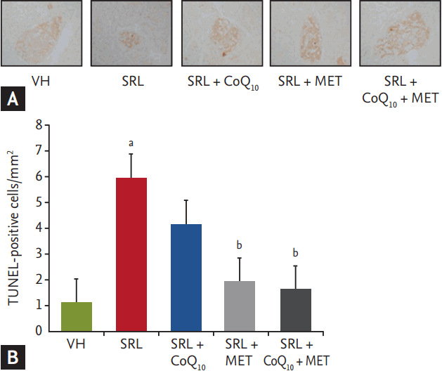
We next evaluated whether CoQ10 treatment affects apoptosis, which is an important mechanism of cell death in SRL-induced pancreatic injury. The number of TUNEL-positive cells in tissue sections was significantly higher in the SRL-group (5.97 ± 0.53 cells/mm2) compared with the VH group (1.13 ± 0.12 cells/mm2). Treatment with both CoQ10 and MET (1.66 ± 0.14 cells/mm2) as well as MET alone (1.94 ± 0.15 cells/mm2) led to a significant decrease (p < 0.05) in the number of TUNELpositive cells in the tissue sections (Fig. 5).

Addition of CoQ10 to MET improves mitochondrial ultrastructure

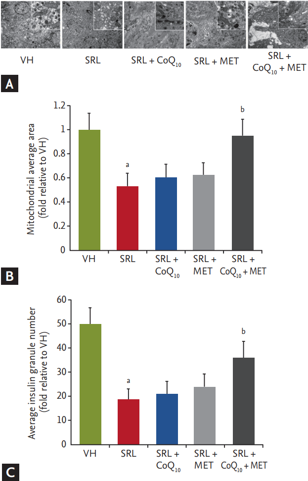
Our findings implied that the addition of CoQ10 to MET protects mitochondrial function, which is strongly associated with reactive oxygen species (ROS) production and cell apoptosis. To evaluate the effects of CoQ10 on mitochondria, electron microscopy was used to detect the ultrastructure and to quantify the number of mitochondria. In islet cells obtained from our animal model, SRL treatment decreased both the average mitochondrial area and number. Although CoQ10 or MET alone did not protect against this decrease, concomitant administration of CoQ10 and MET restored the average mitochondrial area and number. Electron microscopy also showed that the number of insulin granules was significantly reduced by SRL treatment. Only concomitant administration of CoQ10 and MET was effective in attenuating the reduction in granule number (Fig. 6).

DISCUSSION

The results of our study clearly demonstrated that addition of CoQ10 to MET effectively controlled hyperglycemia compared to MET alone and was able to attenuate SRL-induced oxidative injury in pancreatic islets and restore mitochondrial ultrastructure. Importantly, these findings provide a rationale for using CoQ10 as a supplemental therapy in SRL-induced DM in clinical practice.
CoQ10 has been reported to have beneficial effects on insulin sensitivity through modulation of the insulin receptor [12]; however, its effects on NODAT have not been determined. MET is recommended as the first-line drug for NODAT [7], and we previously reported that MET effectively controls SRL-induced hyperglycemia [6]. Therefore, we hypothesized that the combination of MET and CoQ10 provides better control of SRL-induced DM compared with MET alone. In the present study, we found that that addition of CoQ10 to MET significantly decreased blood glucose compared with MET alone. This finding suggests that the combination of CoQ10 and MET provides better control of hyperglycemia in SRL-induced DM.
We found that the combination of CoQ10 and MET decreased HOMA-R index and increased insulin level, which was consistent with the proposed mechanism of hyperglycemia control in SRL-induced DM [2-5]. CoQ10 supplement was reported to have a beneficial effect on insulin sensitivity [17]. We believe that CoQ10 could contribute to the reduction in HOMA-R in presenting study. In general, improvement of insulin sensitivity induces a decline in serum insulin level [18,19]; however, in our study, plasma insulin level increased despite a decreased HOMA-R index. Based on this finding, we suggest that the recovery of insulin secretion through protection of pancreatic islet cell injury in the combined use of CoQ10 and MET therapy could be an important mechanism of glucose control in SRL-induced DM.
To evaluate the effect of the combination of CoQ10 and MET on insulin secretion, we measured plasma insulin and functional islet cell mass. Chronic SRL treatment caused pancreatic islet dysfunction, as demonstrated by the reduced islet size and lower intensity of insulin staining within islets. The addition of CoQ10 to MET significantly restored pancreatic islet size and significantly improved the intensity of insulin staining within islets. Together, these findings suggest that the decreased functional islet mass induced by SRL can be effectively preserved with the combination treatment of MET and CoQ10. In addition, even though treatment with only CoQ10 did not produce a statistically significant improvement, we noted a trend with respect to its benefit on pancreatic islet function and recovery of islet size (SRL < CoQ10 < MET < CoQ10 + MET) (Fig. 2).
To determine the protective effect of the combination of CoQ10 and MET against SRL-induced pancreatic injury, we focused on oxidative stress injury, which is a common pathway of SRL-induced pancreatic injury [20]. There is accumulating evidence indicating that SRL treatment increases oxidative stress in vivo and in vitro [21,22], and that high levels of oxidative stress induced by chronic SRL administration lead to increased production of free radical species in the pancreas and subsequently apoptotic cell death [23]. Our results showed that, while treatment with CoQ10 alone did not alter the level of 8-OHdG (a marker of oxidative DNA damage) in islets or serum, concomitant administration of CoQ10 and MET dramatically decreased the level of this marker. In addition, the rats in the CoQ10 and MET combination treatment group exhibited a significant increase in the levels of antiperoxidative enzymes (MnSOD and catalase) in renal tissue compared to the rats treated with MET alone. Lastly, our results showed that treatment with MET alone or the combination of CoQ10 and MET reduced the number of TUNEL-positive cells. Overall, these findings suggest that the combination of CoQ10 and MET protects against SRL-induced pancreatic islet injury by decreasing oxidative stress and apoptotic cell death.
We next studied whether mitochondrial ROS contribute to SRL-induced oxidative stress. The mitochondrial respiratory chain is the major source of ROS in most mammalian cells [24]. Increases in ROS can alter mitochondrial structure and function, leading to a reduction in total mitochondria number and producing distinct morphological abnormalities [25]. Thus, we evaluated whether the addition of CoQ10 to MET could protect the structure and function of mitochondria. Although MET monotherapy improved the mitochondrial average area and insulin granule number by 10%, the addition of CoQ10 to MET restored these values by approximately 70% and 40%, respectively. Based on these results, we believe that the combination of CoQ10 to MET would be beneficial in the treatment of SRL-induced DM.
AMP-activated protein kinase (AMPK) acts as an energy sensor of the cell and work as a key regulator of mitochondrial biogenesis. MET improves hyperglycemia mainly through AMPK activation [26-28]. Furthermore, CoQ10 activate AMPK indirectly by reducing oxidized low-density lipoprotein [29]. Therefore, it is expected that combination of these two drugs shows an additive or synergistic action on mitochondrial morphology, and this hypothesis may explain further improvement of mitochondrial function than each drug alone in our study.
The guidelines for NODAT management are based on type 2 DM [7], in which MET is recommended as a firstline drug, and a second-line drug can be used to achieve optimal glycemic control. Our study showed that addition of CoQ10 to MET improved glycemic control, which was accompanied by reduction in oxidative stress and improvement of mitochondria morphology. The results of our study can be translated into clinical practice, and we recommend CoQ10 as an option for renal transplant patients with SRL-induced DM who have uncontrolled hyperglycemia despite MET therapy.
In conclusion, the addition of CoQ10 to MET had protective effects on pancreatic islet injury induced by SRL. Our findings suggest that the combination of CoQ10 and MET will be useful in the treatment of SRL-induced DM.

KEY MESSAGE

1. The addition of coenzyme Q10 (CoQ10) to metformin (MET) has a beneficial effect on sirolimus (SRL)-induced diabetes mellitus (DM) compared to MET alone. This effect was related to a protective effect of CoQ10 on SRL-induced pancreatic islet injury.
2. Our findings suggest that the combination of CoQ10 and MET will be useful in the treatment of SRL-induced DM.

Conflict of Interest

No potential conflict of interest relevant to this article was reported.

Acknowledgments

This study was supported by a grant of the Korean Health Technology R&D Project, Ministry for Health and Welfare, Republic of Korea (HI14C3417).

Figure 1.
Effects of combined treatment with coenzyme Q10 (CoQ10) and metformin (MET) on insulin resistance and plasma insulin levels in experimental model of sirolimus (SRL)-induced diabetes mellitus (DM). (A) Homeostatic model assessment of insulin resistance (HOMA-R) index. (B) Plasma insulin levels. Note that all parameters recovered after addition of CoQ10 to MET. VH, vehicle. a p < 0.05 vs. VH, b p < 0.05 vs. SRL.
kjim-2017-004f1.gif
Figure 2.
Effect of addition of coenzyme Q10 (CoQ10) to metformin (MET) on pancreatic islet morphology and size during sirolimus (SRL)-induced injury. Immunohistochemistry for insulin showing pancreatic islet morphology and size in the experimental groups. (A) Representative images of insulin staining of pancreatic sections. (B) Quantitative analysis of islet area. The SRL group showed a smaller size of islet with lower intensity of insulin staining within islet than vehicle (VH) group. In contrast, additional treatment of CoQ10 to MET reversed these changes. Original magnifications, ×400. n = 9 rats per group. a p < 0.05 vs. VH, b p < 0.05 vs. SRL.
kjim-2017-004f2.gif
Figure 3.
Effect of addition of coenzyme Q10 (CoQ10) to metformin (MET) on the expression of 8-hydroxy-2′-deoxyguanosine (8-OHdG) during SRL-induced pancreatic islet injury. Representative images of 8-OHdG staining in each treatment group. (A, B) The strong dark nuclear expression of 8-OHdG in islets was higher in sirolimus (SRL) group compared with groups treated with CoQ10 or MET alone or in combination treatment groups. The strong nuclear expression of 8-OHdG in islets and its area of positive staining were markedly increased in the SRL group compared with the control group; this effect was reversed after addition of CoQ10 to MET therapy. (C) The SRL-induced serum 8-OHdG level is lowered by co-treatment of CoQ10 and MET. Original magnifications, ×400. n = 9 rats per group. VH, vehicle. a p < 0.05 vs. VH, b p < 0.05 vs. SRL, c p < 0.05 vs. SRL + MET.
kjim-2017-004f3.gif
Figure 4.
Effect of addition of coenzyme Q10 (CoQ10) to metformin (MET) on the expression of superoxide dismutase and catalase. (A, B) Representative images of manganese superoxide dismutase (MnSOD) and catalase staining in each treatment group. (C, D) The nuclear expression of MnSOD and catalase in islets was weaker in the sirolimus (SRL) group as compared to normal control group. However, treatment with CoQ10 + MET increased in MnSOD and catalase activities in renal tissue. (E, F) Similar results were also shown in serum MnSOD and catalase activities. Original magnifications, ×400. n = 9 rats per group. VH, vehicle. a p < 0.05 vs. VH, b p < 0.05 vs. SRL, c p < 0.05 vs. CoQ10, d p < 0.05 vs. MET.
kjim-2017-004f4.gif
Figure 5.
Effects of combined treatment with metformin (MET) and coenzyme Q10 (CoQ10) on apoptosis in pancreatic islets in experimental model of sirolimus (SRL)-induced diabetes mellitus (DM). (A) Representative immunohistochemistry of the active form of caspase-3 (caspase-3) in islets and in situ TdT-mediated dUTP–biotin nick end labeling (TUNEL) assay to detect apoptosis in pancreatic islets. (B) Analysis to detect apoptosis in pancreatic islets in the experimental groups. The number of TUNEL-positive cells in tissue sections was significantly higher in the SRL-group compared with the vehicle (VH) group. Addition of coenzyme Q10 (CoQ10) to MET significantly reduced the number of TUNEL-positive cells compared with SRL group. Original magnifications, ×400. n = 9 rats per group. a p < 0.05 vs. VH, b p < 0.05 vs. SRL.
kjim-2017-004f5.gif
Figure 6.
Transmission electron microscopy. (A) Mitochondrial ultrastructure is disordered, and the number of dense core insulin granules is lower in pancreatic β-cells from the sirolimus (SRL) group. However, these findings improved after combination of coenzyme Q10 (CoQ10) and metformin (MET). The scale bar equals 500 nm. Arrowheads indicate mitochondria. Asterisk indicate insulin granules. (B, C) Quantitative analysis of the mitochondrial area and number of insulin granules in the experimental groups. Note the significantly decreased mitochondrial area and number of insulin granules in the SRL group compared with the vehicle (VH) group. Addition of CoQ10 to MET significantly increased the mitochondrial area and number of insulin granules compared with the SRL group. a p < 0.05 vs. VH, b p < 0.05 vs. SRL.
kjim-2017-004f6.gif
Table 1.
Basic parameters in each group
VH (n = 9) SRL (n = 9) SRL + CoQ10 (n = 9) SRL + MET (n = 9) SRL + CoQ10 + MET (n = 9)
ΔBW, g 85 ± 5 19 ± 4a 25 ± 3 41 ± 2b 62 ± 3b,c
UV, mL 14 ± 2 20 ± 3a 17 ± 2 12 ± 3b 13 ± 2b
Water intake, mL 18 ± 4 30 ± 5a 25 ± 6 22 ±5b 19 ± 5b
Scr, mg/dL 0.35 ± 0.04 0.30 ± 0.03 0.34 ± 0.15 0.31 ± 0.06 0.31 ± 0.05
HbA1c, % 3.93 ± 0.12 3.94 ± 0.15 4.02 ± 0.19 3.94 ± 0.20 3.93 ± 0.13
SRL con., ng/mL - 7.26 ± 2.99 6.10 ± 3.11 6.06 ± 2.80 5.88 ± 2.31

Values are presented as mean ± standard error.

VH, vehicle; SRL, sirolimus; CoQ10, coenzyme Q10; MET, metformin; BW, body weight; UV, urine volume; Scr, serum creatinine; HbA1c, hemoglobin A1c; SRC con., sirolimus concentration.

a p < 0.05 vs. VH.

b p < 0.05 vs. SRL.

c p < 0.05 vs. SRL + MET.

Table 2.
Effects of combination treatment of CoQ10 MET on control of SRL-induced hyperglycemia using IPGTT
Group 0 min 30 min 60 min 90 min 120 min AUCg
VH 90 ± 3 210 ± 10 145 ± 5 137 ± 5 108 ± 3 295 ± 9
SRL 83 ± 4 316 ± 20a 250 ± 9a 178 ± 7a 132 ± 2 424 ± 17a
SRL + CoQ10 81 ± 3 312 ± 18a 225 ± 7a 144 ± 4 106 ± 4 386 ± 12
SRL + MET 80 ± 4 275 ± 16a 176 ± 6b 128 ± 3b 106 ± 3 335 ± 14b
SRL + CoQ10 + MET 83 ± 3 242 ± 15a,b,c 144 ± 5b,c 108 ± 2b 93 ± 2b 291 ± 15b,c

Values are presented as mean ± standard error.

CoQ10, coenzyme Q10; MET, metformin; SRL, sirolimus; IPGTT, intraperitoneal glucose tolerance test; AUCg, area under the curve of glucose; VH, vehicle.

a p < 0.05 vs. VH.

b p < 0.05 vs. SRL.

c p < 0.05 vs. SRL + MET.

REFERENCES

1. Kasiske BL, Snyder JJ, Gilbertson D, Matas AJ. Diabetes mellitus after kidney transplantation in the United States. Am J Transplant 2003;3:178–185.
crossref pmid
2. Teutonico A, Schena PF, Di Paolo S. Glucose metabolism in renal transplant recipients: effect of calcineurin inhibitor withdrawal and conversion to sirolimus. J Am Soc Nephrol 2005;16:3128–3135.
crossref pmid
3. Johnston O, Rose CL, Webster AC, Gill JS. Sirolimus is associated with new-onset diabetes in kidney transplant recipients. J Am Soc Nephrol 2008;19:1411–1418.
crossref pmid pmc
4. Gyurus E, Kaposztas Z, Kahan BD. Sirolimus therapy predisposes to new-onset diabetes mellitus after renal transplantation: a long-term analysis of various treatment regimens. Transplant Proc 2011;43:1583–1592.
crossref pmid
5. Shivaswamy V, Bennett RG, Clure CC, et al. Tacrolimus and sirolimus have distinct effects on insulin signaling in male and female rats. Transl Res 2014;163:221–231.
crossref pmid
6. Jin L, Lim SW, Jin J, Chung BH, Yang CW. Effects of addition of a dipeptidyl peptidase IV inhibitor to metformin on sirolimus-induced diabetes mellitus. Transl Res 2016;174:122–139.
crossref pmid
7. Kidney Disease: Improving Global Outcomes (KDIGO) Transplant Work Group. KDIGO clinical practice guideline for the care of kidney transplant recipients. Am J Transplant 2009;9 Suppl 3:S1–S155.
pmid
8. Singh RB, Niaz MA, Rastogi SS, Shukla PK, Thakur AS. Effect of hydrosoluble coenzyme Q10 on blood pressures and insulin resistance in hypertensive patients with coronary artery disease. J Hum Hypertens 1999;13:203–208.
crossref pmid pdf
9. Hodgson JM, Watts GF, Playford DA, Burke V, Croft KD. Coenzyme Q10 improves blood pressure and glycaemic control: a controlled trial in subjects with type 2 diabetes. Eur J Clin Nutr 2002;56:1137–1142.
crossref pmid pdf
10. Overvad K, Diamant B, Holm L, Holmer G, Mortensen SA, Stender S. Coenzyme Q10 in health and disease. Eur J Clin Nutr 1999;53:764–770.
crossref pmid pdf
11. Crane FL. Biochemical functions of coenzyme Q10. J Am Coll Nutr 2001;20:591–598.
crossref pmid
12. Amin MM, Asaad GF, Abdel Salam RM, El-Abhar HS, Arbid MS. Novel CoQ10 antidiabetic mechanisms underlie its positive effect: modulation of insulin and adiponectine receptors, tyrosine kinase, PI3K, glucose transporters, sRAGE and visfatin in insulin resistant/diabetic rats. PLoS One 2014;9:e89169.
crossref pmid pmc
13. Bauerova K, Paulovicova E, Mihalova D, et al. Combined methotrexate and coenzyme Q10 therapy in adjuvant-induced arthritis evaluated using parameters of inflammation and oxidative stress. Acta Biochim Pol 2010;57:347–354.
crossref pmid
14. Lim SW, Doh KC, Jin L, et al. Oral administration of ginseng ameliorates cyclosporine-induced pancreatic injury in an experimental mouse model. PLoS One 2013;8:e72685.
crossref pmid pmc
15. Piao SG, Bae SK, Lim SW, et al. Drug interaction between cyclosporine and mTOR inhibitors in experimental model of chronic cyclosporine nephrotoxicity and pancreatic islet dysfunction. Transplantation 2012;93:383–389.
crossref pmid
16. Lim SW, Hyoung BJ, Piao SG, Doh KC, Chung BH, Yang CW. Chronic cyclosporine nephropathy is characterized by excessive autophagosome formation and decreased autophagic clearance. Transplantation 2012;94:218–225.
crossref pmid
17. Raygan F, Rezavandi Z, Dadkhah Tehrani S, Farrokhian A, Asemi Z. The effects of coenzyme Q10 administration on glucose homeostasis parameters, lipid profiles, biomarkers of inflammation and oxidative stress in patients with metabolic syndrome. Eur J Nutr 2016;55:2357–2364.
crossref pmid pdf
18. Dhalla NS, Takeda N, Rodriguez-Leyva D, Elimban V. Mechanisms of subcellular remodeling in heart failure due to diabetes. Heart Fail Rev 2014;19:87–99.
crossref pmid
19. Velez M, Kohli S, Sabbah HN. Animal models of insulin resistance and heart failure. Heart Fail Rev 2014;19:1–13.
crossref pmid pmc
20. Song HK, Han DH, Song JH, et al. Influence of sirolimus on cyclosporine-induced pancreas islet dysfunction in rats. Am J Transplant 2009;9:2024–2033.
crossref pmid
21. Han DH, Piao SG, Song JH, et al. Effect of sirolimus on calcineurin inhibitor-induced nephrotoxicity using renal expression of KLOTHO, an antiaging gene. Transplantation 2010;90:135–141.
crossref pmid
22. Kofman AE, McGraw MR, Payne CJ. Rapamycin increases oxidative stress response gene expression in adult stem cells. Aging (Albany NY) 2012;4:279–289.
crossref pmid pmc
23. Kunsch C, Medford RM. Oxidative stress as a regulator of gene expression in the vasculature. Circ Res 1999;85:753–766.
crossref pmid
24. Jabs A, Gobel S, Wenzel P, et al. Sirolimus-induced vascular dysfunction: increased mitochondrial and nicotinamide adenosine dinucleotide phosphate oxidase-dependent superoxide production and decreased vascular nitric oxide formation. J Am Coll Cardiol 2008;51:2130–2138.
crossref pmid
25. Ma Z, Wirstrom T, Borg LA, et al. Diabetes reduces β-cell mitochondria and induces distinct morphological abnormalities, which are reproducible by high glucose in vitro with attendant dysfunction. Islets 2012;4:233–242.
crossref pmid pmc
26. Pernicova I, Korbonits M. Metformin: mode of action and clinical implications for diabetes and cancer. Nat Rev Endocrinol 2014;10:143–156.
crossref pmid pdf
27. Bailey CJ, Turner RC. Metformin. N Engl J Med 1996;334:574–579.
crossref pmid
28. Jin J, Lim SW, Jin L, et al. Effects of metformin on hyperglycemia in an experimental model of tacrolimus- and sirolimus-induced diabetic rats. Korean J Intern Med 2017;32:314–322.
crossref pmid pmc pdf
29. Tsai KL, Chen LH, Chiou SH, et al. Coenzyme Q10 suppresses oxLDL-induced endothelial oxidative injuries by the modulation of LOX-1-mediated ROS generation via the AMPK/PKC/NADPH oxidase signaling pathway. Mol Nutr Food Res 2011;55 Suppl 2:S227–S240.
crossref pmid

Editorial Office
101-2501, Lotte Castle President, 109 Mapo-daero, Mapo-gu, Seoul 04146, Korea
Tel: +82-2-2271-6792    Fax: +82-2-790-0993    E-mail: kaim@kams.or.kr                

Copyright © 2024 by Korean Association of Internal Medicine.

Close layer
prev next