


To the Editor,
Thrombocytopenia in cancer patients is caused by several mechanisms: chemotherapy or radiotherapy induce bone marrow hypoplasia, the tumor can infiltrate the bone marrow, or the platelets can be consumed secondary to disseminated intravascular coagulation (DIC) or thrombotic thrombocytopenic purpura. The association between immune thrombocytopenic purpura (ITP) and lymphoid neoplasms, in particular chronic lymphoid leukemia and Hodgkin's disease, was first recognized in 1966 [1] and has been frequently reported since then. However, the association with solid tumors is rare. Five cases of renal cell carcinomas (RCCs) associated with ITP have been reported in the medical literature [2,3,4]. In four cases, the thrombocytopenia was refractory to intravenous immunoglobulin or steroids. However, it completely resolved after curative resection of the underlying RCC.
Here, we report the case of a 63-year-old male presenting with severe ITP and RCC who was successfully treated with danazol and curative nephrectomy.
The patient was admitted to our hospital because of epistaxis and bleeding of the gums during the 2 months prior. He also complained of fever, night sweats, weight loss, and anorexia. He had a history of hypertension and stable angina. The only medication he was taking was an antihypertensive agent. He denied a history of drug abuse or herbal medication, and had no risk factors for human immunodeficiency virus (HIV) or chronic viral hepatitis. The patient reported excessive alcohol intake (approximately 140 g alcohol per day) but had stopped drinking 2 months before presentation.
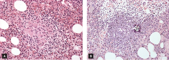
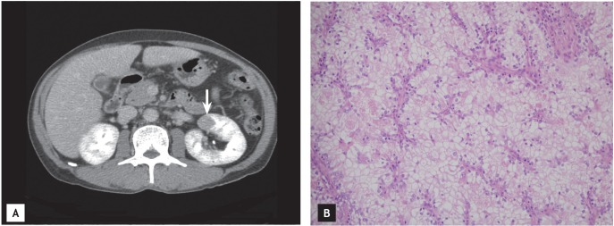
On the initial evaluation, the patient appeared slightly fatigued but not acutely ill. The physical examination detected hepatomegaly to approximately 4 cm. There was no palpable lymphadenopathy. The laboratory evaluation revealed a total leukocyte count of 27,500/mm3 (45.8% neutrophils, 48.7% lymphocytes), hemoglobin 11 g/dL, and a platelet count of 16,000/mm3. The chemistry profile revealed blood urea nitrogen of 10 mg/dL, creatinine 0.8 mg/dL, total protein 7.2 g/dL, albumin 3.7 g/dL, total bilirubin 1.0 mg/dL, aspartate aminotransferase 73 U/L, alanine aminotransferase 23 U/L, alkaline phosphatase 479 U/L (normal range, 30 to 115), and lactate dehydrogenase 284 U/L (normal range, 100 to 225). The fibrinogen level was 424 mg/dL, and the prothrombin time, activated partial thromboplastin time, and bleeding time were normal. The serology tests were all negative for HIV, hepatitis B virus, hepatitis C virus, and Epstein-Barr virus. Rheumatoid factor, antinuclear antibody, double-stranded DNA antibody, and antiplatelet antibody results were negative. A peripheral smear detected normochromic normocytic anemia, anisocytosis, and marked thrombocytopenia. A bone marrow biopsy revealed a normocellular marrow with abundant megakaryocytes. A small granuloma and one lymphoid aggregation were noted (Fig. 1). Angiotensin converting enzyme levels, 24 hours urine calcium levels, and Gallium-67 scans were all normal. The bone marrow cultures for bacterial, fungal, or mycobacterial organisms were negative. Abdominal imaging was performed to rule out a lymphoid malignancy and a 2 cm focal perfusion defect of the mid pole of the left kidney was found in addition to hepatomegaly and a small amount of ascites (Fig. 2A). We did not perform a biopsy of the renal mass due to the risk of bleeding.
A transjugular liver biopsy was performed and there were neither malignant cells nor granuloma found in the specimen. Dense sinusoidal infiltration of Kupffer cells, sinusoidal dilation, and congestion in zone 3, as well as perisinusoidal fibrosis suggested the possibility of chronic venous outflow obstruction or a previous episode of alcoholic steatohepatitis.
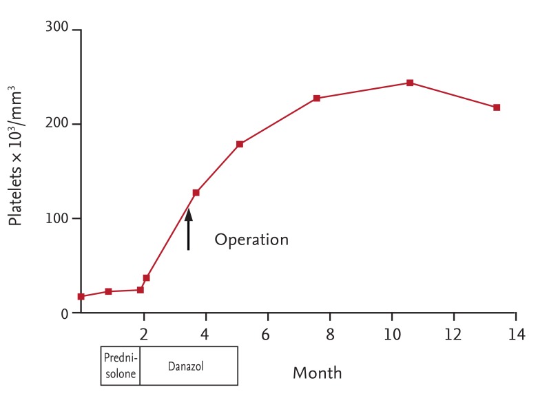
The severe thrombocytopenia was thought to be secondary immune thrombocytopenia associated with RCC. The platelet transfusions were not effective, so prednisolone (1 mg/day for 1 month) was administered before nephrectomy to increase the platelet count. However, this was also not effective. The platelet count was 17,000/mm3 and the patient complained of mood change, which was thought to be a side effect of the prednisolone treatment. Subsequently, danazol (400 mg/day) was prescribed. The patient tolerated the medication and the platelet count increased to 142,000/mm3 after 2 months without transfusion. Then the patient underwent a partial nephrectomy for RCC. There were no apparent extracapsular tumors or enlarged perihilar lymph nodes. The pathology of the nephrectomy specimen revealed a 3.5 ├Ś 1.5 ├Ś 1.5 cm nodular mass with hemorrhage, involving the mid pole of the kidney. Microscopic examination revealed RCC of clear cell type (Fig. 2B); the tumor was a Fuhrman grade 2. The final pathological stage was I (T1N0M0). After continuous treatment with danazol for 2 months, danazol therapy was discontinued. The platelet count increased to 178,000/mm3 at that time and continued to be maintained within the normal range during 1 year of follow-up (Fig. 3).
The association between ITP and solid tumors is rare. Kim and Boggs [4] reported a series of 10 patients with a variety of solid tumors and ITP in 1979. Since then, approximately 20 cases of ITP associated with solid tumors including the breast, gastrointestinal tract, lung, ovary, testis, prostate, urinary bladder, kidney, and vagina have been reported.
The mechanism of platelet destruction seen in secondary ITP remains unclear, but is thought to be identical to that of primary ITP in some aspects. In ITP, platelets coated with autoantibodies react with glycoproteins IIb/IIIa, Ib/IX, Ia/IIa, and other platelet determinants and undergo accelerated clearance via tissue macrophages. In some patients, accelerated platelet clearance does not result in a compensatory increase in platelet production. Platelet production is impaired either through inhibited megakaryocytopoiesis or the intramedullary action of macrophages on antibody-coated platelets and megakaryocytes. Moreover, the production of substances causing platelet aggregation by tumor cells (spleen metastases as well as the intratumor platelet consumption enhanced by their adherence to poorly endothelialized surfaces of abnormal tumor vessels) provides possible explanations for the platelet destruction in patients with thrombocytopenia and solid tumors. Paraneoplastic syndrome has been found in patients with RCC regardless of the tumor burden and occurs with equal frequency among localized and metastatic disease. This suggests that the tumor biology rather than the extent of the tumor plays an important role in the manifestation of paraneoplastic syndrome. Klimberg and Drylie [2] and Kamra et al. [3] reported that patients with ITP were associated with stage II RCC. Yoshinaga et al. [5] reported a patient with paraneoplastic thrombocytopenia associated with stage I RCC. These three patients demonstrated complete recovery of the thrombocytopenia after nephrectomy (with or without splenectomy).
The diagnosis of ITP associated with malignancy is one of exclusion, requiring that other causes of thrombocytopenia be ruled out. In this case, our patient did not use immunosuppressive medication and there was no evidence of infection or DIC. The bone marrow exam revealed a small granuloma and one lymphoid aggregation. However, infection and chronic granulomatous disease such as sarcoidosis or lymphoproliferative disorder were not present. Therefore, the granuloma was thought to be a sarcoid-like reaction associated with RCC. There was no evidence to support the cause of thrombocytopenia, and curative nephrectomy resulted in the patient's recovery from thrombocytopenia. Therefore, we considered this case a secondary ITP associated with RCC.
The treatment of paraneoplastic ITP has included treatments such as corticosteroids, splenectomy, intravenous immunoglobulin, vincristine, and interferon in addition to the specific therapy for the primary cancer. Anticancer treatments such as surgery, chemotherapy, or radiotherapy are effective and ideal strategies for the treatment of paraneoplastic ITP. However, surgery and radiotherapy require adequate platelets for local treatment. Because the only curative treatment for localized RCC is surgery, we continued the ITP treatment with the potential risk of tumor progression. Although recovery of the platelets and the surgery were successful, danazol should be limited to early-stage cancer with a low risk of dissemination.
ITP associated with malignancy can be diagnosed concomitantly with the underlying malignancy and/or can be a presenting sign of the malignancy, regardless of the tumor burden. In cases of refractory ITP it is necessary to consider the possibility of an underlying malignant neoplasm, not only a lymphoid malignancy, but also a solid tumor.
 PDF Links
 PubReader
 ePub Link
 Full text via DOI
 Download Citation
 Print
Print


