


INTRODUCTION
Due to the technological developments of cardiac pacemaker, the ultimate purpose of treatment using this device lies in the optimization of cardiopulmonary function rather than the simple survival of the patient. In other words, the function of the heart should be made to satisfy the metabolic demands of physical activities beyond the maintenance of cardiac output at resting state.
The programmable dual chamber cardiac pacemaker can sense and pace both the atrium and ventricle by controlling the atrioventricular(AV) conduction delay time. The atrioventricular synchrony aims that the atrium can play an assistant role for the cardiac output by contracting atrium and ventricle at proper intervals and therefore, the cardiac output can be improved if the synchrony has been adequately adjusted in comparison with the fixed PR intervals even during exercise as well as at resting state1).
For a normal person, the velocity of AV conduction will improve according to the heart rate increases2,3). The changes of AV conduction can be measured by rating the PR intervals on the electrocardiogram(ECG).
The PR interval on the ECG means the time taken from the activation of the atrium to the His-Purkinje conduction system by the transmission of electrical stimulation. Thus, its changes according to exercise stress will reflect the increase of the intranodal conduction velocity due to physiologic or autonomic nervous system stimulations.
The role of the atrial contraction for cardiac output has been well-known. That is, the role of the atrium will be affected by various factors such as age, autonomic nervous system activity, heart rate, physical activity, AV conduction delay time and contracting condition of atrium and ventricle.
It has been reported that when a DDD-type cardiac pacemaker is used, proper reduction of AV conduction delay, according to heart rates, would be more helpful for the improvement of cardiac output rather than a fixed form of AV nodal delay.
With the above background in mind, this study was aimed at reviewing the physiologic changes or features of the PR intervals, due to exercise stress by means of the exercise treadmill test for patients who visited the hospital because of non-specific cardiovascular symptoms, but who were diagnosed as normal, and the civilian pilots who visited for their regular physical check-up.
MATERIALS AND METHODS
The subjects of this study were those 134 patients(95 males and 39 females, mean age of 47.1±11.7) who visited to complain chiefly of non-specific chest pain, but who were diagnosed as normal on the exercise treadmill test, and 148 civilian pilots(all males, mean age of 42.7±11.7) Who visited for their regular physical examination.
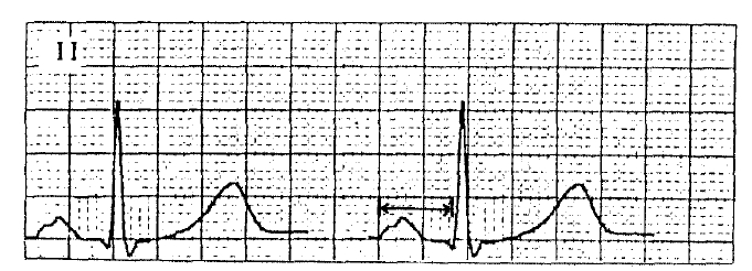
The patients and control groups were all made to do the same exercise treadmill test using 12-lead ECG. The test was conducted according to the standard Bruce protocol and the 12-lead ECG was checked at the speed of 50 mm/sec. The interval from the first deflection of p wave to QRS complex in lead II was measured using the caliper, and its results were indicated with the unit of msec(Fig. 1). In order to repel any differential errors among examiners, the PR intervals were checked by the same examiner. Cases of positive test for myocardial ischemia and less than 5 metabolic equivalents(METs) exercise capacity patients, were excluded from the study.
Results were expressed as mean±standard deviation. Statistical analysis was done using SPSS/C+ program for student’s t-test. A P value of less than 0.05 was regarded as significant.
RESULTS
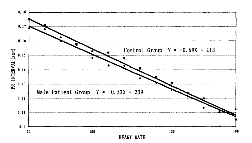
The control group showed 6.9msec reduction rate of the PR interval whenever their heart rate increased by 10 beats per minute(Fig. 2).
The entire patient group showed 5msec reduction rate of the PR interval whenever their heart rate increased by 10 beats per minute.
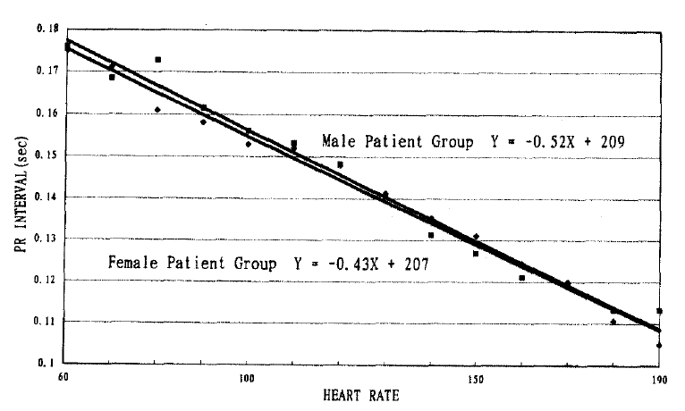
The male patients group showed 5.2msec reduction rate of the PR interval whenever their heart rate increased by 10 beats per minute(Fig. 3).
The female patient group showed 4.3msec reduction rate of the PR interval whenever their heart rate increased by 10 beats per minute(Fig. 4).
There were significant differences of the PR interval changes between the entire or male patient group and the control group within the same range of heart rates.
DISCUSSION
As the conduction delay time can be adjusted by the heart rate in DDD-type cardiac pacing, due to the recent technological development of the pacemaker system, the co-relationship between cardiac output and AV conduction delay time is being actively studied. Up to now, in order to determine the most proper AV conduction delay time, according to heart rate in the application of the artificial cardiac pacemaker, studies have been conducted using such means as systolic time interval4,5), echocardiogram6), Doppler echocardiogram7,8), radionuclide ventriculogram9) and hemodynamic study10,11).
The programmable dual chamber cardiac pacemaker can sense and pace both the atrium and ventricle by controlling the AV conduction delay time in many ways. The artioventricular synchrony aims that the atrium can play an assistant role for the cardiac output by contracting atrium and ventricle at proper intervals and therefore, the cardiac output can be improved if the synchrony has been adequately adjusted in comparison with the fixed PR intervals even during exercise as well as at resting state1,12,13). For a normal person, the velocity of AV conduction will be increased according to the heart rate increases2,3). The changes of AV conduction can be measured by rating the PR intervals on the ECG.
The PR interval on the ECG means the time taken from the activation of the atrium to the His-Purkinje conduction system by the transmission of electrical stimulation. Thus, its changes according to exercise stress will reflect the increase of the intranodal conduction velocity due to physiologic or autonomic nervous system stimulations.
It has been known from the studies so far that heart rate, AV conduction delay time and exercise stress are linearly co-related1,14–16). In 1990, Barbieri et al.17) reported that heart rate and AV conduction delay times are reversely co-related in linear terms within the range of 55–155 beats per minute of heart rate, but PR interval seldom changes beyond this range.
But in our study the linear co-relationship could be observed even within the range of 155–195 beats per minute of heart rate. In 1973, Tail et al.4) reported in their study using ECG, carotid arterial phonogram and apex phonogram that the PR interval would reach its peak of stroke volume at 180–200 msec within the range of 70±20 beats per minute of heart rate.
In 1975, Karlof et al.1) reported that the rate-matched VVI cardiac pacing would increase the cardiac output by 18% more at resting state and 8% more during exercise than the atrial synchronous ventricular(VAT) pacing. These differences may be attributed to the atrioventricular synchrony, although they are not significantly, wide enough. In 1979, Greenberg et al.18) reported that the atrial contraction would not affect the stroke volume when the left ventricular filling pressure is increased
In order to know the most desirable cardiac pacing in physiologic terms, the heart rate should be properly increased first according to the exercise stress and secondly, the velocity of AV conduction delay time should be adequately reduced, for which the left ventricular function is normal. What is important here is to choose the algorithms which can determine the change of PR interval according to the heart rate in applying the cardiac pacemaker. In 1985, Leman and Kratz19), reported that 100 msec of AV conduction delay time would lead to an increase of more end-diastolic and stroke volumes, within the range of 92–115 beats per minutes of heart rate, in comparison with 150 msec of AV conduction delay time within the same heart rate by using the bicycle ergometer test. In this study, it was disclosed that these patient groups differ much from those of normal control group(150–134 msec of PR intervals at 92–115 beats per minute of heart rate). In 1989, Mehta et al.20) reported in their study by using the Doppler echocardiogram, that 100–200 msec of AV conduction delay time at resting state and 75–80 msec during exercise would improve the cardiac output.
In 1990, Margaret et al.21) said in their study for 32 healthy men, that the AV conduction delay time should be reduced to 3.5 msec per 10 beats, which differs much from the reduction rate of 6.9 msec per 10 beats for the healthy male group in our study. This difference may attributable to the racial factor or the size of the subjects. This study has found the desirable equation y=−0.69×+213 at the rate of 60–190 beats per minute of heart rate for the normal control group, which differs from the equation y=−0.50×+208 for the entire patient group. For the male patient group the equation is y=−0.52×+209, while it is y=−0.43×+207 for the female patient group. As can be seen, there are little differences between both sexes, but there can be found some significant differences between normal and male patient group at more than 100 beats per minutes of heart rate. Although we conducted the exercise treadmill test for the entire patient group who complained about nonspecific cardiovascular symptoms, they were all diagnosed as normal. Considering the mean sensitivity of 68% and specificity of 77% of this exercise treadmill test, the possibility of their cardiovascular disease could not be excluded22). Upon reviewing the previous literature, there has been no study about the PR interval distinguishing males from females. In this study there was no significant difference between both sexes of the patient group.

 PDF Links
 PubReader
 ePub Link
 Full text via DOI
 Download Citation
 Print
Print


