Korean J Intern Med > Volume 37(3); 2022 > Article
Lee and Jang: Role of T cells in ischemic acute kidney injury and repair

Abstract

Ischemic acute kidney injury (AKI) is a common medical problem with significant mortality and morbidity, affecting a large number of patients globally. Ischemic AKI is associated with intrarenal inflammation as well as systemic inflammation; thus, the innate and adaptive immune systems are implicated in the pathogenesis of ischemic AKI. Among various intrarenal immune cells, T cells play major roles in the injury process and in the repair mechanism affecting AKI to chronic kidney disease transition. Importantly, T cells also participate in distant organ crosstalk during AKI, which affects the overall outcomes. Therefore, targeting T cell-mediated pathways and T cell-based therapies have therapeutic promise for ischemic AKI. Here, we review the major populations of kidney T cells and their roles in ischemic AKI.

INTRODUCTION

Ischemic acute kidney injury (AKI) is an important clinical disorder affecting a large number of patients worldwide and is a major risk factor for chronic kidney disease (CKD) and end-stage renal disease [1]. Any clinical conditions that can result in an interruption of renal blood flow—such as intravascular volume depletion, reduced cardiac output, and vasodilatory or vasoconstrictive disorders—can cause ischemic AKI [2,3]. Given its high prevalence and mortality, despite continued and substantial improvement in critical care and dialysis techniques, improving ischemic AKI outcomes is challenging [4,5]. Therapies that reduce injury or enhance adaptive repair are not available [5]. Among various cellular and molecular pathways involved in its pathogenesis, inflammation orchestrated by kidney immune cells affects both injury and repair processes [6]. Normal kidneys of human and animals possess considerable amounts of resident immune cells [7,8]. Indeed, even germ-free (GF) mice that have not been exposed to microbiota have abundant kidney resident immune cells, which are related to ischemic AKI pathogenesis, indicating that they play pathogenic roles under sterile conditions [9]. In ischemic AKI, endogenous molecules released from stressed or injured cells trigger activation and proliferation of kidney immune cells, contributing to the progression of injury. This process resembles immune responses to microbial pathogens and is thus termed sterile inflammation [10,11]. Among the immune cells participating in this process, T cells, a major component of the adaptive immune system, play crucial roles as major effectors and regulators [12]. Although most evidence of their role in AKI is from animal studies [13], continued clinical experience of AKI in patients receiving T cell-targeted immune check point inhibitor or chimeric antigen receptor T cell therapy highlights their pathogenic roles in human AKI [14,15]. A rigorous mechanistic understanding of AKI would facilitate development of novel therapeutic targets. Here we review the mechanisms of ischemic AKI, focusing on the role of T cells during the injury phase and recovery process based on experimental studies, emphasizing recent literature.

ROLE OF T CELLS DURING EARLY RENAL INJURY IN ISCHEMIC AKI

Studies using murine ischemia-reperfusion injury (IRI) models have provided the most mechanistic evidence of T cells in ischemic AKI (Table 1) [7,9,1642]. In contrast to the classical concept of immunity, which describes adaptive immune cells as relatively late responders in the injured site, T cells traffic into post-ischemic kidneys at a very early time point and coordinate innate immunity [16,17]. An in vitro study of hypoxia-reoxygenation demonstrated T cell adhesion to tubular epithelial cells [18].
During the early phase of IRI, T cells play pathogenic roles by contributing to the injury process, as demonstrated using mice lacking T cells. Both T cell-deficient mice (nu/nu mice) and CD4, CD8 double-knockout (KO) mice were protected from IRI [18,19]. A study using a whole body IRI model, by inducing cardiac arrest followed by resuscitation, reproduced these findings, showing significant protection in T cell-deficient mice with reduced expression of intercellular adhesion molecule 1 (Icam1) [20]. There was substantial functional and structural protection from IRI in T cell receptor (TCR) αβ-deficient mice, with a reduction of tumor necrosis factor α (TNF-α) and interleukin 6 (IL-6) expression in the post-ischemic kidneys [21]. A subsequent study using genetically engineered DO11.10 transgenic mice demonstrated that diverse TCR repertoires are essential for the initiation of T cell activation but, once activated, TCR diversity is likely to have less impact on their pathogenic effect [22].
Importantly, T cell costimulatory molecules, emerging therapeutic targets in oncology, play important roles in the pathogenesis of ischemic AKI. When T cells recognize antigens through TCR, binding of CD28 to B7 (also known as CD80 and CD86 for B7-1 and B7-2, respectively) on antigen-presenting cells functions as a costimulatory pathway, amplifying the TCR signal to activate T cells [43]. A negative costimulatory molecule on T cells, cytotoxic T lymphocyte-associated protein 4 (CTLA-4), outcompetes B7 binding, dampening T cell activation [43]. Blockade of the CD28-B7 costimulatory pathway by CTLA-4-Ig administration in cold IRI reduced renal dysfunction and inhibited T cell infiltration into the post-ischemic kidneys [44]. In line with this finding, adoptive transfer of CD28-deficient T cells into T cell-deficient mice (nu/nu mice) did not reinduce the injury phenotype [19]. Programmed death-1 (PD-1) expressed on T cells is another negative costimulatory molecule, which has two ligands, programmed death ligand 1 (PD-L1) and PD-L2 [43]. Blocking of the PD-1 pathway using an antibody against PD-L1 or PD-L2, or genetic deficiency of either, led to deterioration of structural and functional renal injury in post-ischemic kidneys [23]. Blocking both PD-L1 and PD-L2 led to upregulation of proinflammatory gene expression in post-ischemic kidneys, including Il6, C-X-C motif chemokine ligand 1 (Cxcl1), and Icam1 [23]. Importantly, PD-L1 is expressed on renal tubular epithelial cells as well as on immune cells, and interferon γ (IFN-γ) induces upregulation of PD-L1 on renal tubular epithelial cells [45]. Because bone marrow transfer from wild-type (WT) mice to PD-L1 KO mice failed to induce protection, the PD-L1 expression on renal tubular epithelial cells, rather than immune cells, may serve as a major protective mechanism in ischemic AKI [23].
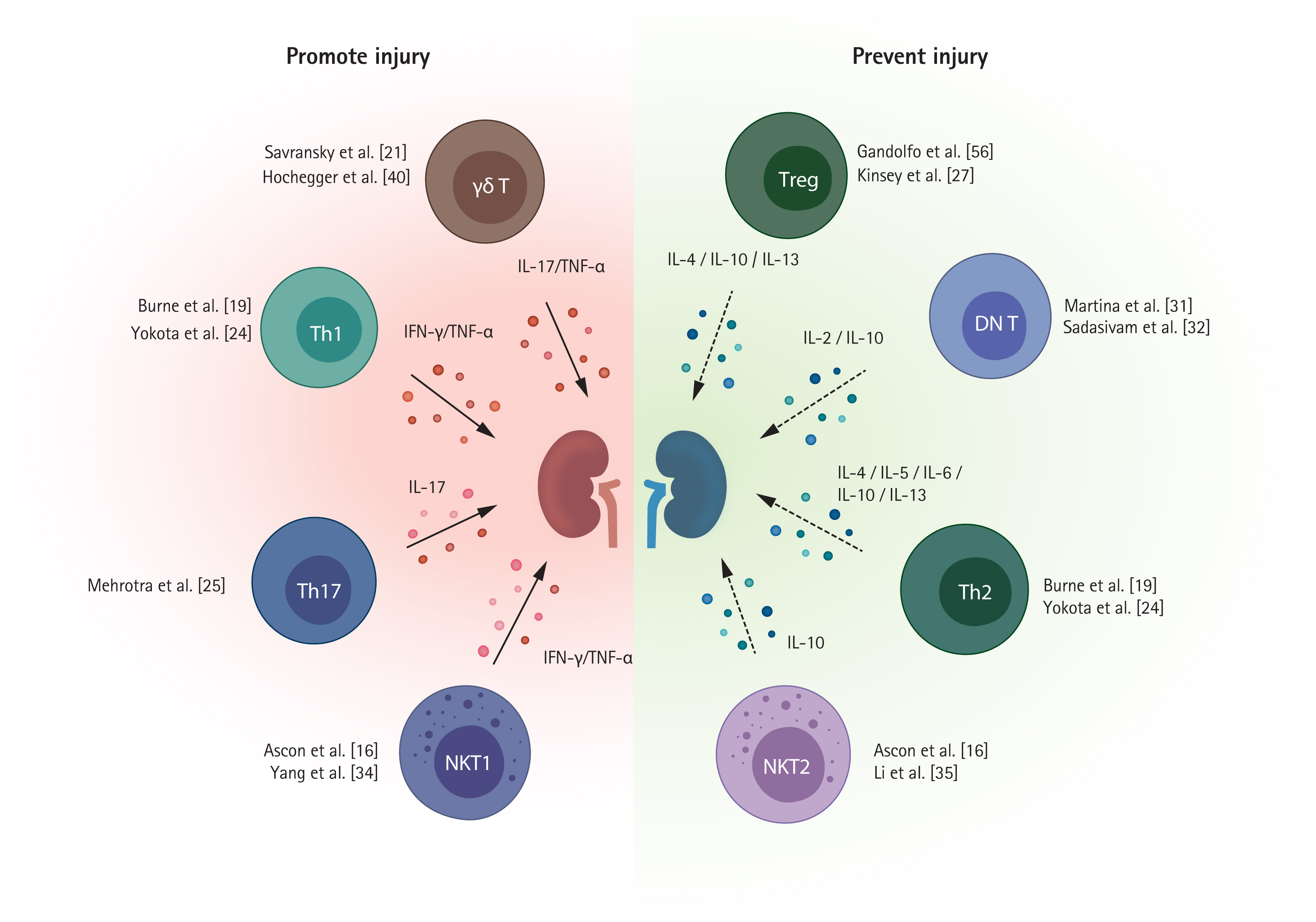
Several T cell subpopulations exhibit distinct roles during the injury process (Fig. 1). Below we discuss the major subpopulations of T cells.

CD4+ T cells

CD4+ T cells are major mediators of early renal injury, supported by reports that mice lacking CD4+ T cells are more protected from IRI than those lacking CD8+ T cells [19]. CD4+ T cell adoptive transfer to CD4+ T cell-deficient mice reconstituted renal injury [19]. Sphingosine-1-phosphate (S1P) and S1P receptor (S1PR) signaling is required for T cell trafficking to peripheral tissues from lymphoid organs, and S1PR1 selective agonists block T cell egress from lymphoid tissue [46]. S1PR1 selective agonist pretreatment before IRI exerted a renoprotective effect by inhibiting CD4+ T cell infiltration into post-ischemic kidneys [17]. Renal CD4+ T cells can undergo polarization into T helper (Th) cells, which comprise three major subsets—Th1, Th2, and Th17.

Th1/Th2 cells

Th1 cells secrete IFN-γ and TNF-α and are regulated by the transcription factor T-bet. Th2 cells produce IL-4, IL-5, IL-10, and IL-13 and are regulated by the transcription factor GATA binding protein 3 (GATA3) [47]. Th1 cells and Th2 cells are likely to have opposite effects on the pathogenesis of ischemic AKI: Th1 cells play a pathogenic, and Th2 cells a protective, role [24,48]. The signal transducer and activator of transcription 4 (STAT4) gene and STAT6 gene are responsible for Th1 differentiation and Th2 differentiation, respectively. Mice lacking STAT4 were protected from IRI, whereas mice lacking STAT6 showed more severe injury after IRI [24]. The dendritic cell sphingosine 1-phosphate 3 (S1P3) pathway is involved in Th1 polarization, and adoptive transfer of S1P3-deficient dendritic cells protected against IRI by inducing Th2 polarization [48].

Th17 cells

Th17 cells are regulated by the transcription factors STAT3 and retinoic acid receptor-related orphan receptor-γt (RORγt) and produce IL-17, which recruits innate immune cells [49,50]. Th17 cell polarization is mainly driven by the STAT3 activating cytokines IL-1 and IL6, and transforming growth factor β (TGFβ) contributes by suppressing Th1 or Th2 polarization [51,52]. Along with their detrimental roles in autoimmune disorders and transplantation, they play pathogenic roles in ischemic AKI [25,53]. IL-1 secreted by renal dendritic cells and macrophages promotes Th17 differentiation and activation following kidney injury [54]. Increased kidney Th17 cells were observed within the first 3 days after IRI and decreased to a comparable steady state within 7 days of renal recovery [25]. More specifically, calcium channel Orai1-expressing Th17 cells are involved in IL-17 production in IRI, contributing to tissue damage [26].

CD8+ T cells

CD8+ T cells can differentiate into cytotoxic effector cells with upregulation of TNF-α and IFN-γ [7], but they have been less studied than CD4+ T cells in AKI. CD8+ T cells may contribute to renal injury by producing INF-γ in renal IRI [7]. However, compared to CD4+ T cells, CD8+ T cells are likely to have a minor effect on renal IRI. Unlike CD4-deficient mice, mice lacking CD8+ T cells showed a comparable level of injury to WT mice after IRI [19]. The roles of CD8+ T cells were evaluated using GF mice. GF status was linked to a reduced proportion of effector memory phenotypes (CD44high CD62Llow) of CD8+ T cells and worsened kidney injury after IRI with enhanced CD8+ T cell trafficking into post-ischemic kidneys. Therefore, naive CD8+ T cells associated with underexposure to microbial stimuli appear to have a pathogenic role in post-ischemic kidneys [9].

Regulatory T cells

Regulatory T cells (Tregs) have CD4 and CD25 as surface markers and upregulation of nuclear transcription factor forkhead box P3 (FoxP3) [55]. A small percentage of CD4+ T cells are Tregs in normal mouse kidneys, accounting for only 3% to 4% of total CD4+ T cells [7,56]. Contrary to the deleterious role of conventional CD4+ T cells, Tregs exert protective effects by suppressing proinflammatory responses despite their small numbers [57].
Partial Treg depletion with an anti-CD25 antibody (PC61) in WT mice before IRI resulted in more severe injury, with increased neutrophil and macrophage trafficking and upregulation of Il6, Tnfα, and Tgfβ in post-ischemic kidneys [27]. Adoptive Treg transfer to mice lacking lymphocytes (Rag1 KO mice) prior to IRI showed a protective effect. An anti-inflammatory cytokine, IL-10 produced by Tregs, appears to be an important mediator for their protective mechanism. IL-10-deficient Treg adoptive transfer did not show a protective effect in Rag1 KO mice [27]. A study using a more complex IRI model to simulate ischemic preconditioning demonstrated that Tregs are involved in the protective mechanism of the ischemic preconditioning [28].
An important mechanism by which Tregs exert an anti-inflammatory effect on AKI is adenosine generation by CD73, to which adenosine 2A receptor (A2AR) on Tregs contributes in an autocrine manner [29]. Notably, PD-1 expression on Tregs is likely to be vital for their anti-inflammatory function. Activation of the A2AR signaling pathway enhanced PD-1 expression on Tregs, and PD-1 depletion on Tregs abolished the protective effect [29]. Blocking PD-1 ligands using an anti-PD-L1 or PD-L2 antibody before Treg adoptive transfer also negated its protective effect on IRI [23].
Because the complex of IL-2 and a particular form of IL-2 antibody (JES6-1) reportedly preferentially expanded Tregs, an IL-2/anti-IL-2 complex (IL-2C) has been tested in renal IRI [30]. IL-2C induced expansion of Tregs in kidneys and ameliorated renal injury. Treg depletion by anti-CD25 antibody abolished the protective effect of IL-2C [30].
The pro-reparative role of Tregs during the recovery phase of ischemic AKI will be reviewed separately in the next section.

Double-negative αβ T cells

Double-negative (DN) T cells (TCRαβ+, CD4, CD8 T cells) are an important subset of kidney resident αβ T cells [7]. Although rare in lymphoid organs, DN T cells represent approximately 20% to 25% of kidney αβ T cells in normal mouse kidneys and post-ischemic kidneys [7,31]. Considerable proportions of DN T cells were also found in human ischemic kidneys [7,31]. DN T cells were previously thought to originate from CD4+ or CD8+ T cells by losing their CD4 or CD8 receptor [58,59], but are now considered a discrete subset distinguished from conventional CD4+ or CD8+ T cells in kidneys because DN T cells were present in β2m-deficient mice and major histocompatibility complex (MHC) II-deficient mice [32]. IL-2 is required for activation and proliferation of DN T cells [32]. Kidney DN T cells contain PD-1+ subset and NK1.1+ subset in both human and mice [32]. DN T cells have different immunophenotypic profiles with higher expression of CD28, CD44, and CD69 compared to CD4+ and CD8+ T cells in a steady state and significantly higher proliferation capacities [31]. Thus, DN T cells are expected to play distinct roles in AKI pathophysiology.
Mouse anti-thymocyte globulin treatment increased the DN T cell proportion in the post-ischemic kidney [33]. DN T cells increase rapidly within 24 hours after IRI and likely have an IL-10-dependent protective function in the early injury process [31]. An adoptive transfer of DN T cells from Faslgld mice before IRI showed a protective effect, and IL-10 neutralization with an anti-IL-10 receptor antibody abrogated the protective effect [31].
The PD-1+ DN T cell subset was predominant following IRI, whereas the NK1.1+ subset decreased; thus, PD-1+ DN T cells may be important in the pathogenesis of ischemic AKI. Based on the higher expression of PD-1 on DN T cells from human kidney tumor tissue, DN T cells may be relevant targets of immune checkpoint inhibitor therapy [32]. Few studies have addressed DN T cells, so further research is needed.

Unconventional T cells

Unconventional T cells—including γδ T cells and natural killer (NK) T cells—are different types of T cells from conventional CD4+ and CD8+ T cells in their TCR usage. Unconventional T cells present as minor T cell subsets in the kidney [60]. Because unconventional T cells can act in a non-MHC-restricted manner, a feature of innate immunity, they are also referred to as ‘innate-like T cells’ in the literature [60]. These unconventional T cells are much less understood and have extensive interspecies differences unlike conventional T cells, hampering studies using experimental animal models.

Natural killer T cells

NK T cells can directly recognize antigens presented on CD1d molecules from antigen-presenting cells, thus are defined as CD1d-restricted T cells. NK T cells reportedly infiltrate post-ischemic kidneys at a very early time point [16]. NK T cells were also found in renal tissue of patients with acute tubular necrosis, but are rare in normal human kidney specimens [34]. Given the capability of NK T cells to become rapidly activated, resulting in cytokine production (including INF-γ) and recruitment of innate immune cells [35], they are considered a major effector and modulator cell type in the very early phase of IRI. NK T cells are likely to mainly have a deleterious effect on ischemic AKI [35]. NK T cell depletion using an anti-NK1.1 antibody and mice lacking NK T cells were protective against ischemic AKI [35]. Blocking NK T cell activation using an anti-CD1d antibody ameliorated renal injury [35]. Tolerogenic dendritic cells treated with an A2AR agonist exerted a protective effect on IRI by suppressing NK T cell production of IFN-γ [36]. In addition, IL-33 is a mediator of NK T cell recruitment and activation in post-ischemic kidneys [37], whereas the hypoxia-inducible factor (HIF)-2α and A2AR pathways exert protective effects by restricting NK T cell infiltration and activation [38].
There are two subsets of NK T cells, type I NK T cells and type II NK T cells [61]. Because type I NK T cells have an invariant TCR α chain, they are also referred to as ‘invariant NK T cells,’ whereas type II NK T cells are called ‘diverse NK T cells’ based on their more diverse TCR α- and β-chain expression [61].
Type I NK T cells can be stimulated by α-galactosylceramide (α-GalCer), whereas type II NK T cells cannot [61]. α-GalCer injection in normal mice induced AKI by activating type I NK T cells in the kidney [39]. The TNF-α/FAS ligand pathway and perforin-mediated pathway are related to NK T cell-mediated glomerular injury and tubular injury, respectively [39].
Type II NK T cells are likely to have opposite roles to type I NK T cells in ischemic AKI. They can be stimulated by sulfatide and nonlipid antigens. Sulfatide injection into WT mice and type I NK T cell-deficient mice ameliorated renal injury following IRI. Sulfatide injection to CD1d-deficient mice (mice lacking both type I and II NK T cells) did not show a protective effect [34]. This renoprotective function in renal IRI is mediated via the HIF-1α and IL-10 pathways [34].

γδ T cells

γδ T cells exacerbate ischemic AKI—γδ T cell deficiency or depletion has a protective effect on renal IRI [21,40]. γδ T cells were identified in human healthy kidney tissues as a minor fraction of T cells, and their number in the kidney was correlated negatively with the estimated glomerular filtration rate and positively with the degree of fibrosis in patients with CKD [62]. γδ T cells infiltrate damaged kidneys from the blood following ischemic AKI; a rapid drop in circulating γδ T cell levels followed by increased γδ T cells in the kidneys was observed in human and mouse AKI [40,41]. Although the significance of γδ T cells in the pathogenesis of ischemic AKI is unclear, given their minor fraction and less protective effect on γδ T cell deficiency than αβ T cell deficiency, they are likely to have less influence on AKI pathophysiology compared to conventional αβ T cells [40,41].

ROLE OF T CELLS IN RECOVERY OR AKI TO CKD TRANSITION

Given the lack of established real-time biomarkers for AKI, clinical interventions mostly become available after the establishment of renal injury as well as in the recovery phase [63]. Moreover, therapeutic approaches that inhibit inflammatory processes during the injury phase may disrupt the adaptive repair process [6]. Thus, understanding and targeting the T cell-mediated repair process has high clinical importance, but few studies have focused on the recovery phase. Here, we review studies that reported long-term renal outcomes or conducted interventions after renal injury (Table 2) [28,30,56,6470].

CD4+ and CD8+ T cells

Chronic T cell infiltration into post-ischemic kidneys was investigated using IRI models. The study based on immunohistochemistry findings demonstrated increased CD4+ T cells at 6 weeks after IRI, and there was enhanced expression of IL-1 and C-C motif chemokine ligand 5 (CCL5) [64]. Subsequent studies based on flow cytometry with isolated kidney infiltrating mononuclear cells showed consistent findings [65]. Activated phenotype (CD69+) and effector-memory phenotypes (CD44high CD62L) of T cells were the dominant phenotypes during the long-term recovery phase [65]. Moreover, expression of inflammatory cytokines and chemokines was enhanced in post-ischemic kidneys until 6 weeks [65]. Thus, T cell activation and an inflammatory microenvironment following ischemic AKI are likely to be maintained chronically and play important roles in tubular regeneration or the AKI to CKD transition. A study using single-cell RNA-sequencing (scRNA-seq) showed a marked increase in the T cell compartment at the transcriptomic level during the recovery phase. Among T cell subtypes, Th17 cells were gradually increased after IRI until 4 weeks [66].
Transcriptomic analyses of sorted T cells from post-ischemic kidney tissue showed that their Ccl5 gene expression was consistently upregulated until 6 weeks after IRI [67]. Also, C-C motif chemokine receptor 5 (CCR5) blockade using an anti-CCR5 antibody improved renal outcomes functionally and structurally following ischemic AKI [67]. This finding was reproduced by scRNA-seq data showing the Ccl5-Ccr5 interaction was significantly associated with failed repair of tubular cells following IRI [71].
Extrarenal T cells are also activated until the recovery phase of ischemic AKI. Enhanced INF-γ secretion was observed from splenic T cells at 6 weeks after IRI [64]. Adoptive transfer of splenic T cells isolated from post-IRI donor mice at 6 weeks after IRI induced albuminuria in naïve recipient mice at 12 weeks after the injection [68]. This finding suggests that the inflammatory property of extrarenal T cells is chronically maintained following renal IRI, which may affect renal recovery.

Regulatory T cells

Tregs not only prevent damage in the early phase but also have a pro-reparative function by preventing fibrosis development during the recovery phase. A late Treg (CD4+ CD25+ FoxP3+) trafficking into post-ischemic kidneys were demonstrated [56], and IL-10 expression on Tregs were unregulated during recovery phase [28]. A scRNA-seq based study reproduced the finding of late Treg accumulation at the transcriptomic level [66]. Notably, it was also revealed that a set of reparative genes in Tregs were expressed at the highest levels at later time points following IRI, indicating their important roles in the repair process [66].
Adoptive transfer of Tregs isolated from spleen into post-IRI mice at 24 hours after IRI mitigated structural renal injury and enhanced tubular regeneration at 10 days after IRI, accompanied by a reduction of the CD4+ T cell TNF-α production [56]. Treg depletion using an anti-CD25 antibody starting at 24 hours after IRI suppressed tubular regeneration and increased T cell TNF-α and IFN-γ production [56]. Treg expansion using an IL-2C reduced renal fibrosis at 4 weeks after IRI. Also, Treg depletion with an anti-CD25 antibody abrogated the beneficial effect of IL-2C [30]. A combination of IL-2C and IL-33 administration markedly increased Tregs in post-ischemic kidneys and reduced renal injury with less fibrosis at 4 weeks after IRI [66].
Although theoretically promising, the use of immunosuppressive agents in AKI has failed to achieve favorable outcomes. However, our understanding of the role of Tregs has increased. Mycophenolate mofetil worsened tubular injury and reduced Treg expansion in post-ischemic kidneys at 10 days after IRI, highlighting the reparative role of Tregs during renal recovery [69]. Because early initiation of mammalian target of rapamycin (mTOR) inhibitors can retard wound healing and delay renal recovery [72,73], delayed administration of rapamycin was evaluated. However, it also increased albuminuria following IRI [74]. Notably, rapamycin induces Treg expansion and IL-10 upregulation in vitro. Thus, rapamycin ex vivo-treated Tregs were adoptively transferred at 24 hours after IRI. There was less tubular injury and lower expression of fibrosis markers in the rapamycin ex vivo-treated Treg transfer group at 14 days after IRI [70].
There was a clear transcriptional difference between Tregs in a regenerative environment and those in a profibrotic environment. Tregs seem to have complicated tissue specific functions, warranting further research [66].

T cell and distant organ crosstalk during ischemic AKI

Although renal failure can be managed with renal replacement therapy, unlike other organ failures, AKI-associated mortality remains high even when prompt dialysis is available [75,76]. Thus, increased mortality in patients with AKI cannot be solely explained by reduced renal clearance per se, and mechanisms beyond that are postulated [77]. One is the inflammatory response by kidney T cells and other immune cells, which has promise as a therapeutic target to reduce AKI-associated mortality. In this section, we briefly review the role of T cell-mediated systemic inflammatory effects on distant organs.
Pulmonary inflammation is a common AKI-related distant organ damage [77]. AKI induces acute lung injury or acute respiratory distress syndrome (ARDS) [78]. Beyond volume overload due to reduced renal clearance, the systemic inflammatory effect of AKI contributes to AKI-associated pulmonary complications [79]. Higher levels of circulating inflammatory cytokines were found in patients with AKI-related ARDS than in ARDS patients without AKI [80]. A landmark experimental study demonstrated that renal IRI generated distinguishable lung transcriptomic profiles (including upregulated proinflammatory and proapoptotic pathways) from those induced by bilateral nephrectomy [81]. T cells traffic into the lungs and cause pulmonary endothelial apoptosis through the caspase 3 pathway during ischemic AKI [82]. T cell-deficient mice did not show caspase-3 activation, and adoptive transfer of T cells from WT mice into T cell-deficient mice reconstituted pulmonary endothelial apoptosis [82]. Circulating IL-6 is a major pathogenic mediator of lung injury following AKI and induces upregulation of pulmonary CXCL1 expression [83].
AKI is also associated with adverse cardiac events, and interactions between heart and kidney diseases are clinically defined as cardiorenal syndrome [84]. Among the five subtypes of cardiorenal syndrome, the pathophysiology of cardiorenal syndrome type 3, acute cardiac dysfunction caused by an abrupt and primary worsening of kidney function, is less studied [78]. T cell-mediated mechanisms including systemic inflammation and activation of neurohormonal pathways may be involved [78,85]. This concept is supported by experimental evidence showing that renal IRI induced cardiac cell apoptosis but bilateral nephrectomy did not [86]. This cardiac injury was accompanied by increased levels of TNF-α and IL-1 in heart tissue and the systemic circulation [86]. Importantly, the activation of neurohormonal pathways such as the renin-angiotensin-aldosterone system (RAAS) in AKI promotes cardiac inflammation and fibrosis [87]. RAAS is strongly related to T cell-mediated inflammation—renal T cells not only express type 1 angiotensin II receptor but also synthesize angiotensin II [8890]. Thus, RAAS activation in AKI-heart cross talk may amplify the deleterious effect on cardiac inflammation by a T cell-mediated mechanism.
Neurologic manifestations are well known complications of AKI. The blood-brain barrier (BBB) interacts with circulating immune cells, leading to loss of the endothelial lining [91], and AKI-related systemic inflammation can induce BBB disruption [92]. Ischemic AKI can increase levels of soluble inflammatory proteins in the brain, which is associated with abnormal neuropsychiatric behaviors [93,94]. As an additional key mechanism, neurohormonal changes have been reported to contribute to kidney-brain crosstalk [95]. Renocerebral sympathetic reflex accompanied by both renal and cerebral RAAS activation play a crucial role in AKI-induced brain inflammation [96].
Gut microbiota dysbiosis is related to kidney-gut crosstalk [97]. AKI was linked to altered gut microbial populations at the genus and family levels [98]. The bidirectional relationship between the gut microbiota and AKI was discovered using GF mice [9]. The GF mice exhibited worse renal outcomes following IRI and had more NK T cells in the kidney. Reconstitution of the gut microbiota by stool transfer from WT mice before IRI alleviated their susceptibility to IRI [9]. Also, transfer of stool from WT post-IRI mice led to exacerbation of renal injury in GF mice [99]. The gut microbiota exerts immunomodulatory effects via its metabolites [100,101]. These include short-chain fatty acids (SCFAs), which have an anti-inflammatory effect by modulating immune cell function. SCFAs promote Treg proliferation in the intestine and reduce dendritic cell accumulation in the kidney [102,103]. A lack of a microbiota in GF mice affects T cell differentiation and function due to the absence of SCFAs [104].

NOVEL TECHNIQUES FOR STUDYING T CELLS IN AKI

Opportunities to apply novel techniques to improve understanding of kidney T cells in AKI pathophysiology are increasing. Below we briefly discuss recent notable technological advances, including spectral flow cytometry, scRNA-seq, and spatial transcriptomics.
Although fluorescence-based flow cytometers are commonly used to study kidney immune cells, their low resolution and limited multiplexing capacity have discouraged more sophisticated analyses. The recently developed spectral cytometers record full emission patterns across the entire wavelength range, whereas conventional flow cytometers detect only the peak emission signals [105]. A spectral unmixing algorithm enables differentiation of fluorophores with similar peak signals and extraction of autofluorescence contributions, allowing highly complex multicolor panels [106]. The combination of spectral flow cytometry and machine learning-based high-dimensional analysis enables exploration of the roles of immune cells in kidney diseases [107,108].
Although microarray or bulk RNA sequencing data provide the average transcriptomic profiles of predominant cell populations, scRNA-seq assesses gene expression at individual cell levels. This enables to exploit changes in specific cell types independently of predefined hierarchies [109,110]. As such, scRNA-seq enables identification of unrecognized cell populations or discovery of candidate markers for previously defined cell populations. In the past few years, many sensitive and accurate scRNA-seq protocols have been developed [109,110].
Spatial transcriptomic sequencing combines the benefits of histology with high throughput RNA sequencing [111]. RNAs released from intact tissue sections are captured by arrayed oligo-dT spots and bar codes with spatial information. Because the major limitation of single-cell based approaches is loss of spatial information within the tissue, integrating data from spatial transcriptomics and scRNA-seq allow mapping of transcriptomically defined cell types onto a tissue [112]. Although the spatial transcriptomic techniques need more improvement due to their limited resolution, further development of spatial transcriptomic techniques can potentially clarify the spatiotemporal fingerprints of immune cells.

NOVEL THERAPEUTIC APPROACHES

Based on the important roles of T cells in AKI, therapeutic approaches targeting T cells have shown promising outcomes. Below we briefly discuss interventions using adoptive cell transfer, stem cell-based therapy, and approaches targeting gut-kidney crosstalk.

T cell-based therapy

Given the anti-inflammatory properties of Tregs and DN T cells, adoptive transfer of these cells has been investigated in preclinical studies. Adoptive transfer of Tregs before AKI attenuated structural and functional renal injury in both IRI and cisplatin AKI models [27,113]. Moreover, Treg transfer after AKI enhanced tubular regeneration and improved renal outcomes during the recovery phase [56]. Adoptive transfer of ex-expanded Tregs by ex vivo rapamycin treatment attenuated tubular injury and reduced expression of fibrosis markers [70]. DN T cell transfer ameliorated renal injury in both IRI and cisplatin AKI models [31,114], but the effect on renal recovery is unknown. Given that clinical trials of Treg adoptive transfer in kidney transplant recipients showed feasibility and a favorable safety profile [115], cell-based therapies show promise for AKI. However, the preparation required, such as cell collection, isolation, and ex vivo expansion, hamper clinical application because the clinical course of AKI is unpredictable in most patients.

Stem cell-based approaches

Despite the promising results from preclinical trials involving direct injection of mesenchymal stem cells (MSCs) [116,117], this approach has been questioned as the studies failed to achieve even modest survival durations for transplanted cells. Therefore, the beneficial effects did not rely on differentiation of the injected cells [116,117]. Rather, transplanted cells appear to promote tissue regeneration by passing signals in exosomes or microparticles to surrounding cells [118]. Although the mechanism of their paracrine effect is unclear, much evidence supports the immunomodulatory properties of stem cells and the critical role of T cells [119]. MSC treatment in mice attenuated renal injury by facilitating Treg trafficking to the injured kidneys in both IRI and cisplatin AKI. And the protective effect was blunted by anti-CD25 antibody-mediated depletion of Tregs [120,121]. MSC treatment after reperfusion in rat IRI reduced the expression of inflammatory cytokines, including IL-1β, TNF-α, and IFN-γ, and increased levels of anti-inflammatory cytokines, including IL-10 and basic fibroblast growth factor [116]. Stem cell-based therapies appear to be safe and promising for AKI [118], and a clinical trial is underway (NCT03015623).

Therapeutic strategies targeting gut-kidney crosstalk

The mechanistic evidence supporting a bidirectional relationship between the gut microbiota and AKI led to interventional studies targeting the gut microbiota. Gut microbiota depletion using broad-spectrum antibiotics (ampicillin, neomycin, vancomycin, and metronidazole) showed a protective effect on AKI, and stool transplantation from untreated mice abolished their renoprotective effect [122]. Importantly, a reduced Th1 and Th17 response and Treg expansion were suggested as the mechanism by which antibiotic microbiota depletion attenuates renal injury [99]. By contrast, GF mice showed deteriorated renal outcomes following IRI [9], and another combination of antibiotics (ampicillin, vancomycin, and levofloxacin) resulted in more severe structural injury [123]. Therefore, selective deletion of deleterious microbiota populations may exert a protective effect on AKI [124].
Because the gut microbiota controls immune cells via metabolites [124], microbiota-derived products were tested in AKI models. SCFA ameliorated IRI and inhibited dendritic cell maturation, thereby reducing CD4+ and CD8+ T cell proliferation [102]. Similarly, a gut microbial D-amino acid product, D-serine, protected against IRI [125], which warrants further investigation.
The microbial taxa responsible for the protective or exacerbating effects on AKI are unknown. Studies of interventions targeting the gut-kidney axis and sequencing analyses of the gut microbiota may lead to the development of AKI-specific prebiotic or probiotic treatment approaches.

CONCLUSIONS

T cells are important in the pathogenesis of ischemic AKI. Recent advances in our understanding of their roles in repair and regeneration have provided insight into the AKI to CKD transition. Thus, immunomodulatory therapies targeting the T cell mediated pathophysiology show promise for improving AKI outcomes. Other types of immune cells such as mononuclear phagocytic cells, neutrophils, B cells, and innate lymphoid cells are also involved in ischemic AKI pathogenesis, although we did not review them here. Future studies using cutting-edge technologies will facilitate the development of precision-directed therapies.

Acknowledgments

Kyungho Lee was supported by the Korea Health Industry Development Institute (HI19C1337) and the National Research Foundation of Korea (NRF-2021R1A6A3A03039863).
Hye Ryoun Jang was supported by the Korea Health Industry Development Institute (HC20C0085), the National Research Foundation Korea (2016R1A2B4008235 & 2019R1A2B5B01069346, NRF-2019R1A5A2027340), and the Korean Health Technology R&D Project through the Korean Health Industry Development Institute (KHIDI), funded by the Ministry of Health & Welfare, Republic of Korea (grant number: HI14C3484).
The illustration was created using BioRender.com Software.

Conflict of Interest

No potential conflict of interest relevant to this article was reported.

Figure 1
Proinflammatory and anti-inflammatory T cells in ischemic acute kidney injury. T helper 1 cell (Th1), Th17, γδ T cells, and type I natural killer (NK) T cells (NKT1) promote renal injury and exert a proinflammatory effect in ischemic acute kidney injury, whereas regulatory T cells (Tregs), double-negative (DN) T cells, Th2 cells, and type II NK T cells (NKT2) play protective roles and have anti-inflammatory properties. Numbers of key landmark studies for each subset of T cells are presented. IL, interleukin; TNF-α, tumor necrosis factor α; IFN-γ, interferon γ.
kjim-2021-526f1.jpg
Table 1
Role of T cells during the injury phase of ischemic acute kidney injury
Study Major findings
T cells
 Rabb et al. (2000) [18] CD4/CD8-deficient mice were protected with reduced neutrophil trafficking
T cell-renal tubular epithelial cell adhesion is enhanced after hypoxia-reoxygenation in vitro
 Burne et al. (2001) [19] T cell-deficient mice (nu/nu mice) were protected
Adoptive transfer of WT T cells to T cell-deficient mice reconstituted renal injury
CD28-deficient T cell transfer to T cell-deficient mice did not reinduce injury phenotype
 Burne-Taney et al. (2003) [20] T cell-deficient mice (nu/nu mice) were protected from whole body IRI
T cell-deficient mice (nu/nu mice) showed attenuated expressions of Icam1 after whole body IRI
 Burne-Taney et al. (2005) [42] T cell adoptive transfer to Rag1-deficient mice (mice lacking both T and B cells) led to protection
 Ascon et al. (2006) [16] CD4+ and CD8+ T cells traffic into post-ischemic kidneys during the early injury phase
 Savransky et al. (2006) [21] TCR αβ-deficient mice were protected
TCR αβ-deficient mice showed reduced TNF-α and IL-6 expression in post-ischemic kidneys at 24 hours
 Satpute et al. (2009) [22] T cell-deficient mice (nu/nu mice) reconstituted with T cells having limited TCR repertoire (DO11.10 T cells) were protected from IRI compared to those reconstituted with WT T cells
Antigen specific activation of DO11.10 T cells using OVA-CFA abolished its protective effect
 Jaworska et al. (2015) [23] Blocking or genetical deficiency of PD-L1 or PD-L2 was protected
CD4+ T cells
 Burne et al. (2001) [19] CD4+ T cell-deficient mice were protected
Adoptive transfer of WT CD4+ T cells to T cell-deficient mice reconstituted renal injury
Adoptive transfer of CD4+ T cells lacking CD28 or IFN-γ to T cell-deficient mice did not restore injury phenotype
 Yokota et al. (2003) [24] STAT4-deficient mice were protected, whereas STAT6-deficient mice had more severe injury
STAT4-deficient T cells showed higher IL-4 expression and lower IFN-γ expression
IL-4-deficient mice showed more severe renal injury
 Lai et al. (2007) [17] CD4+ T cells infiltrated post-ischemic kidneys as early as 1 hour
S1PR1 agonist treatment reduced CD4+ T cell infiltration and renal injury
 Mehrotra et al. (2015) [25] Th17 cells (CD4+ IL-17+ T cells) increased during early injury phase thereafter deceased to the control level at 7 days
 Mehrotra et al. (2019) [26] Store-operative calcium entry channel, Orai1 contributed to IL-17 expression in CD4+ T cells
Pharmacologic Orai1 blockade attenuated IL-17+ cell activation and mitigated renal injury
 CD8+ T cells
 Burne et al. (2001) [19] Mice lacking CD8+ T cells mice were not protected from IRI
 Jang et al. (2009) [9] GF mice have lower percentage of effector memory (CD44high CD62Llow) CD8+ T cells
GF mice showed more severe injury with enhanced CD8+ T cell trafficking
Tregs
 Kinsey et al. (2009) [27] Treg depletion using anti-CD25 antibody (PC61) before IRI worsened renal injury with increased IFN-γ producing neutrophil infiltration
Rag1 KO mice (mice lacking both T and B cells) that received lymphocytes from Sf mice (mice lacking FoxP3) showed more severe renal injury compared to those that received WT lymphocytes at 24 hours
Co-transfer of Sf lymphocytes and Tregs alleviated susceptibility to IRI in Rag1 KO mice
IL-10-deficient Treg adoptive transfer did not show protective effect in Rag1 KO mice, whereas WT Treg transfer was protective.
 Kinsey et al. (2010) [28] Adoptive transfer of Tregs prior to IRI showed protective effect
Treg depletion in preconditioned mice with anti-CD25 antibody (PC61) abolished protective effect of ischemic preconditioning
 Kinsey et al. (2012) [29] CD73 or A2AR deficiency on Tregs abolished protective effect
Pharmacologic A2AR activation on Tregs augmented protective effect and upregulated PD-1 expression on Tregs
Adoptive transfer of PD-1 blocking antibody treated Tregs did not show protective effect
 Kim et al. (2013) [30] Treg expansion by IL-2C treatment before IRI ameliorated renal injury
Treg depletion by anti-CD25 antibody (PC61) treatment abolished the protective effect of IL-2C
 Jaworska et al. (2015) [23] Blocking PD-1 ligands with anti-PD-L1 or PD-L2 antibody abolished protective effect of Treg adoptive transfer
DN T cells
 Ascon et al. (2008) [7] DN T cells existed in normal kidneys and post-ischemic kidneys
 Jang et al. (2009) [33] Mouse anti-thymocyte globulin treatment increased percentage of DN T cells among total T cells
 Martina et al. (2016) [31] DN T cells have higher expression of IL-10 and IL-27 than CD4+ T cells in steady state
DN T cells rapidly increased within 24 hours after IRI
IL-10 expression on DN T cells was upregulated following IRI, whereas IL-27 expression was decreased
DN T cell adoptive transfer before IRI attenuated renal injury, and IL-10 blocking with anti-IL-10 receptor antibody abrogated protective effect of DN T cells
DN T cells were present in human ischemic kidney tissue with considerable proportion
 Sadasivam et al. (2019) [32] PD-1+ and NK1.1+ subsets are two major DN T cell subsets in both human and mice
DN T cells required IL-2 for their activation and function in vitro and in vivo
PD-1+ subset predominated kidney DN T cells in post-ischemic kidneys
γδ T cells
 Savransky et al. (2006) [21] γδ TCR-deficient mice had less structural renal injury
 Hochegger et al. (2007) [40] γδ T cell-deficiency and γδ T cell depletion using anti-γδ TCR antibody showed protective effect
γδ T cell-deficiency reduced αβ T cell trafficking to post-ischemic kidneys during early injury phase
 Gocze et al. (2018) [41] Decline of γδ T cells in circulating blood correlated with urinary cell stress biomarker levels in human (TIMP-2 and IGFBP7)
γδ T cells increased in post-ischemic kidneys, while circulating γδ T cells decreased at 24 hours after IRI
NK T cells
 Ascon et al. (2006) [16] Traffic as early as 3 hours and reduced to baseline level at 24 hours after IRI
 Li et al. (2007) [35] NK T cell depletion using anti-NK1.1 antibody or blocking NK T cell activation by anti-CD1d antibody attenuated renal injury
Type I NK T cell-deficient mice (Jα 18−/−) were protected from IRI
NK T cells mediated neutrophil infiltration and IFN-γ production in IRI
 Yang et al. (2011) [34] Type II NK T cell activation by sulfatide injection reduced renal injury in WT mice and type I NK T cell deficient mice (Jα 18−/−), but not in NK T cell-deficient mice (CD1d −/−)
Adoptive transfer of sulfatide treated NK T cells attenuated renal injury in WT, type I NK T cell-deficient mice, and NK T cell-deficient mice
Type II NK T cell mediated protective mechanisms involved in HIF-1α and IL-10 pathways
NK T cells (CD3+ Vα24+) were found in kidney tissue from patients with acute tubular necrosis
 Zhang et al. (2016) [38] HIF-2α/A2AR axis served protective mechanism by restricting NK T cell infiltration and activation
 Ferhat et al. (2018) [37] IL-33 drove recruitment of type I NK T cells and induced INF-γ/IL-17 production
 Uchida et al. (2018) [39] α-Galactosylceramide administration induced AKI and hematuria via NK T cell activation
Perforin-mediated pathway and TNF-α/Fas ligand pathways are related to glomerular injury and tubular injury, respectively

IRI, ischemia-reperfusion injury; WT, wild-type; ICAM-1, intercellular adhesion molecule 1; TCR, T cell receptor; TNF-α, tumor necrosis factor α; IL, interleukin; OVA-CFA, ovalbumin in complete Freund’s adjuvant; PD-L, programmed death ligand; IFN-γ, interferon γ; STAT, signal transducer and activator of transcription; S1PR1, sphingosine-1-phosphate receptor 1; GF, germ-free; Treg, regulatory T cell; KO, knockout; Sf, scurfy; FoxP3, forkhead box P3; A2AR, adenosine 2A receptor; PD-1, programmed death-1; IL-2C, IL-2/anti-IL-2 complex; DN, double-negative; TIMP-2, tissue inhibitor of metalloproteinase 2; IGFBP7, insulin-like growth factor-binding protein 7; NK, natural killer; HIF, hypoxia-inducible factor; AKI, acute kidney injury.

Table 2
Role of T cells during the repair phase of ischemic acute kidney injury
Study Major findings
T cells
 Burne-Taney et al. (2005) [64] CD4+ T cells infiltration in post-ischemic kidneys at 6 weeks after IRI
Upregulation of IL-1 and CCL5 expression in post-ischemic kidneys at 6 weeks after IRI
Enhanced IFN-γ secretion of splenic T cells at 6 weeks after IRI
 Burne-Taney et al. (2006) [68] Adoptive transfer of splenic T cells obtained from post-IRI donor mice resulted in albuminuria in normal recipient mice at 12 weeks after transfer
Increased activated (CD3+ CD25+) or memory phenotypes (CD4+ CD44+) of T cells in recipient spleens
 Ascon et al. (2009) [65] Activated phenotypes (CD69+) of T cells were expanded at late recovery phase (6 weeks from BIRI, 11 weeks from UIRI)
Effector memory (CD44high CD62L) phenotype T cells were major T cell phenotype at 6 weeks after BIRI or UIRI
Inflammatory cytokines and chemokines (Il1b, Il6, Tnfa, Ifng, Cxcl2, and Ccl5) were upregulated in post-ischemic kidneys at 6 weeks after UIRI
T cell depletion reduced Ifng expression
 Ko et al. (2012) [67] Upregulation of Ccr5 gene expression on T cells was maintained from early injury to late recovery (4 weeks)
CCR5 blockade with anti-CCR5 antibody after IRI attenuated renal injury and reduced renal trafficking of activated phenotype (CD69+) T cells on day 3 after IRI
 Duraes et al. (2020) [66] scRNA-seq analyses of kidney CD45+ cells showed increased T cell cluster at 4 weeks after IRI
scRNA-seq analyses of kidney CD4+ T cells showed substantial expansion of Th17 cell cluster during the recovery phase
Regulatory T cells
 Gandolfo et al. (2009) [56] Marked Treg (CD4+ CD25+ FoxP3+) expansion in post-ischemic kidneys on day 3 and day 10 after IRI
Adoptive transfer of Tregs (CD4+ CD25+) at 24 hours after IRI reduced structural injury, enhanced tubular regeneration, and reduced TNF-α production in CD4+ T cells on day 10
Treg depletion with anti-CD25 antibody treatment at 24 hours after IRI inhibited tubular regeneration and increased TNF-α and IFN-γ production in CD4+ and CD8+ T cells
 Kinsey et al. (2010) [28] IL-10 producing Tregs increased at 7 days after IRI
 Gandolfo et al. (2010) [69] MMF administration at 48 hours post-ischemia reduced Treg expansion and worsened tubular injury on day 10 after IRI
MMF administration in T cell-deficient mice did not alter renal outcome
 Kim et al. (2013) [30] Treg expansion by IL-2C administration reduced renal fibrosis on day 28 after IRI
Treg depletion with anti-CD25 antibodies abrogated the beneficial effect of IL-2C treatment.
 Chen et al. (2016) [70] mTOR inhibition by rapamycin treatment in CD4+ T cells induced Treg expansion and enhanced expression of IL-10 and TGF-β1 in vitro.
Adoptive transfer of rapamycin ex vivo treated Tregs at 24 hours post-ischemia reduced structural renal injuries and a fibrosis marker expression on day 14 after IRI
 Duraes et al. (2020) [66] A set of gene expression in Tregs are highest at late recovery phase
Treg expansion by a combination of IL-2C and IL-33 treatment before IRI reduced kidney fibrosis on day 28 from IRI
Tregs in fibrotic and regenerative environments had distinct signatures of gene expression

IRI, ischemia-reperfusion injury; IL, interleukin; CCL5, C-C motif chemokine ligand 5; BIRI, bilateral ischemia-reperfusion injury; UIRI, unilateral ischemia-reperfusion injury; IFN-γ, interferon γ; CCR5, C-C motif chemokine receptor 5; scRNA-seq, single cell RNA-sequencing; Th17, T helper 17 cell; Treg, regulatory T cell; FoxP3, forkhead box P3; TNF-α, tumor necrosis factor α; MMF, mycophenolate mofetil; IL-2C, IL-2/anti-IL-2 complex; mTOR, mammalian target of rapamycin; TGF-β1, transforming growth factor β1.

REFERENCES

1. Chawla LS, Eggers PW, Star RA, Kimmel PL. Acute kidney injury and chronic kidney disease as interconnected syndromes. N Engl J Med 2014;371:58–66.
crossref pmid
2. Sharfuddin AA, Molitoris BA. Pathophysiology of ischemic acute kidney injury. Nat Rev Nephrol 2011;7:189–200.
crossref pmid
3. Han SJ, Lee HT. Mechanisms and therapeutic targets of ischemic acute kidney injury. Kidney Res Clin Pract 2019;38:427–440.
crossref pmid pmc
4. Park WY, Hwang EA, Jang MH, Park SB, Kim HC. The risk factors and outcome of acute kidney injury in the intensive care units. Korean J Intern Med 2010;25:181–187.
crossref pmid pmc
5. Kellum JA, Romagnani P, Ashuntantang G, Ronco C, Zarbock A, Anders HJ. Acute kidney injury. Nat Rev Dis Primers 2021;7:52.
crossref pmid
6. Rabb H, Griffin MD, McKay DB, et al. Inflammation in AKI: current understanding, key questions, and knowledge gaps. J Am Soc Nephrol 2016;27:371–379.
crossref pmid
7. Ascon DB, Ascon M, Satpute S, et al. Normal mouse kidneys contain activated and CD3+CD4− CD8− double-negative T lymphocytes with a distinct TCR repertoire. J Leukoc Biol 2008;84:1400–1409.
crossref pmid pmc
8. Park JG, Na M, Kim MG, et al. Immune cell composition in normal human kidneys. Sci Rep 2020;10:15678.
crossref pmid pmc
9. Jang HR, Gandolfo MT, Ko GJ, Satpute S, Racusen L, Rabb H. Early exposure to germs modifies kidney damage and inflammation after experimental ischemia-reperfusion injury. Am J Physiol Renal Physiol 2009;297:F1457–F1465.
crossref pmid pmc
10. Chen GY, Nunez G. Sterile inflammation: sensing and reacting to damage. Nat Rev Immunol 2010;10:826–837.
crossref pmid pmc
11. Kundert F, Platen L, Iwakura T, Zhao Z, Marschner JA, Anders HJ. Immune mechanisms in the different phases of acute tubular necrosis. Kidney Res Clin Pract 2018;37:185–196.
crossref pmid pmc
12. Gharaie Fathabad S, Kurzhagen JT, Sadasivam M, et al. T lymphocytes in acute kidney injury and repair. Semin Nephrol 2020;40:114–125.
crossref pmid
13. Jang HR, Rabb H. Immune cells in experimental acute kidney injury. Nat Rev Nephrol 2015;11:88–101.
crossref pmid
14. Rosner MH, Perazella MA. Acute kidney injury in the patient with cancer. Kidney Res Clin Pract 2019;38:295–308.
crossref pmid pmc
15. Hong MH. Nephrotoxicity of cancer therapeutic drugs: focusing on novel agents. Kidney Res Clin Pract 2021;40:344–354.
crossref pmid pmc
16. Ascon DB, Lopez-Briones S, Liu M, et al. Phenotypic and functional characterization of kidney-infiltrating lymphocytes in renal ischemia reperfusion injury. J Immunol 2006;177:3380–3387.
crossref pmid
17. Lai LW, Yong KC, Igarashi S, Lien YH. A sphingosine-1-phosphate type 1 receptor agonist inhibits the early T-cell transient following renal ischemia-reperfusion injury. Kidney Int 2007;71:1223–1231.
crossref pmid
18. Rabb H, Daniels F, O’Donnell M, et al. Pathophysiological role of T lymphocytes in renal ischemia-reperfusion injury in mice. Am J Physiol Renal Physiol 2000;279:F525–F531.
crossref pmid
19. Burne MJ, Daniels F, El Ghandour A, et al. Identification of the CD4(+) T cell as a major pathogenic factor in ischemic acute renal failure. J Clin Invest 2001;108:1283–1290.
crossref pmid pmc
20. Burne-Taney MJ, Kofler J, Yokota N, Weisfeldt M, Traystman RJ, Rabb H. Acute renal failure after whole body ischemia is characterized by inflammation and T cell-mediated injury. Am J Physiol Renal Physiol 2003;285:F87–F94.
crossref pmid
21. Savransky V, Molls RR, Burne-Taney M, Chien CC, Racusen L, Rabb H. Role of the T-cell receptor in kidney ischemia-reperfusion injury. Kidney Int 2006;69:233–238.
crossref pmid
22. Satpute SR, Park JM, Jang HR, et al. The role for T cell repertoire/antigen-specific interactions in experimental kidney ischemia reperfusion injury. J Immunol 2009;183:984–992.
crossref pmid
23. Jaworska K, Ratajczak J, Huang L, et al. Both PD-1 ligands protect the kidney from ischemia reperfusion injury. J Immunol 2015;194:325–333.
crossref pmid
24. Yokota N, Burne-Taney M, Racusen L, Rabb H. Contrasting roles for STAT4 and STAT6 signal transduction pathways in murine renal ischemia-reperfusion injury. Am J Physiol Renal Physiol 2003;285:F319–F325.
crossref pmid
25. Mehrotra P, Patel JB, Ivancic CM, Collett JA, Basile DP. Th-17 cell activation in response to high salt following acute kidney injury is associated with progressive fibrosis and attenuated by AT-1R antagonism. Kidney Int 2015;88:776–784.
crossref pmid pmc
26. Mehrotra P, Sturek M, Neyra JA, Basile DP. Calcium channel Orai1 promotes lymphocyte IL-17 expression and progressive kidney injury. J Clin Invest 2019;129:4951–4961.
crossref pmid pmc
27. Kinsey GR, Sharma R, Huang L, et al. Regulatory T cells suppress innate immunity in kidney ischemia-reperfusion injury. J Am Soc Nephrol 2009;20:1744–1753.
crossref pmid pmc
28. Kinsey GR, Huang L, Vergis AL, Li L, Okusa MD. Regulatory T cells contribute to the protective effect of ischemic preconditioning in the kidney. Kidney Int 2010;77:771–780.
crossref pmid pmc
29. Kinsey GR, Huang L, Jaworska K, et al. Autocrine adenosine signaling promotes regulatory T cell-mediated renal protection. J Am Soc Nephrol 2012;23:1528–1537.
crossref pmid pmc
30. Kim MG, Koo TY, Yan JJ, et al. IL-2/anti-IL-2 complex attenuates renal ischemia-reperfusion injury through expansion of regulatory T cells. J Am Soc Nephrol 2013;24:1529–1536.
crossref pmid pmc
31. Martina MN, Noel S, Saxena A, et al. Double-negative αβ T cells are early responders to AKI and are found in human kidney. J Am Soc Nephrol 2016;27:1113–1123.
crossref pmid
32. Sadasivam M, Noel S, Lee SA, et al. Activation and proliferation of PD-1+ kidney double-negative T cells is dependent on nonclassical MHC proteins and IL-2. J Am Soc Nephrol 2019;30:277–292.
crossref pmid pmc
33. Jang HR, Gandolfo MT, Ko GJ, Racusen L, Rabb H. The effect of murine anti-thymocyte globulin on experimental kidney warm ischemia-reperfusion injury in mice. Transpl Immunol 2009;22:44–54.
crossref pmid
34. Yang SH, Lee JP, Jang HR, et al. Sulfatide-reactive natural killer T cells abrogate ischemia-reperfusion injury. J Am Soc Nephrol 2011;22:1305–1314.
crossref pmid pmc
35. Li L, Huang L, Sung SS, et al. NKT cell activation mediates neutrophil IFN-gamma production and renal ischemia-reperfusion injury. J Immunol 2007;178:5899–5911.
crossref pmid
36. Li L, Huang L, Ye H, et al. Dendritic cells tolerized with adenosine A2AR agonist attenuate acute kidney injury. J Clin Invest 2012;122:3931–3942.
crossref pmid pmc
37. Ferhat M, Robin A, Giraud S, et al. Endogenous IL-33 contributes to kidney ischemia-reperfusion injury as an alarmin. J Am Soc Nephrol 2018;29:1272–1288.
crossref pmid pmc
38. Zhang J, Han C, Dai H, et al. Hypoxia-inducible factor-2α limits natural killer T cell cytotoxicity in renal ischemia/reperfusion injury. J Am Soc Nephrol 2016;27:92–106.
crossref pmid
39. Uchida T, Nakashima H, Ito S, et al. Activated natural killer T cells in mice induce acute kidney injury with hematuria through possibly common mechanisms shared by human CD56+ T cells. Am J Physiol Renal Physiol 2018;315:F618–F627.
crossref pmid pmc
40. Hochegger K, Schatz T, Eller P, et al. Role of alpha/beta and gamma/delta T cells in renal ischemia-reperfusion injury. Am J Physiol Renal Physiol 2007;293:F741–F747.
pmid
41. Gocze I, Ehehalt K, Zeman F, et al. Postoperative cellular stress in the kidney is associated with an early systemic γδ T-cell immune cell response. Crit Care 2018;22:168.
crossref pmid pmc
42. Burne-Taney MJ, Yokota-Ikeda N, Rabb H. Effects of combined T- and B-cell deficiency on murine ischemia reperfusion injury. Am J Transplant 2005;5:1186–1193.
crossref pmid
43. Waldman AD, Fritz JM, Lenardo MJ. A guide to cancer immunotherapy: from T cell basic science to clinical practice. Nat Rev Immunol 2020;20:651–668.
crossref pmid pmc
44. Takada M, Chandraker A, Nadeau KC, Sayegh MH, Tilney NL. The role of the B7 costimulatory pathway in experimental cold ischemia/reperfusion injury. J Clin Invest 1997;100:1199–1203.
crossref pmid pmc
45. Schoop R, Wahl P, Le Hir M, Heemann U, Wang M, Wuthrich RP. Suppressed T-cell activation by IFN-gamma-induced expression of PD-L1 on renal tubular epithelial cells. Nephrol Dial Transplant 2004;19:2713–2720.
crossref pmid
46. Rivera J, Proia RL, Olivera A. The alliance of sphingosine-1-phosphate and its receptors in immunity. Nat Rev Immunol 2008;8:753–763.
crossref pmid pmc
47. Abbas AK, Murphy KM, Sher A. Functional diversity of helper T lymphocytes. Nature 1996;383:787–793.
crossref pmid
48. Bajwa A, Huang L, Ye H, et al. Dendritic cell sphingosine 1-phosphate receptor-3 regulates Th1–Th2 polarity in kidney ischemia-reperfusion injury. J Immunol 2012;189:2584–2596.
crossref
49. Acosta-Rodriguez EV, Rivino L, Geginat J, et al. Surface phenotype and antigenic specificity of human interleukin 17-producing T helper memory cells. Nat Immunol 2007;8:639–646.
crossref pmid
50. Chung BH, Yang CW, Cho ML. Clinical significance of Th17 cells in kidney transplantation. Korean J Intern Med 2018;33:860–866.
crossref pmid pmc
51. Iwasaki A, Medzhitov R. Control of adaptive immunity by the innate immune system. Nat Immunol 2015;16:343–353.
crossref pmid pmc
52. Littman DR, Rudensky AY. Th17 and regulatory T cells in mediating and restraining inflammation. Cell 2010;140:845–858.
crossref pmid
53. Mehrotra P, Collett JA, McKinney SD, Stevens J, Ivancic CM, Basile DP. IL-17 mediates neutrophil infiltration and renal fibrosis following recovery from ischemia reperfusion: compensatory role of natural killer cells in athymic rats. Am J Physiol Renal Physiol 2017;312:F385–F397.
crossref pmid
54. Pindjakova J, Hanley SA, Duffy MM, et al. Interleukin-1 accounts for intrarenal Th17 cell activation during ureteral obstruction. Kidney Int 2012;81:379–390.
crossref pmid
55. Hori S, Nomura T, Sakaguchi S. Control of regulatory T cell development by the transcription factor Foxp3. Science 2003;299:1057–1061.
crossref pmid
56. Gandolfo MT, Jang HR, Bagnasco SM, et al. Foxp3+ regulatory T cells participate in repair of ischemic acute kidney injury. Kidney Int 2009;76:717–729.
crossref pmid
57. D’Alessio FR, Kurzhagen JT, Rabb H. Reparative T lymphocytes in organ injury. J Clin Invest 2019;129:2608–2618.
crossref pmid pmc
58. Grishkan IV, Ntranos A, Calabresi PA, Gocke AR. Helper T cells down-regulate CD4 expression upon chronic stimulation giving rise to double-negative T cells. Cell Immunol 2013;284:68–74.
crossref pmid pmc
59. Erard F, Wild MT, Garcia-Sanz JA, Le Gros G. Switch of CD8 T cells to noncytolytic CD8-CD4− cells that make TH2 cytokines and help B cells. Science 1993;260:1802–1805.
crossref pmid
60. Kaminski H, Couzi L, Eberl M. Unconventional T cells and kidney disease. Nat Rev Nephrol 2021;17:795–813.
crossref pmid
61. Brennan PJ, Brigl M, Brenner MB. Invariant natural killer T cells: an innate activation scheme linked to diverse effector functions. Nat Rev Immunol 2013;13:101–117.
crossref pmid
62. Law BM, Wilkinson R, Wang X, et al. Effector γδ T cells in human renal fibrosis and chronic kidney disease. Nephrol Dial Transplant 2019;34:40–48.
crossref pmid
63. Thomas ME, Blaine C, Dawnay A, et al. The definition of acute kidney injury and its use in practice. Kidney Int 2015;87:62–73.
crossref pmid
64. Burne-Taney MJ, Yokota N, Rabb H. Persistent renal and extrarenal immune changes after severe ischemic injury. Kidney Int 2005;67:1002–1009.
crossref pmid
65. Ascon M, Ascon DB, Liu M, et al. Renal ischemia-reperfusion leads to long term infiltration of activated and effector-memory T lymphocytes. Kidney Int 2009;75:526–535.
crossref pmid
66. do Valle Duraes F, Lafont A, Beibel M, et al. Immune cell landscaping reveals a protective role for regulatory T cells during kidney injury and fibrosis. JCI Insight 2020;5:e130651.
crossref pmc
67. Ko GJ, Linfert D, Jang HR, et al. Transcriptional analysis of infiltrating T cells in kidney ischemia-reperfusion injury reveals a pathophysiological role for CCR5. Am J Physiol Renal Physiol 2012;302:F762–F773.
crossref pmid
68. Burne-Taney MJ, Liu M, Ascon D, Molls RR, Racusen L, Rabb H. Transfer of lymphocytes from mice with renal ischemia can induce albuminuria in naive mice: a possible mechanism linking early injury and progressive renal disease? Am J Physiol Renal Physiol 2006;291:F981–F986.
crossref pmid
69. Gandolfo MT, Jang HR, Bagnasco SM, et al. Mycophenolate mofetil modifies kidney tubular injury and Foxp3+ regulatory T cell trafficking during recovery from experimental ischemia-reperfusion. Transpl Immunol 2010;23:45–52.
crossref pmid pmc
70. Chen G, Dong Z, Liu H, et al. mTOR signaling regulates protective activity of transferred CD4+Foxp3+ T cells in repair of acute kidney injury. J Immunol 2016;197:3917–3926.
crossref pmid
71. Kirita Y, Wu H, Uchimura K, Wilson PC, Humphreys BD. Cell profiling of mouse acute kidney injury reveals conserved cellular responses to injury. Proc Natl Acad Sci U S A 2020;117:15874–15883.
crossref pmid pmc
72. Lieberthal W, Fuhro R, Andry CC, et al. Rapamycin impairs recovery from acute renal failure: role of cell-cycle arrest and apoptosis of tubular cells. Am J Physiol Renal Physiol 2001;281:F693–F706.
crossref pmid
73. Fuller TF, Freise CE, Serkova N, Niemann CU, Olson JL, Feng S. Sirolimus delays recovery of rat kidney transplants after ischemia-reperfusion injury. Transplantation 2003;76:1594–1599.
crossref pmid
74. Liu M, Agreda P, Crow M, Racusen L, Rabb H. Effects of delayed rapamycin treatment on renal fibrosis and inflammation in experimental ischemia reperfusion injury. Transplant Proc 2009;41:4065–4071.
crossref pmid
75. Hoste EA, Kellum JA, Selby NM, et al. Global epidemiology and outcomes of acute kidney injury. Nat Rev Nephrol 2018;14:607–625.
crossref pmid
76. Medina-Liabres KR, Kim S. Continuous renal replacement therapy in elderly with acute kidney injury. Korean J Intern Med 2020;35:284–294.
crossref pmid pmc
77. Lee SA, Cozzi M, Bush EL, Rabb H. Distant organ dysfunction in acute kidney injury: a review. Am J Kidney Dis 2018;72:846–856.
crossref pmid pmc
78. Doi K, Rabb H. Impact of acute kidney injury on distant organ function: recent findings and potential therapeutic targets. Kidney Int 2016;89:555–564.
crossref pmid
79. Joannidis M, Forni LG, Klein SJ, et al. Lung-kidney interactions in critically ill patients: consensus report of the Acute Disease Quality Initiative (ADQI) 21 Workgroup. Intensive Care Med 2020;46:654–672.
crossref pmid
80. Liu KD, Glidden DV, Eisner MD, et al. Predictive and pathogenetic value of plasma biomarkers for acute kidney injury in patients with acute lung injury. Crit Care Med 2007;35:2755–2761.
crossref pmid pmc
81. Hassoun HT, Grigoryev DN, Lie ML, et al. Ischemic acute kidney injury induces a distant organ functional and genomic response distinguishable from bilateral nephrectomy. Am J Physiol Renal Physiol 2007;293:F30–F40.
crossref pmid
82. Lie ML, White LE, Santora RJ, Park JM, Rabb H, Hassoun HT. Lung T lymphocyte trafficking and activation during ischemic acute kidney injury. J Immunol 2012;189:2843–2851.
crossref pmid
83. Ahuja N, Andres-Hernando A, Altmann C, et al. Circulating IL-6 mediates lung injury via CXCL1 production after acute kidney injury in mice. Am J Physiol Renal Physiol 2012;303:F864–F872.
crossref pmid pmc
84. Ronco C, Bellasi A, Di Lullo L. Cardiorenal syndrome: an overview. Adv Chronic Kidney Dis 2018;25:382–390.
crossref pmid
85. Legrand M, Rossignol P. Cardiovascular consequences of acute kidney injury. N Engl J Med 2020;382:2238–2247.
crossref pmid
86. Kelly KJ. Distant effects of experimental renal ischemia/reperfusion injury. J Am Soc Nephrol 2003;14:1549–1558.
crossref pmid
87. Crowley SD, Rudemiller NP. Immunologic effects of the renin-angiotensin system. J Am Soc Nephrol 2017;28:1350–1361.
crossref pmid pmc
88. Zhang JD, Patel MB, Song YS, et al. A novel role for type 1 angiotensin receptors on T lymphocytes to limit target organ damage in hypertension. Circ Res 2012;110:1604–1617.
crossref pmid pmc
89. Hoch NE, Guzik TJ, Chen W, et al. Regulation of T-cell function by endogenously produced angiotensin II. Am J Physiol Regul Integr Comp Physiol 2009;296:R208–R216.
crossref pmid
90. De Miguel C, Das S, Lund H, Mattson DL. T lymphocytes mediate hypertension and kidney damage in Dahl salt-sensitive rats. Am J Physiol Regul Integr Comp Physiol 2010;298:R1136–R1142.
crossref pmid pmc
91. Neuwelt EA, Bauer B, Fahlke C, et al. Engaging neuroscience to advance translational research in brain barrier biology. Nat Rev Neurosci 2011;12:169–182.
crossref pmid pmc
92. Nongnuch A, Panorchan K, Davenport A. Brain-kidney crosstalk. Crit Care 2014;18:225.
crossref pmid pmc
93. Liu M, Liang Y, Chigurupati S, et al. Acute kidney injury leads to inflammation and functional changes in the brain. J Am Soc Nephrol 2008;19:1360–1370.
crossref pmid pmc
94. Rahati M, Nozari M, Eslami H, Shabani M, Basiri M. Effects of enriched environment on alterations in the prefrontal cortex GFAP- and S100B-immunopositive astrocytes and behavioral deficits in MK-801-treated rats. Neuroscience 2016;326:105–116.
crossref pmid
95. Hasegawa S, Inoue T, Inagi R. Neuroimmune interactions and kidney disease. Kidney Res Clin Pract 2019;38:282–294.
crossref pmid pmc
96. Cao W, Li A, Li J, et al. Reno-cerebral reflex activates the renin-angiotensin system, promoting oxidative stress and renal damage after ischemia-reperfusion injury. Antioxid Redox Signal 2017;27:415–432.
crossref pmid pmc
97. Gong J, Noel S, Pluznick JL, Hamad AR, Rabb H. Gut microbiota-kidney cross-talk in acute kidney injury. Semin Nephrol 2019;39:107–116.
crossref pmid pmc
98. Noel S, Mohammad F, White J, Lee K, Gharaie S, Rabb H. Gut microbiota-immune system interactions during acute kidney injury. Kidney360 2021;2:528–531.
crossref pmid pmc
99. Yang J, Kim CJ, Go YS, et al. Intestinal microbiota control acute kidney injury severity by immune modulation. Kidney Int 2020;98:932–946.
crossref pmid
100. Kim SM, Song IH. The clinical impact of gut microbiota in chronic kidney disease. Korean J Intern Med 2020;35:1305–1316.
crossref pmid pmc
101. Kim MG, Yang J, Jo SK. Intestinal microbiota and kidney diseases. Kidney Res Clin Pract 2021;40:335–343.
crossref pmid pmc
102. Andrade-Oliveira V, Amano MT, Correa-Costa M, et al. Gut bacteria products prevent AKI induced by ischemia-reperfusion. J Am Soc Nephrol 2015;26:1877–1888.
crossref pmid pmc
103. Smith PM, Howitt MR, Panikov N, et al. The microbial metabolites, short-chain fatty acids, regulate colonic Treg cell homeostasis. Science 2013;341:569–573.
crossref pmid
104. Arpaia N, Campbell C, Fan X, et al. Metabolites produced by commensal bacteria promote peripheral regulatory T-cell generation. Nature 2013;504:451–455.
crossref pmid pmc
105. Saeys Y, Van Gassen S, Lambrecht BN. Computational flow cytometry: helping to make sense of high-dimensional immunology data. Nat Rev Immunol 2016;16:449–462.
crossref pmid
106. Bonilla DL, Reinin G, Chua E. Full spectrum flow cytometry as a powerful technology for cancer immunotherapy research. Front Mol Biosci 2021;7:612801.
crossref pmid pmc
107. Louis K, Macedo C, Bailly E, et al. Coordinated circulating T follicular helper and activated B cell responses underlie the onset of antibody-mediated rejection in kidney transplantation. J Am Soc Nephrol 2020;31:2457–2474.
crossref pmid pmc
108. Brummelman J, Haftmann C, Nunez NG, et al. Development, application and computational analysis of high-dimensional fluorescent antibody panels for single-cell flow cytometry. Nat Protoc 2019;14:1946–1969.
crossref pmid
109. Wu H, Humphreys BD. The promise of single-cell RNA sequencing for kidney disease investigation. Kidney Int 2017;92:1334–1342.
crossref pmid pmc
110. Kolodziejczyk AA, Kim JK, Svensson V, Marioni JC, Teichmann SA. The technology and biology of single-cell RNA sequencing. Mol Cell 2015;58:610–620.
crossref pmid
111. Andersson A, Bergenstrahle J, Asp M, et al. Single-cell and spatial transcriptomics enables probabilistic inference of cell type topography. Commun Biol 2020;3:565.
crossref pmid pmc
112. Stuart T, Satija R. Integrative single-cell analysis. Nat Rev Genet 2019;20:257–272.
crossref pmid
113. Lee H, Nho D, Chung HS, et al. CD4+CD25+ regulatory T cells attenuate cisplatin-induced nephrotoxicity in mice. Kidney Int 2010;78:1100–1109.
crossref pmid
114. Gong J, Noel S, Hsu J, et al. TCR+CD4-CD8− (double negative) T cells protect from cisplatin-induced renal epithelial cell apoptosis and acute kidney injury. Am J Physiol Renal Physiol 2020;318:F1500–F1512.
crossref pmid pmc
115. Sawitzki B, Harden PN, Reinke P, et al. Regulatory cell therapy in kidney transplantation (The ONE Study): a harmonised design and analysis of seven non-randomised, single-arm, phase 1/2A trials. Lancet 2020;395:1627–1639.
crossref pmid
116. Togel F, Hu Z, Weiss K, Isaac J, Lange C, Westenfelder C. Administered mesenchymal stem cells protect against ischemic acute renal failure through differentiation-independent mechanisms. Am J Physiol Renal Physiol 2005;289:F31–F42.
crossref pmid
117. Duffield JS, Park KM, Hsiao LL, et al. Restoration of tubular epithelial cells during repair of the postischemic kidney occurs independently of bone marrow-derived stem cells. J Clin Invest 2005;115:1743–1755.
crossref pmid pmc
118. Perico N, Casiraghi F, Remuzzi G. Clinical translation of mesenchymal stromal cell therapies in nephrology. J Am Soc Nephrol 2018;29:362–375.
crossref pmid
119. Aurora AB, Olson EN. Immune modulation of stem cells and regeneration. Cell Stem Cell 2014;15:14–25.
crossref pmid pmc
120. Park JH, Jang HR, Kim DH, et al. Early, but not late, treatment with human umbilical cord blood-derived mesenchymal stem cells attenuates cisplatin nephrotoxicity through immunomodulation. Am J Physiol Renal Physiol 2017;313:F984–F996.
crossref pmid
121. Hu J, Zhang L, Wang N, et al. Mesenchymal stem cells attenuate ischemic acute kidney injury by inducing regulatory T cells through splenocyte interactions. Kidney Int 2013;84:521–531.
crossref pmid pmc
122. Emal D, Rampanelli E, Stroo I, et al. Depletion of gut microbiota protects against renal ischemia-reperfusion injury. J Am Soc Nephrol 2017;28:1450–1461.
crossref pmid
123. Osada Y, Nakagawa S, Ishibe K, et al. Antibiotic-induced microbiome depletion alters renal glucose metabolism and exacerbates renal injury after ischemia-reperfusion injury in mice. Am J Physiol Renal Physiol 2021;321:F455–F465.
crossref pmid
124. Rabb H, Pluznick J, Noel S. The microbiome and acute kidney injury. Nephron 2018;140:120–123.
crossref pmid
125. Nakade Y, Iwata Y, Furuichi K, et al. Gut microbiota-derived D-serine protects against acute kidney injury. JCI Insight 2018;3:e97957.
crossref pmc

Editorial Office
101-2501, Lotte Castle President, 109 Mapo-daero, Mapo-gu, Seoul 04146, Korea
Tel: +82-2-2271-6792    Fax: +82-2-790-0993    E-mail: kaim@kams.or.kr                

Copyright © 2024 by Korean Association of Internal Medicine.

Close layer
prev next