


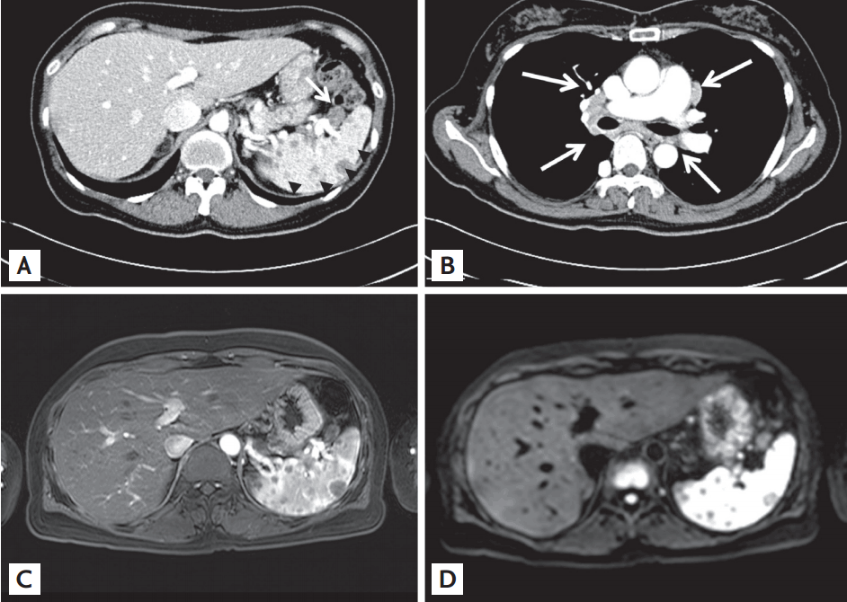
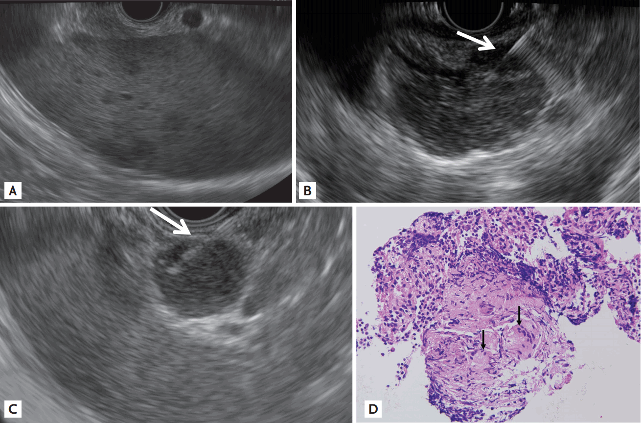
A 48-year-old woman presented with dyspepsia, a 5-kg weight loss, and night sweating for 2 months. She had no underlying disease and denied alcohol consumption or smoking. Her vital signs, physical examination, and laboratory results were unremarkable. Abdominal computed tomography (CT) showed multiple low-density lesions of variable sizes in the spleen, with perisplenic lymph node enlargement (Fig. 1A). Chest CT revealed enlargement of the right upper and bilateral lower paratracheal, para-aortic, aortopulmonary, subcarinal, bilateral hilar, and bilateral interlobar lymph nodes without lung parenchymal abnormalities (Fig. 1B). Abdominal magnetic resonance imaging showed multiple low-signal splenic lesions in all sequences, with hypointensity relative to the background parenchyma (Fig. 1C and 1D). On endoscopic ultrasound (EUS), multiple, ill-defined hypoechoic splenic lesions were seen (Fig. 2A), and EUS-guided fine-needle biopsy (FNB) with a 22-gauge needle (Echotip ProCore, Cook Endoscopy Inc., Limerick, Ireland), targeting the lymph nodes of the subcarinal and perisplenic regions, was performed (Fig. 2B and 2C). Pathologic examination showed noncaseating epithelioid cell granulomas with multinucleated giant cells (Fig. 2D). The results of the polymerase chain reaction for Mycobacterium tuberculosis and special stains for acid-fast bacilli and fungi in tissue samples were negative. The findings indicated sarcoidosis with splenic involvement.
Sarcoidosis with splenic nodular lesions is uncommon and difficult to diagnose in the absence of clinical suspicion. EUS-FNB for sarcoidosis is a minimally invasive technique, enabling a more accurate tissue acquisition under real-time visualization, avoiding cutaneous infection, and allowing evaluation of lymph nodes measuring < 1 cm, compared with transbronchial lung biopsy, mediastinoscopy, or percutaneous biopsy.
Unlike conventional needles, ProCore needles have a reverse bevel at the tip, which promotes the collection of core tissue samples, enabling a definitive diagnosis of malignant or benign conditions.
When a clinician encounters a patient with multiple enlarged lymph nodes and splenic nodular lesions, sarcoidosis should be considered in the differential diagnosis and EUS-FNB may be a useful diagnostic modality.
 PDF Links
 PubReader
 ePub Link
 Full text via DOI
 Download Citation
 Print
Print


