To the Editor,
Synchronous malignancy of primary lymphoma and adenocarcinoma of the gastrointestinal tract is not common and a careful diagnostic approach is needed when it is suspected. Moreover, the treatment plan should be made considering the stage of both diseases and the performance status of the patient. We present a rare case of synchronous diffuse large B-cell lymphoma of the small intestine and adenocarcinoma of the colon.
A 73-year-old man with an 8-year history of hypertension and type 2 diabetes mellitus on medical control presented to our emergency department with abdominal pain. He complained of intermittent right lower quadrant abdominal pain since the previous day. He did not exhibit fever, night sweating, or weight loss. He also denied anorexia, vomiting, constipation, diarrhea, melena, and hematochezia.
On physical examination, his vital signs were stable including blood pressure of 140/80 mmHg, pulse rate of 80 beats/min, respiratory rate of 20 breaths/min, and body temperature of 36°C. His abdomen was soft and flat, and there was mild tenderness in the right lower quadrant but no rebound tenderness, peritoneal irritation, or palpable mass. He did not present palpable cervical, supraclavicular, axillary, or inguinal lymphadenopathy. His the Eastern Cooperative Oncology Group performance score was grade 1.
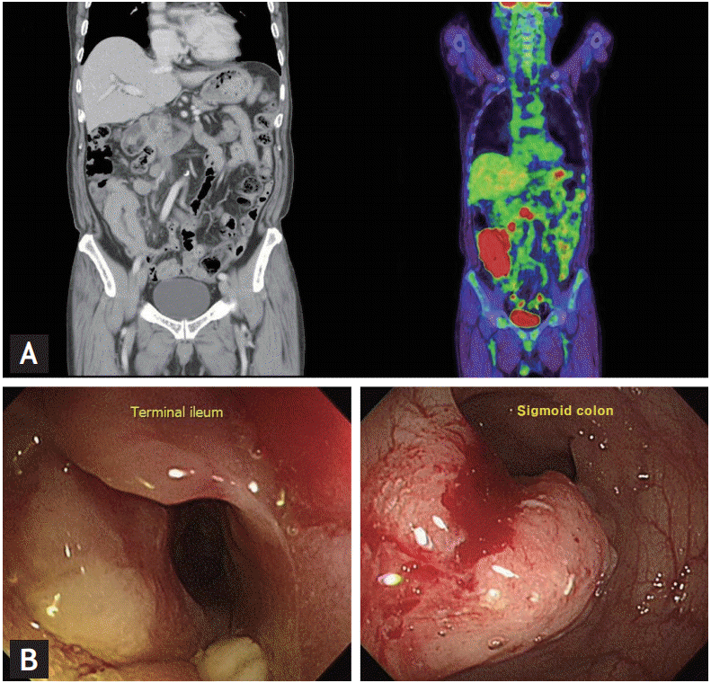
A computed tomography (CT) scan of the abdomen showed 10-cm long segmental concentric wall thickening of the terminal ileum showing homogeneous enhancement with contrast and a few enlarged adjacent lymph nodes. A positron emission tomography (PET) scan of the whole body was performed for staging. There was intense fluorodeoxyglucose (FDG) uptake in the terminal ileum, cecum, and sigmoid colon with focal FDG uptake in the right paraaortic and mesenteric lymph nodes (Fig. 1A). Colonoscopy showed extensive ulceration and marked narrowing of the terminal ileum with resultant spontaneous opening of the ileocecal valve. A 3.5-cm fungating mass with easy bleeding was found at the sigmoid colon, 30 cm from the anal verge (Fig. 1B).
From the result of colonoscopic biopsies, the diagnosis of diffuse large B-cell lymphoma of the terminal ileum and synchronous well differentiated adenocarcinoma of the sigmoid colon was established.
Bone marrow aspiration and biopsy showed no lymphoma involvement. His serum carcinoembryonic antigen (CEA) level was 1.22 ng/mL. The non-Hodgkin’s lymphoma was determined to be stage II by the Lugano Modification of the Ann Arbor staging system. The international prognostic index score was 2. For the colon cancer, clinical stage T2–T3N0M0 was suspected.
We decided to treat the patient by surgical resection for the double primary tumor followed by chemotherapy with curative intent for the diffuse large B-cell lymphoma. He also underwent anterior resection with curative intent for the sigmoid colon tumor. R2 resection was performed for lymphoma-involved lymph nodes including the paraaortic lymph nodes. His postoperative course was uneventful and pathologic staging of the sigmoid colon adenocarcinoma was T3N0 (stage IIA) with negative resection margins. Three resected regional lymph nodes were consistent with malignant lymphoma. In our case, the stage II lymphoma was more likely to affect survival than the stage IIA colon cancer, and we decided to perform 6 cycles of R-CHOP (rituximab, cyclophosphamide, doxorubicin, vincristine and prednisolone). On postoperative day 30, he was readmitted for the first cycle of chemotherapy. After completing the third cycle of R-CHOP, the patient suffered from grade IV neutropenia so we performed the fourth cycle of R-CHOP at a dose reduction to 75%. After the patient was recovered from Pneumocystis jiroveci pneumonia, he refused any further chemotherapy, so we carried out regular follow-up and observation.
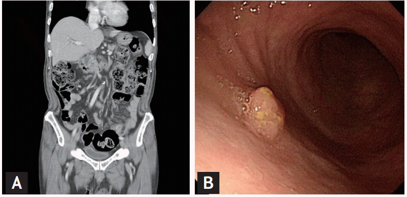
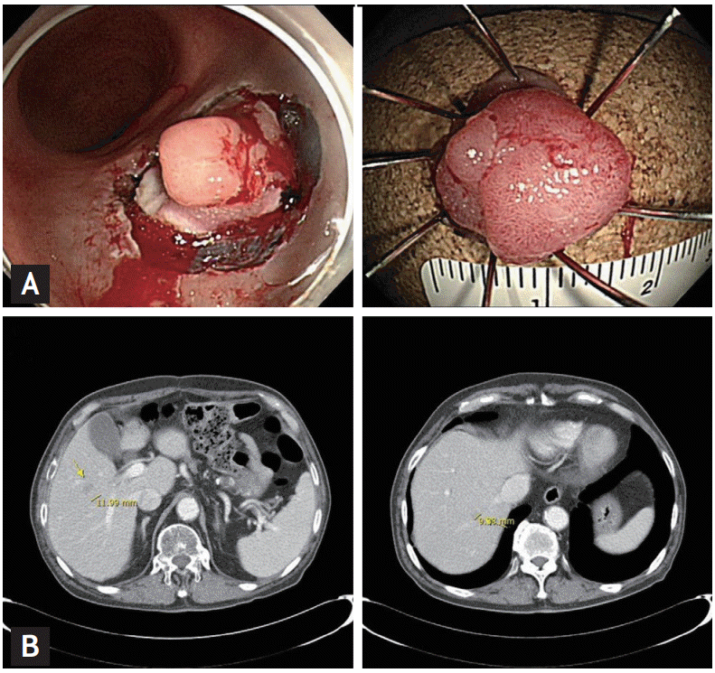
A follow-up CT scan after completion of 4 cycle of chemotherapy showed complete response of the lymphoma (Fig. 2A), and a PET scan also showed no discernible FDG uptake in the previously involved area. However, edematous wall thickening around the rectum was noted with haziness and streaky density in the mesorectal space. The follow-up serum CEA level had increased to 4.19 ng/mL. Sigmoidoscopy showed a 7-mm sized rectal polyp (Fig. 2B). The previous anastomosis site was intact. Biopsy of the polyp revealed adenocarcinoma. We performed endoscopic submucosal dissection for the localized rectal lesion (Fig. 3A).
On a follow-up CT scan after 3-month, two new small hepatic nodules (12-mm sized and 9-mm sized, at segment 5 and segment 7, respectively) were found (Fig. 3B). Liver biopsy of the nodule at segment 5 confirmed metastatic adenocarcinoma. Considering the location of the metastases and the poor performance of the patient, radiofrequency ablation was performed for the two hepatic nodules. We recommended palliative 5-fluorouracil based chemotherapy for recurrent colon cancer, but the patient did not want further chemotherapy. He is continuing regular follow-up at our out-patient department and has been disease-free for 4 months.
Although primary malignant lymphomas of the gastrointestinal tract are uncommon comprising only 1% to 4% of all malignant tumors of the gastrointestinal tract [1], the gastrointestinal tract is the most frequently involved extranodal site in non-Hodgkin’s lymphoma, with the stomach being the most common site (50% to 75%) [2]. Malignant lymphomas of the small intestine and colorectum are less common and account for 8% to 30% and 5.8% to 7% of all gastrointestinal tract lymphomas, respectively [2].
Synchronous adenocarcinoma and lymphoma of the gastrointestinal tract in the same patient is extremely rare with an estimated incidence of approximately 0.0002%and only a few cases have been reported in the literature. Furthermore, a case of synchronous adenocarcinoma and diffuse large B-cell lymphoma of the small intestine and colon has never been reported in Korea to our knowledge.
Diagnosis of such double primary malignancies may be difficult and one of the malignancies could be missed by the practitioner in some clinical situations. In this case, the patient was found to have suspicious lymphoma in the small intestine at the first imaging evaluation by CT scan, and a thorough work-up including colonoscopy led us to incidentally discover the synchronous adenocarcinoma of the colon.
The management of synchronous small bowel lymphoma and colon adenocarcinoma is controversial [3]. Surgery, chemotherapy or both have been the mainstay of treatment, but there has been no definitive evidence regarding the benefits of any strategy and related studies have been limited due to low incidence of the disease.
Most of the recent literature suggests that chemotherapy has a survival benefit over surgery alone for synchronous gastrointestinal lymphoma and adenocarcinoma. However, surgery still has a role in diagnosis in many cases and should be performed first due to the potential of local complications such as intestinal obstruction, perforation, and bleeding during chemotherapy [3].
Less than 20 cases of synchronous lymphoma and adenocarcinoma of the GI tract have been reported in the literature worldwide. Most of the case reports predominantly described the course from the initial presentation to the diagnosis of the double malignancy. Only a few cases present the treatment modality and follow-up course of the patient.
Bhanote et al. [4] reported a case of a 55-year-old man with follicular lymphoma of the ileum combined with extensive mesenteric and retroperitoneal lymph node involvement and a simultaneous adenocarcinoma of the cecum. He underwent right hemicolectomy with lymph node dissection and the pathologic staging of the colon cancer was T4N1M0. He was managed with 6 cycles of adjuvant chemotherapy with 5-fluorouracil and leucovorin and also treated with rituximab for low grade lymphoma after 2 cycles of adjuvant chemotherapy. He also underwent 4 cycles of fludarabine due to lymphoma progression. The lymphoma showed complete remission, but the colon cancer recurred 1.5 years later [4].
Tseng et al. [5] reported a case of a 63-year-old man with synchronous adenocarcinoma of the sigmoid colon and extranodal natural killer/T-cell lymphoma of the ascending colon. He was managed with right hemicolectomy and sigmoidectomy, followed by CHOP chemotherapy. However, there was rapid progression of the lymphoma in the bilateral lung and he died of respiratory failure [5].
In our case, we performed right hemicolectomy for resection of the lymphoma-involved lesion to prevent local complications and anterior resection for the adenocarcinoma of the sigmoid colon with curative intent. There was a remnant lymphoma-involved lesion after the surgery. Since it is known that stage II lymphoma is generally more aggressive than resected stage II colon cancer, we decided to perform chemotherapy combined with monoclonal antibody therapy after operation, which showed excellent response.
The patient should have been treated for recurrent adenocarcinoma more aggressively, but the least invasive management was performed considering the patient’s general condition and personal willingness. Moreover, this management was feasible because metastasis was confined to liver.
In conclusion, the coexistence of double primary malignancy of the gastrointestinal tract is not common but clinicians should not ignore the possibility of such conditions and careful investigations and proper management of both diseases are required.


 PDF Links
 PubReader
 ePub Link
 Full text via DOI
 Download Citation
 Print
Print


