To the Editor,
Acute invasive fungal rhinosinusitis (AIFRS) is a rare but frequently fatal infection that occurs primarily in immunocompromised individuals. The causes of immunosuppression include hematologic malignancy, diabetes, immunosuppressive medications, and chemotherapy [1]. The most frequent causes of AIFRS are Aspergillus and Mucor species. Because of rapid spread of the fungi from the nasal mucosa and sinus to the orbit and the brain, AIFRS is associated with high mortality and morbidity. Early medical and surgical treatment is essential to improve the prognosis of AIFRS. Although diabetes is one of the most common underlying disease for AIFRS, to our knowledge, so far there have been no reports of hyperglycemic hyperosmolar state (HHS) associated with invasive rhino-orbital aspergillosis in a patient with diabetes-related end-stage renal disease (ESRD). Here, we present a case of HHS associated with invasive rhino-orbital aspergillosis in a patient with ESRD.
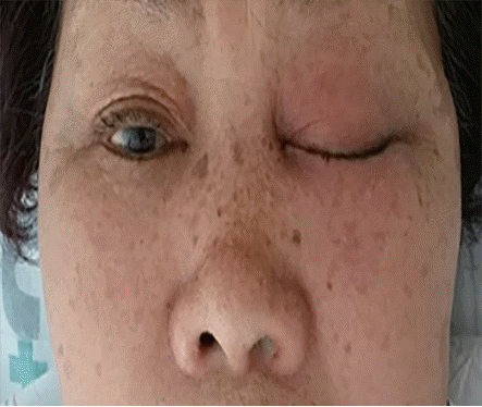
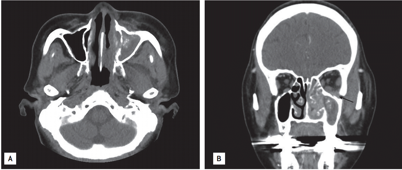
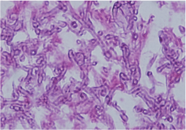
A 71-year-old woman presented with a 1-week history of swelling and discomfort around the left eye, general weakness, and poor oral intake. She had also had a tendency to fall towards the left for 2 days. Her medical history included hypertension, diabetes, and ESRD on hemodialysis. Hemodialysis for ESRD caused by diabetes had been initiated 16 months before. She had been prescribed a twice-daily dose of Humalog Mix (75/25 14 U before breakfast and 50/50 15 U before dinner, Eli Lilly Nederland B.V., Houten, The Netherlands), which had brought her blood glucose under control. Physical examination showed dehydration of the tongue, a whitish discharge from the left nasal cavity, and erythematous swelling and tenderness in the left periorbital area without ocular discharge (Fig. 1). The patientŌĆÖs vital signs were as follows: blood pressure, 120/60 mmHg; heart rate, 72 beats per minute; and body temperature, 36.5┬░C. Laboratory findings were as follows: white blood cell count, 8,500 ├Ś 109/L (82.5% neutrophils); hemoglobin, 10.0 g/dL; platelet count, 258 ├Ś 109/L; arterial blood gas analysis results, pH 7.342, HCO3 - 22.1 mEq/L, anion gap 11.9 mEq/L; serum glucose, 932 mg/dL; glycated hemoglobin, 10.7%; blood urea nitrogen, 40.5 mg/dL; creatinine, 6.32 mg/dL; sodium, 139 mEq/L; potassium, 4.3 mEq/L; C-reactive protein (CRP), 3.75 mg/dL; effective serum osmolality, 329 mOsm/kg; serum ketone, 0.5 mmol/L; ketone-negative urine. Computed tomography (CT) of the paranasal sinuses showed unilateral invasive maxillary sinusitis with cellulitis in the left preseptal and malar area (Fig. 2A), which extended beyond the ipsilateral intraconal space (Fig. 2B). Treatment was started with hydration, continuous intravenous insulin infusion, and antibiotics. Endoscopic surgical debridement was performed immediately. On histopathologic examination, the maxillary sinus tissue was found to contain septated fungal hyphae with acute angle branching, typical of Aspergillus spp., and hence indicative of aspergillosis (Fig. 3). Based on the laboratory findings and the results of histopathologic examinations, AIFRS-associated HHS was diagnosed. Intravenous administration of amphotericin B was initiated and serum glucose levels were strictly controlled by continuous insulin infusion. Two weeks later, periorbital edema and redness were almost resolved and a follow-up CT scan showed remarkably decreased opacity in the maxillary sinus. Fungal cultures of the debrided tissue were negative. The patient was prescribed an oral antifungal agent and discharged. A month after discharge, nasal endoscopy showed few crusts and a nearly clear sinus.
HHS is a syndrome characterized by severe hyperglycemia, hyperosmolarity, and dehydration in the absence of ketoacidosis. Current diagnostic criteria include a plasma glucose level more than 600 mg/dL, and effective plasma osmolality more than 320 mOsm/kg in the absence of significant ketoacidosis. The overall mortality rate is estimated to be as high as 20% and the prognosis is determined by the severity of dehydration, presence of comorbidities, and age of the patient (advanced age is generally associated with a poor prognosis) [2]. Infection represents the commonest precipitating factor for HHS, with the most common precipitating infections being pneumonia (40% to 60%) and urinary tract infection (5% to 16%) [2]. In our case, because there was no identifiable medical illness and infection other than sinusitis, AIFRS was thought to be the predisposing factor for HHS with uncontrolled hyperglycemia. Infection provokes the release of counterregulatory hormones of insulin and/or compromises the access to water, resulting in severe dehydration and consequently, HHS. Fungal rhinosinusitis (FRS) can be classified into invasive and noninvasive subtypes, based on histopathological evidence of tissue invasion by fungi. The invasive type includes AIFRS, granulomatous invasive FRS, and chronic invasive FRS [3]. AIFRS is FRS of less than 4 weeks duration in immunocompromised patients and the most frequent causes are Aspergillus and Mucor species. Aspergillus species are thinner, septate, with regular branching and branch at acute angles when compared with the organisms of mucormycosis (45┬░ as opposed to 90┬░). It seems to be important to distinguish invasive mucormycosis from aspergillosis since zygomycetes are resistant in vitro to the newer antifungal agents such as voriconazole. The initial symptoms are fever, nasal discharge, nasal obstruction, and nasal crusting; however, these symptoms are not characteristic of the disease. Although our patient had AIFRS, she had no fever or leukocytosis but only elevated CRP levels. It was understood that the impaired immunity of our patient was associated with diabetes and ESRD. Immunological abnormalities in patients with ESRD include alterations in the two major branches of the immune system, innate, and adaptive immune systems. Functional abnormalities of monocytes, neutrophils, and dendritic cells are directly linked with risk of infection; impaired maturation of TH lymphocytes also leads to a disabled immune response and susceptibility to infection [4]. Immune dysfunction associated with uremia significantly contributes to the high premature mortality in patients who have ESRD along with infectious complications. Therefore, intensive evaluation should be performed in patients with ESRD with suspected systemic infection, even if they are afebrile. AIFRS should be suspected in immunocompromised patients with new-onset, rapidly progressing sinusitis or facial discomfort. Despite improvements in both medical and surgical treatments for AIFRS, mortality remains high. Survival rates reported have generally varied from 20% to 80%. Patients who have intracranial involvement, or who do not receive surgery as part of their therapy, have a poor prognosis [5]. Amphotericin B is the mainstay of medical treatment of AIFRS and endoscopic sinus surgery is important for the adjunct management of AIFRS. It is likely that debriding necrotic areas may lead to improved antimicrobial penetration and that surgically decreasing the fungal burden may reduce the risk of regional and systemic spread of the disease [5].
In conclusion, we describe a successfully treated case of HHS associated with invasive rhino-orbital aspergillosis in a patient with diabetes-related ESRD. Although AIFRS in diabetes is fatal, early diagnosis and aggressive treatment with surgical debridement and intravenous administration of amphotericin B may lead to good prognosis.


 PDF Links
 PubReader
 ePub Link
 Full text via DOI
 Download Citation
 Print
Print


