Jeong, Kim, Kim, Ki, Choi, and Park: Too much synchrony? Left bundle branch area pacing-induced systolic anterior motion and left ventricular outflow tract obstruction managed by intentional atrioventricular dyssynchrony
Too much synchrony? Left bundle branch area pacing-induced systolic anterior motion and left ventricular outflow tract obstruction managed by intentional atrioventricular dyssynchrony
1Department of Cardiovascular Medicine, Won Kwang University School of Medicine, Iksan,
Korea
2Department of Cardiovascular Medicine, Chosun University School of Medicine, Gwangju,
Korea
Correspondence to: Sung Soo Kim, M.D., Ph.D. Department of Cardiovascular Medicine, Chosun University Medical School, 309 Pilmun-daero, Dong-gu, Gwangju 61452, Korea, Tel: +82-62-220-3025, Fax: +82-62-228-7174, E-mail: kholywater@gmail.com, https://orcid.org/0000-0002-5190-227X
Received July 20, 2025; Revised September 16, 2025; Accepted September 22, 2025;
This is an Open Access article distributed under the terms of the Creative Commons Attribution Non-Commercial License (http://creativecommons.org/licenses/by-nc/4.0/) which permits unrestricted noncommercial use, distribution, and reproduction in any medium, provided the original work is properly cited.
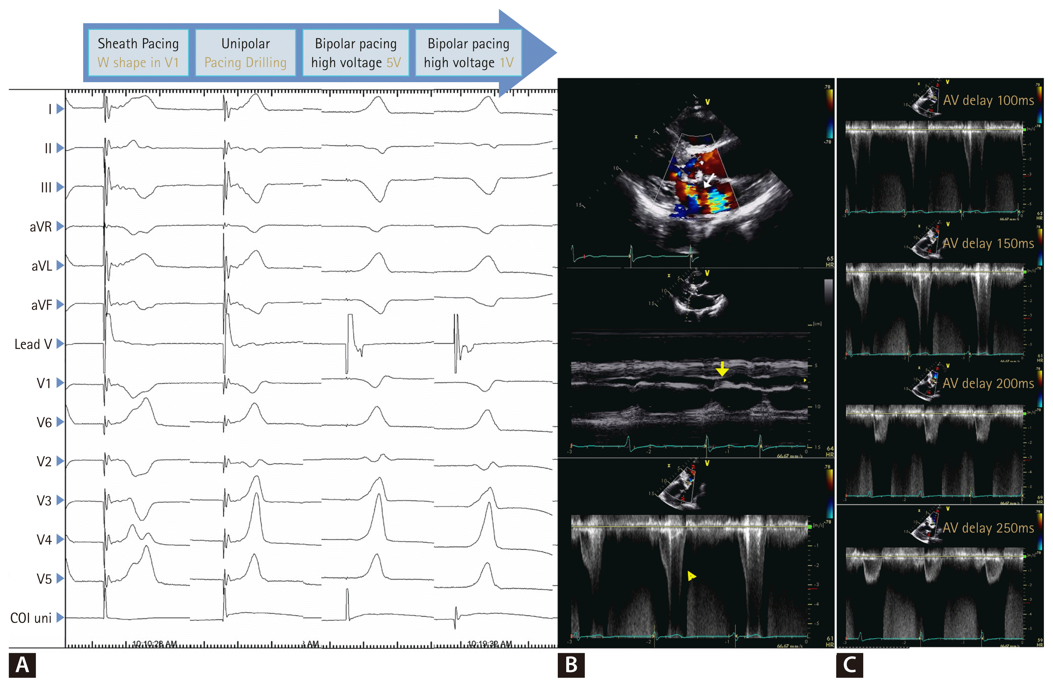
An 86-year-old woman with hypertension presented with recurrent syncope. Electrocardiography revealed first-degree atrioventricular (AV) block with intermittent high-grade block. Echocardiography showed preserved left ventricular ejection fraction (LVEF), mild aortic stenosis, and septal hypertrophy (Supplementary Video 1, 3). Anticipating high pacing burden, a dual-chamber pacemaker was implanted using a Medtronic 3830 lead. Left bundle branch area pacing (LBBAP) was confirmed by qR morphology in V1, short left ventricular activation time, and a paced QRS of 124 ms (Fig. 1A). The device was programmed in DDD mode with sensed AV delay of 150 ms.
Several hours later, she developed hypotension and cardiogenic shock. Repeat echocardiography revealed preserved LVEF but new systolic anterior motion (SAM), severe left ventricular outflow tract obstruction (LVOTO, gradient 120 mmHg), and acute mitral regurgitation (Fig. 1B, Supplementary Video 2, 4). Extending the AV delay to 250 ms promptly reduced the gradient to 30 mmHg (Fig. 1C), with rapid recovery. She was discharged in stable condition.
This case highlights a rare hemodynamic complication of LBBAP in a patient with latent LVOTO. With short AV intervals, ventricular contraction may occur before mitral valve closure, producing SAM and obstruction. Unlike right ventricular pacing, which delays septal contraction and may blunt LVOT gradients [1], LBBAP preserves near-normal septal timing and can aggravate obstruction in susceptible patients. Prolonging the AV delay allows AV filling and complete mitral coaptation before systole, thereby relieving obstruction. Prior studies in hypertrophic cardiomyopathy also show that LVOT gradients vary with pacing interval [2]. We maintained LBBAP rather than switching to RV pacing, as AV optimization alone abolished SAM while preserving physiologic synchrony and avoiding dyssynchrony. Careful echocardiographic monitoring and individualized programming are essential when performing conduction system pacing in patients with septal hypertrophy or potential latent LVOTO.
Written informed consent was obtained from the patient for the publication of this case report (IRB-CHOSUN 2025-06-033).
Parasternal long-axis view before pacemaker implantation demonstrates preserved left ventricular ejection fraction, mild aortic stenosis, and septal hypertrophy.
Supplementary Video 2.
Parasternal long-axis view after pacemaker implantation shows preserved left ventricular ejection fraction but reveals newly developed systolic anterior motion of the mitral valve and acute mitral regurgitation.
Supplementary Video 3.
Apical three-chamber view before pacemaker implantation demonstrates preserved left ventricular ejection fraction, mild aortic stenosis, and septal hypertrophy.
Supplementary Video 4.
Apical three-chamber view after pacemaker implantation reveals left ventricular outflow tract obstruction caused by systolic anterior motion of the mitral valve.
This study was supported by research funds from the Chosun University, 2025.
Figure 1
Electrocardiographic and echocardiographic findings before and after LBBAP with stepwise AV delay adjustment. (A) Twelve-lead electrocardiogram recorded during stepwise pacing maneuvers for LBBAP. Initial sheath pacing shows a W-shaped QRS complex in lead V1, followed by unipolar pacing during “drilling,” and subsequent bipolar pacing at 5 V and 1 V outputs. The final paced morphology demonstrates a qR pattern in lead V1 with a narrow QRS duration, consistent with successful left bundle branch capture. (B) Transthoracic echocardiographic findings following LBBAP. Color Doppler (top), M-mode (middle), and continuous-wave Doppler (bottom) images demonstrate systolic anterior motion of the anterior mitral leaflet (yellow arrow), acute mitral regurgitation (white arrow), and a LVOT gradient of 120 mmHg with a characteristic “dagger-shaped” profile (yellow triangle). (C) Continuous-wave Doppler images (right column) show progressive reduction in LVOT gradient with stepwise prolongation of AV delay from 100 ms to 250 ms. The final setting (AV delay 250 ms) results in near-complete resolution of dynamic LVOT obstruction. LBBAP, left bundle branch area pacing; AV, atrioventricular; LVOT, left ventricular outflow tract.


 , Hyun Kuk Kim2, Young Jae Ki2, Dong Hyun Choi2, Keun Ho Park2