Polysplenia syndrome with truncal pancreas presenting as diabetic ketoacidosis
-
Hea Min Yu
 , Jun Choul Lee
- Received June 23, 2022; Revised July 31, 2022; Accepted August 16, 2022;
A 17-year-old girl with no past medical history was referred to our hospital for vomiting and abdominal pain. At presentation, she was well-oriented and had stable vital signs. Her laboratory results showed: random blood glucose 410 mg/dL, hemoglobin A1c 12.3%, venous blood gas analysis (pH 7.17, PO2 42 mmHg, PCO2 32 mmHg, HCO3 11.7 mEq/L, lactate 1.1 mg/dL, base excess −15.7, anion gap 19.3), serum ketone body 1,000 μmol/L, serum insulin 13.1 μU/mL, C-peptide 0.7 ng/mL, sodium 135 mEq/L, potassium 4.4 mEq/L, anti-glutamic acid decarboxylase antibody 0.06 U/mL, insulin antibody 4.7%, islet cell antibody-negative, and insulin receptor antibody-negative.
With a diagnosis of diabetic ketoacidosis (DKA), she was shifted to the medical intensive care unit and received treatment with intravenous fluids, intravenous insulin infusion, and potassium replacement.
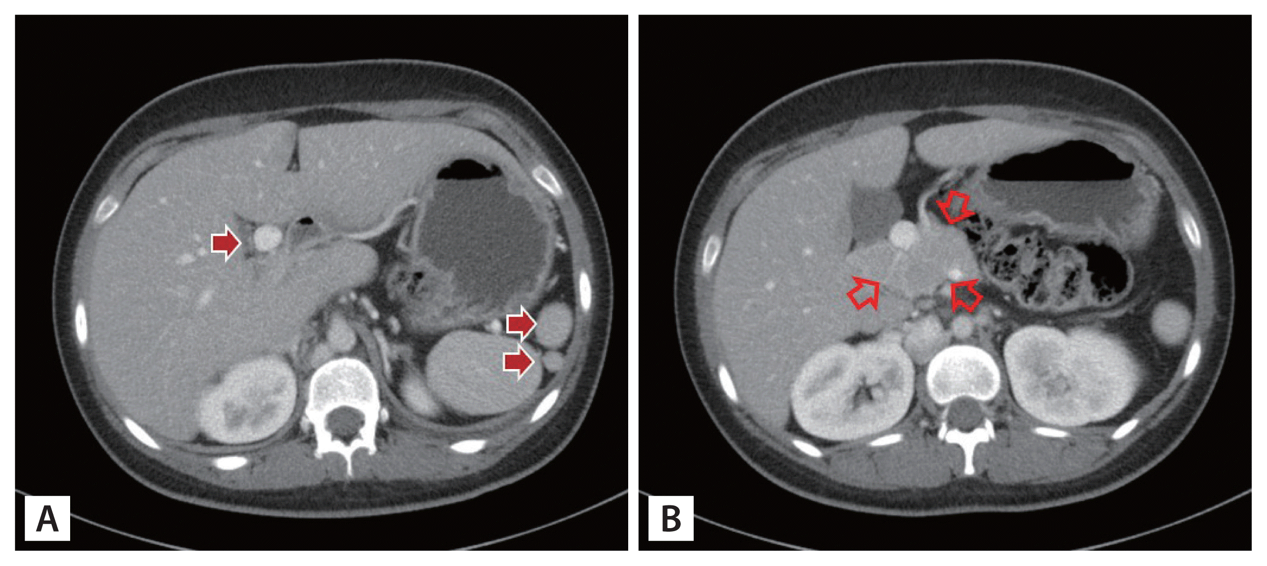
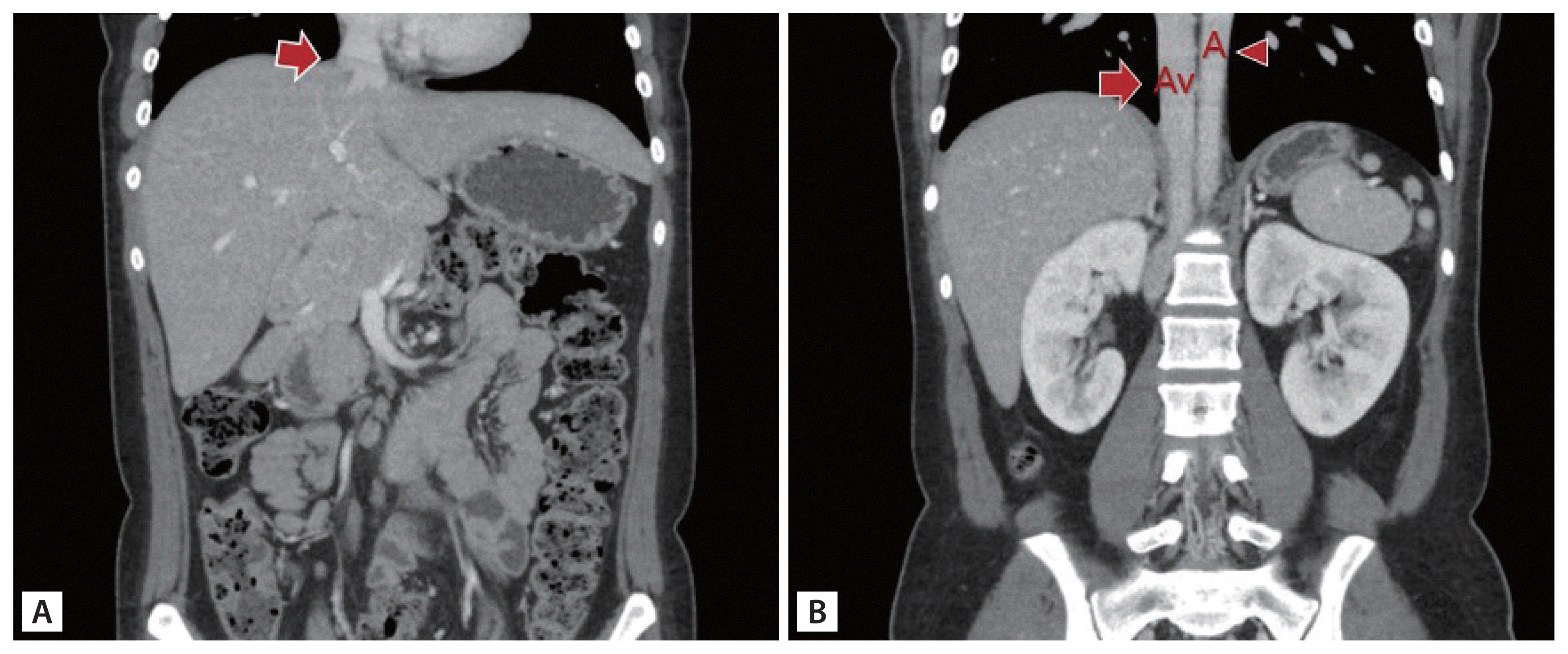
Enhanced abdominal computed tomography (CT) revealed polysplenia in the left upper quadrant, truncated pancreas, preduodenal portal vein (Fig. 1), and absence of the hepatic segment of the inferior vena cava (IVC) and azygos continuation of the IVC (Fig. 2). No cardiac anomalies were observed on contrast-enhanced chest CT.
Polysplenia syndrome (PS) is a rare condition defined as the presence of multiple spleens and is associated with various visceral abnormalities. Although there is no characteristic pathognomonic anomaly, one of the most common features associated with PS is the vascular malformation of the IVC with azygos or hemiazygos continuation. Pancreatic anomalies in patients with PS are related to the increased incidence of diabetes mellitus. However, it is not clear to what extent PS affects diabetes development, and little has been reported.
Our patient was a rare case of DKA with coincident truncated pancreas and PS, requiring continuous insulin treatment even after discharge.
- Conflicts of Interest
- Conflicts of Interest
No potential conflict of interest relevant to this article was reported.