Successful treatment of tracheoesophageal fistula using a covered esophageal stent
- Jung Hun Kim, Young Koog Cheon
- Received October 11, 2017; Revised February 12, 2018; Accepted February 14, 2018;
We report herein a case of superficial esophageal cancer not eligible for endoscopic submucosal dissection. This patient developed a tracheo-esophageal fistula after radiation therapy followed by photodynamic therapy (PDT).
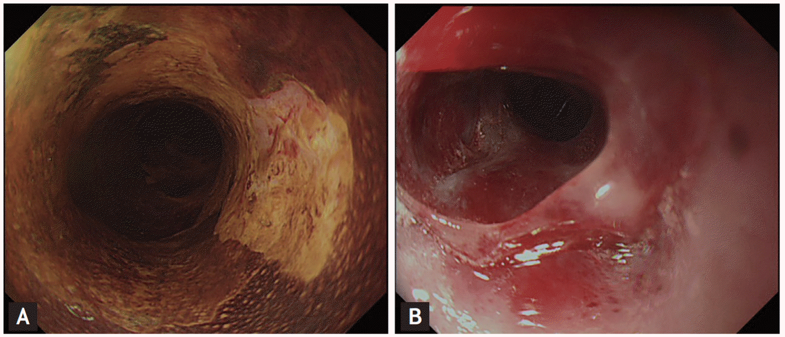
A 74-year-old male underwent total gastrectomy due to advanced gastric cancer in 2014. In 2017, a flat discolored lesion encircling the lumen at 30 to 35 cm from the incisors was found by gastroscopy. Lugol’s solution showed an unstained mucosal lesion (Fig. 1A). Radiotherapy was performed at a different hospital, because the patient did not consent to surgery. Follow-up gastroscopy showed squamous cancer at the radiotherapy site. The patient was transferred to our hospital for PDT, which was performed four times using a 2-cm light diffuser and 160 J/cm. The duration of light exposure was 440 seconds each time (Fig. 1B).
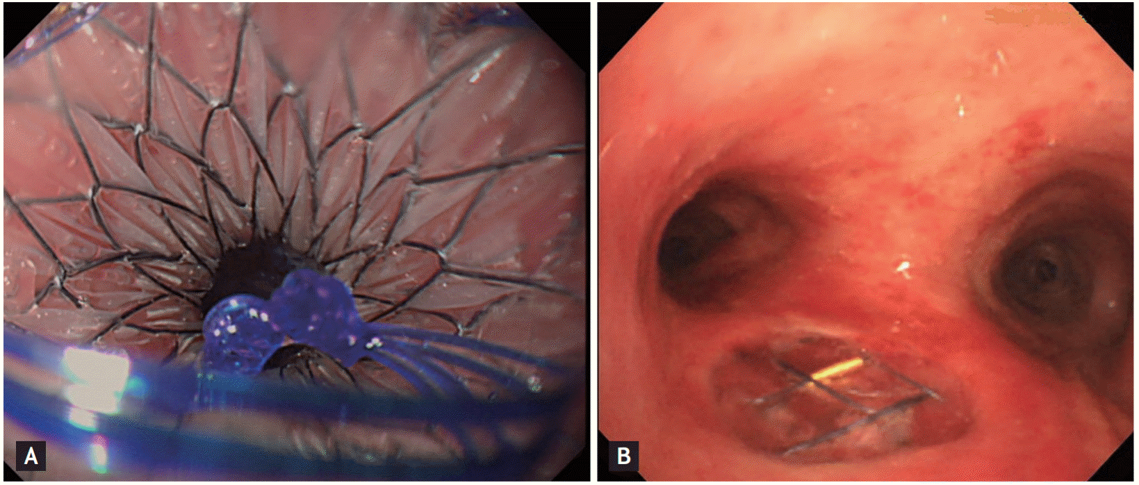
A follow-up esophagogastroduodenoscopy at 1 day after PDT showed esophageal swelling and spontaneous bleeding at the PDT site. One month later, gastroscopy showed necrotic material and luminal narrowing at the PDT site. Two months after PDT, the patient visited another hospital because of dyspnea; he was transferred to our hospital because of pneumonia and a tracheoesophageal fistula. Gastroscopy revealed a tracheoesophageal fistula at the PDT site, which was covered using a metal stent (Fig. 2A). One month later, the tracheoesophageal fistula was sealed by the stent (Fig. 2B). At the time of writing 6 months later, the patient is well with no other complications.
In summary, we treated a tracheoesophageal fistula that developed after PDT by insertion of a covered esophageal stent.
Written informed consents were obtained.
- Conflicts of Interest
- Conflicts of Interest
No potential conflict of interest relevant to this article was reported.