Subacute respiratory symptoms in a patient with Crohn’s disease and ankylosing spondylitis
Article information

A 30-year-old man was diagnosed with ankylosing spondylitis five years ago and was treated with non-steroidal anti-inflammatory drugs and sulfasalazine. Three years later, the patient underwent small bowel resection due to chronic abdominal pain and was diagnosed with ileal Crohn’s disease with multifocal strictures, microperforation, and an abscess (Fig. 1). After the surgery, the patient was treated with azathioprine, followed by methotrexate. Approximately nine months ago, the patient’s back pain worsened, leading to the introduction of adalimumab. The patient had no history of tuberculosis, a negative interferon-gamma release assay (IGRA) test result, and unremarkable chest radiography result. During follow-up, the patient remained clinically stable on combination maintenance therapy.
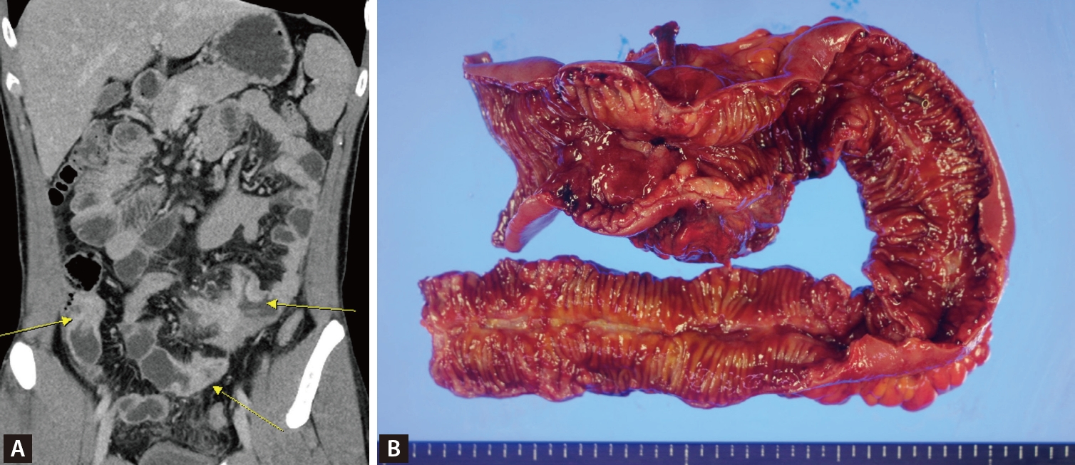
(A) Computed tomography enteroscopy findings. On coronal view, multiple small bowel strictures were seen (yellow arrow). (B) Pathologic findings. Multiple strictures of the ileum were seen.
However, the patient developed persistent cough, dyspnea, and shortness of breath over one month, showing no improvement with conventional management, along with several palpable cervical lymph nodes. Chest computed tomography revealed a large pericardial effusion with impending tamponade physiology and diffuse parenchymal infiltration in the left lung field (Fig. 2A). Ultrasound-guided biopsy of the cervical lymph nodes revealed granulomatous inflammation with necrosis (Fig. 2B). Pericardiocentesis drainage was performed, showing positive AFB with M. tuberculosis culture, and adenosine deaminase (ADA) 11.3 IU/L, which was compatible with tuberculous pericarditis, and IGRA result were positive. Standard anti-tuberculosis medication (HREZ) was initiated for six months with rapid symptom improvement (Fig. 2C, D).
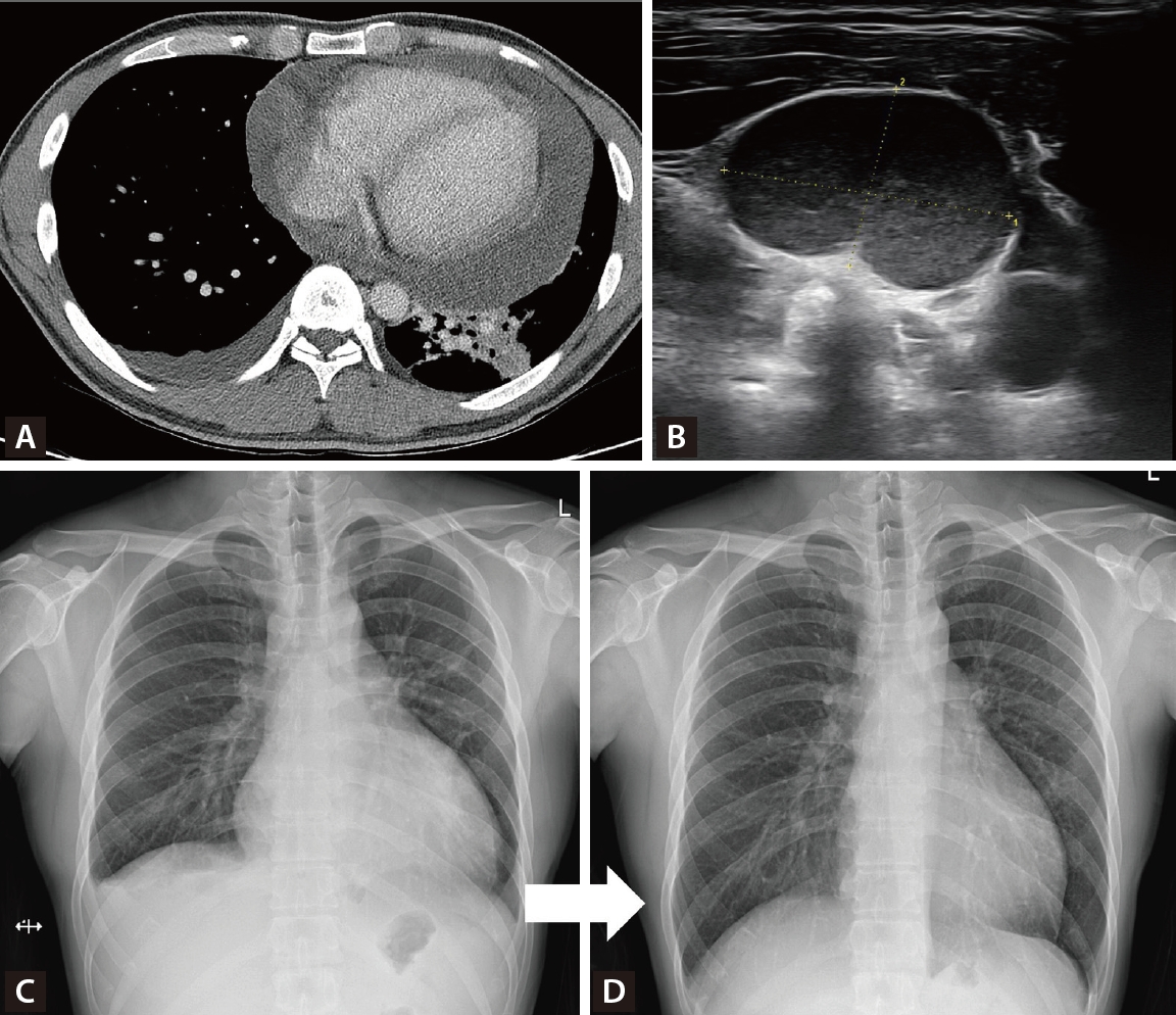
(A) Chest computed tomography findings. On axial view, massive pericardial effusion with left lower lung infiltration were seen. (B) Neck ultrasound findings. About 2.1 cm-sized enlarged lymph node was seen. (C) Initial X-ray findings. Cardiomegaly due to pericardial effusion and pleural effusion were seen. (D) Follow-up X-ray findings. One month after pericardiocentesis, pericardial effusion was completely disappeared.
Pericarditis is an important extra-pulmonary presentation of tuberculosis. Its late diagnosis may result in serious complications, such as constrictive pericarditis, cardiac tamponade, and increased mortality rate [1]. Tuberculous pericarditis can be diagnosed by examining the pericardial exudate or by detecting a good response after anti-tuberculous drug use in patients with pericarditis [2]. High ADA levels have also been associated with tuberculous pericarditis [3]. Early suspicion and prompt management in patients with immunosuppressive treatment are crucial for improving treatment outcome of tuberculous pericarditis [4,5].
Notes
Acknowledgments
The patient provided informed consent and agreed to publication of the images.
CRedit authorship contributions
Doohyuck Lee: methodology, investigation, writing - original draft; Kwangwoo Nam : conceptualization, methodology, resources, investigation, writing - original draft, writing - review & editing, supervision; Ho Jin Yong: conceptualization, resources, investigation, writing - review & editing; Juntae Kim: conceptualization, resources, investigation, writing - review & editing; Miil Kang: conceptualization, resources, writing - review & editing
Conflicts of interest
The authors disclose no conflicts.
Funding
None