Findings suggestive of Fanconi syndrome and multiple myeloma
Article information

A 52-year-old man was referred to the nephrology department following a health examination showing decreased renal function with proteinuria. He had no significant underlying disease except for intermittent analgesic use for back pain. Initial laboratory findings were as follows: Na+-K+-Cl--, 139-3.6-109 mmol/L; HCO3- 21mmol/L; blood urea nitrogen/creatinine, 11/1.75 mg/dL; protein/albumin, 7.0/4.4 g/dL; calcium, 9.1 mg/dL; uric acid, 1.6 mg/dL; phosphorus, 1.2 mg/dL; glucose, 79 mg/dL; HbA1c, 5.2 %.
Urinalysis revealed 3+ proteinuria 3+ glucose, with a spot urine protein/creatinine ratio of 10.5 g/g. These results indicated normal anion gap acidosis, normoglycemic glycosuria, hypophosphatemia, and hypouricemia, consistent with Fanconi syndrome (FS).
Additional blood and urine tests and kidney biopsy were performed to determine the potential causes of renal insufficiency with FS. Urine protein electrophoresis revealed an M-spike in the beta-globulin region, whereas serum immunoglobulin electrophoresis revealed an abnormal sedimentation line in the kappa region. The free kappa/lambda ratio was elevated to 1,133.86. The kidney biopsy showed abundant granular/elongated, and weakly eosinophilic crystals in the proximal tubular cell cytoplasm and the tubular lumen (Fig. 1). Ultrastructural examination clearly demonstrated electron-dense crystals with various shapes, ranging from rhomboid to pentagonal and elongated (Fig. 2). The bone marrow biopsy included 18% plasma cells, leading to the definitive diagnosis of multiple myeloma, light chain disease, kappa type.
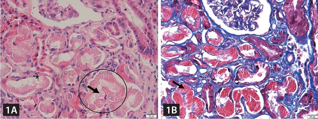
Light microscopic findings of kidney biopsy specimen. (A) Proximal tubular cells contain abundant weakly eosinophilic crystals in which are granular and elongated that distort the cell architecture (circle), obscure the apical cell membrane (asterisk) and fill the tubular lumen (arrow) (hematoxylin and esosin stain, original magnification × 400). (B) A trichrome stain highlights distinct crystals (arrow) that are needle shaped and brightly red-stained (Masson’s trichrome stain, original magnification × 400).
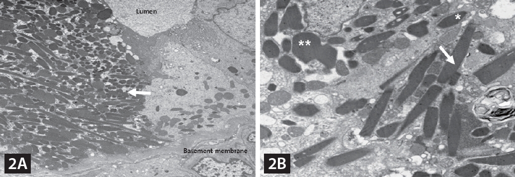
Electron microscopic findings of kidney biopsy specimen. (A) On low power view, a proximal tubular cell is engorged with randomly oriented crystals (arrow) (original magnification, × 1,000). (B) On higher power view, individual crystals exhibit vague striation and a range of shapes from rhomboid (asterisk) to pentagonal (double asterisk) and elongated (arrow) (original magnification, × 3,000).
FS is commonly caused by genetic disorders, exposure to heavy metals, or medications, but it can rarely present as a latent manifestation of light chain disease. The crystalline inclusions produced by light chains can lead to cytotoxicity in proximal tubular epithelial cells, resulting in proximal tubulopathy, whereas crystal-laden phagocytes can lead to tubular atrophy and interstitial fibrosis through tissue injury, as shown here. In patients with proximal tubulopathy accompanied by renal insufficiency and proteinuria, multiple myeloma, particularly light chain disease, should be considered in the differential diagnosis.
Notes
Acknowledgments
The authors would like to extend their sincere gratitude to Ryan Hosung Lee of Cornerston Collegiate Academy for his invaluable assistance in proofreading and editing this manuscript.
CRedit authorship contri
Hwa-Young Lee: conceptualization, methodology, resources, investigation, writing – original draft; Sung Gyul Lim: review & editing, visualization; Han Sang Lee: conceptualization, methodology, review & editing; Won-Ae Lee: methodology, resources, writing – original draft; So Mi Kim: review & editing, visualization, supervision
Conflicts of interest
The authors disclose no conflicts.
Funding
None