Metabolic musculoskeletal disorders in patients with inflammatory bowel disease
Article information

Abstract
Inflammatory bowel disease (IBD), which includes Crohn’s disease and ulcerative colitis, is a chronic inflammatory disorder that affects not only the gastrointestinal tract but also extraintestinal organs, leading to various extraintestinal manifestations and complications. Among these, musculoskeletal disorders such as osteoporosis, sarcopenia, and axial and peripheral spondyloarthritis are the most commonly observed. These conditions arise from complex mechanisms, including chronic inflammation, malnutrition, gut dysbiosis, and glucocorticoid use, all of which contribute to reduced bone density, muscle loss, and joint inflammation. Osteoporosis and sarcopenia may co-occur as osteosarcopenia, a condition that heightens the risk of fractures, impairs physical performance, and diminishes quality of life, particularly in elderly patients with IBD. Holistic management strategies, including lifestyle modifications, calcium, and vitamin D supplementation, resistance training, and pharmacological interventions, are essential for mitigating the impact of these conditions. Spondyloarthritis, which affects both axial and peripheral joints, further complicates disease management and significantly compromises joint health. Timely diagnosis and appropriate medical interventions, such as administration of nonsteroidal anti-inflammatory drugs and biologics, are critical for preventing chronic joint damage and disability. Moreover, a multidisciplinary approach that addresses both metabolic and inflammatory aspects is essential for optimizing physical function and improving treatment outcomes in patients who have IBD with musculoskeletal involvement.
INTRODUCTION
Inflammatory bowel disease (IBD), which includes the subtypes Crohn’s disease (CD) and ulcerative colitis (UC), is a chronic, systemic inflammatory disorder of the gastrointestinal (GI) tract. This debilitating condition is marked by periods of relapse and remission [1,2]. Although the exact etiology of IBD remains unclear, it is believed to result from a dysregulated immune response to microbial and environmental triggers in genetically predisposed individuals [3,4]. This immune-mediated inflammation primarily affects the GI tract but also extends to extraintestinal organs, including the joints, skin, hepatobiliary system, and eyes, giving rise to extraintestinal manifestations [5]. Moreover, the chronic nature of IBD and its lifelong treatment are associated with extraintestinal complications such as malabsorption, nutritional deficiencies, osteoporosis, sarcopenia, peripheral neuropathies, and treatment-related adverse effects [6,7].
Among the extraintestinal symptoms of IBD–encompassing both extraintestinal manifestations and complications–musculoskeletal (MS) involvement is the most frequently observed. This includes peripheral and axial spondyloarthritis (SpA), fibromyalgia, and metabolic MS complications such as osteoporosis and sarcopenia. These MS disorders typically emerge after the diagnosis or onset of IBD but may occasionally precede it [8]. Furthermore, MS symptoms are often nonspecific and may be overlooked when the primary focus is on managing GI symptoms, potentially leading to a reduced quality of life, rare but severe complications, and missed opportunities to address modifiable factors contributing to metabolic MS disorders.
The growing proportion of elderly patients with IBD, including both adult-onset elderly IBD and elderly-onset IBD, further highlights the importance of addressing metabolic MS disorders in IBD management. Elderly individuals are inherently at higher risk for metabolic MS disorders, and the presence of IBD introduces additional risk factors, exacerbating negative outcomes and imposing a significant social and economic burden on this population [9–11]. Therefore, physicians managing IBD must develop a comprehensive understanding of MS manifestations and complications to provide effective treatment. This includes adopting a multidisciplinary approach in collaboration with rheumatologists and endocrinologists to enhance quality of life and improve treatment satisfaction.
The present review summarizes the epidemiology and pathogenesis of key MS disorders, specifically osteoporosis, sarcopenia, and axial and peripheral SpA in patients with IBD, and proposes strategies for their management and prevention.
METABOLIC MS COMPLICATIONS IN IBD
Osteoporosis and osteoporotic fracture
Osteopenia and osteoporosis are common metabolic bone disorders in IBD. Osteopenia is defined by a T-score between −1.0 and −2.5 standard deviations, while osteoporosis is defined by a T-score below −2.5 standard deviations. The reported prevalence of osteopenia and osteoporosis in patients with IBD ranges widely, from 4.4% to 41% for osteopenia and from 20.1% to 77% for osteoporosis, depending on the study population and the site of bone mineral density (BMD) measurement [12]. Patients with CD have significantly lower BMD at diagnosis and are more frequently diagnosed with osteoporosis than those with UC. This may result from longer disease duration before diagnosis, greater glucocorticoid exposure, and small bowel (SB) involvement or resection in patients with CD [13–15].
Osteoporosis increases the risk of pathologic fractures, with IBD patients, particularly those with CD, experiencing a higher global fracture risk. For example, the incidence of spine fractures is doubled in this population, although no significant increase is observed for other fracture sites [16]. A recent Korean study that used the national health insurance claims database showed that the fracture risk in patients with CD is elevated regardless of corticosteroid exposure [17]. However, while the risk of osteoporosis and related fractures is higher in patients with IBD, not all are affected. Identifying risk factors for osteoporosis and pathologic fractures is therefore essential to managing high-risk individuals.
A recent meta-analysis highlighted potential risk factors for low BMD in patients with IBD, including systemic inflammation, frequent glucocorticoid use, low body mass index, CD diagnosis, smoking, malnutrition, reduced physical activity, genetic predisposition, and malabsorption of vitamin D, vitamin K, and calcium [15]. Notably, age and glucocorticoid use are associated with lower BMD only in patients with CD. In addition, recent nationwide cohort studies have identified obesity, diabetes, hypertension, physical inactivity, CD itself, and cumulative glucocorticoid doses exceeding 4,500 mg as risk factors for fractures in patients with IBD [18,19].
Pathogenesis of osteoporosis in IBD
The development of osteoporosis in IBD involves complex pathophysiological processes that are not yet fully understood. Proposed mechanisms include chronic inflammation, glucocorticoid use, and nutritional deficiencies due to malabsorption and/or inadequate oral intake. Bone remodeling is a tightly regulated process that balances bone resorption, which is mediated by osteoclasts, and bone formation. This balance relies on the interaction between receptor activator of nuclear factor kappa B ligand (RANKL) and its receptor RANK, which promotes bone resorption, and osteoprotegerin (OPG), which inhibits RANKL [20,21].
In systemic inflammation, pro-inflammatory cytokines such as tumor necrosis factor-α (TNF-α), interleukin-1β (IL-1β), and interleukin-6 (IL-6) promote RANKL secretion by osteoblasts, leading to osteoclastogenesis and increased bone resorption [22,23]. Although individual cytokines may negatively affect bone health, the overall impact on bone metabolism is determined by their combined action. For instance, Blaschke et al. [24] demonstrated that while individual application of IL-6, TNF-α, or IL-1β had no significant effect on RANKL or OPG expression, their combined application shifted the RANKL/OPG ratio in favor of bone resorption, contributing to osteoporosis.
Glucocorticoids are another major contributor to osteoporosis in IBD. They impair osteoblast function by inhibiting differentiation and maturation and by inducing apoptosis in both osteoblasts and osteocytes, leading to low-turnover bone loss [25,26]. Glucocorticoids also disrupt calcium metabolism by reducing calcium absorption and increasing its excretion [25,27]. This glucocorticoid-induced osteoporosis is dose-dependent, with both the dose and duration of glucocorticoid treatment playing a role. A meta-analysis found that a daily glucocorticoid dose exceeding 5 mg is associated with reduced BMD and increased fracture risk [28].
Malnutrition further contributes to reduced BMD, particularly through deficiencies in essential micronutrients such as calcium, magnesium, potassium, and vitamin D, all of which are critical for bone metabolism. Because these nutrients are primarily absorbed in the SB, chronic SB inflammation or resection in patients with IBD can lead to malabsorption and subsequent deficiencies. Moreover, patients with IBD often restrict or eliminate certain foods because of concerns about disease aggravation or discomfort, such as lactose intolerance. One study showed that approximately one-third of patients with IBD have insufficient calcium intake [29].
Vitamin B12 deficiency, resulting from terminal ileum inflammation or resection, may impair the absorption of fat-soluble vitamins such as vitamin D. Reduced physical activity and limited sunlight exposure in patients with IBD further exacerbate vitamin D deficiency. Vitamin D is critical for bone health because it maintains calcium balance, enhances calcium absorption in the SB, and promotes osteoclast differentiation through RANKL, which increases bone resorption [30,31].
Management of osteoporosis in patients with IBD
For screening metabolic bone disorders in patients with IBD, dual-energy X-ray absorptiometry (DXA) is the gold standard for evaluating BMD and is widely used in clinical practice. It is recommended that serum calcium and vitamin D levels be evaluated at least annually in patients with IBD [32]. The European Crohn’s and Colitis Organisation (ECCO) guidelines recommend measuring BMD using DXA scans in patients at high risk for osteoporosis [5].
More specifically, the American College of Gastroenterology guidelines suggest DXA screening for patients with IBD who have risk factors for abnormal BMD. These include advanced age (> 65 years for women and >70 years for men), preexisting fragility fractures, and medical conditions or medications that influence BMD, such as glucocorticoid use for more than 3 consecutive months at doses exceeding 7.5 mg/day of prednisone-equivalent. Screening should be done at the time of diagnosis and periodically thereafter [33].
The primary goal in managing osteoporosis in patients with IBD is to improve bone health and reduce fracture risk. Lifestyle modifications, including outdoor physical exercise for sun exposure, smoking cessation, and limiting alcohol consumption, are generally encouraged [5]. Calcium and vitamin D supplementation is recommended to prevent bone loss, particularly in patients treated with glucocorticoids at a dose ≥ 2.5 mg/day for ≥ 3 months, including those with or without fracture risk, known osteopenia, or other risk factors for low BMD [5,34]. The recommended dose is 1,000 to 1,200 mg/day of calcium and 600 to 800 IU/day of vitamin D to ensure sufficient bioavailability in patients taking prednisolone at a dose ≥ 2.5 mg/day for ≥ 3 months with normal vitamin D levels [34,35].
If vitamin D deficiency is identified, 25,000 to 50,000 IU/ month of vitamin D3 (cholecalciferol) is recommended for patients with serum levels of 25-OH vitamin D < 20 ng/mL. For patients with vitamin D levels < 10 ng/mL, 50,000 IU per week for 6 to 8 weeks, followed by 800 IU daily, is suggested [36]. If osteoporosis is diagnosed, particularly corticosteroid-induced or postmenopausal osteoporosis, bisphosphonate treatment is recommended to reduce bone loss and fracture risk [5]. Bisphosphonates are also advised for patients with moderate to high fracture risk [34].
A recent network meta-analysis of 12 randomized controlled trials (RCTs) that evaluated treatments for low BMD, including anti-resorptive drugs (bisphosphonates) and bone-anabolic drugs (sodium fluoride and phylloquinone), found that zoledronate was the most effective for increasing lumbar spine BMD while risedronate was the safest option for patients with IBD [37]. Given the influence of pro-inflammatory cytokines on bone health, anti-TNF-α agents or ustekinumab, which inhibits IL-12 and IL-23, may have beneficial effects on bone metabolism in IBD. While previous studies have shown improvements in biochemical parameters of bone metabolism with infliximab treatment, the protective effects of other IBD medications on bone metabolism have not yet been fully established in clinical studies [38,39].
Sarcopenia
Skeletal muscle is increasingly recognized as a metabolically active endocrine organ that interacts with other organs through cytokines and peptides. Sarcopenia, defined as low muscle mass accompanied by decreased muscle strength or low physical performance, has recently gained attention as a significant metabolic disorder [40]. While sarcopenia is typically considered an age-associated process (primary sarcopenia), it can also occur earlier in life due to chronic inflammation, malignancy, infection, and malnutrition (secondary sarcopenia). In patients with IBD, sarcopenia is frequently observed, with prevalence rates ranging from 36.7% to 65.0%, significantly higher than the global prevalence in the general population (approximately 10%) [41].
Muscle mass in patients with IBD is influenced by both disease subtype and activity [42]. A previous meta-analysis reported that sarcopenia was more common in patients with CD (52%) than in those with UC (37%) [43]. Furthermore, muscle mass is significantly lower in patients with active to severe CD compared to those in remission, although sarcopenia is also observed during remission [44]. Recent studies have highlighted the clinical implications of sarcopenia for the disease course and outcomes in patients with IBD. For instance, a retrospective study showed that 57.7% of patients with primary non-response to anti-TNF-α therapy had sarcopenia, suggesting that sarcopenia may serve as a predictor of treatment response [45]. Other studies have shown that sarcopenia is associated with poor outcomes, including disease flares, medical treatment failure, hospitalization, and colectomy [46–48]. In addition, sarcopenia has been linked to worsened postoperative complications in patients with IBD [49,50].
Sarcopenia and osteoporosis are often observed together in patients with IBD, reflecting a close relationship between the two conditions. One study reported that 91% of patients with sarcopenic CD also had osteopenia and that these patients had significantly lower BMD compared to non-sarcopenic patients [51]. In that study, the appendicular skeletal muscle index was also found to correlate significantly with BMD. Low lean mass and sarcopenia are independent predictors of osteopenia and osteoporosis, whereas body mass index does not show a significant association with osteopenia [52]. When sarcopenia and osteoporosis coexist, the risks of falls, fractures, reduced quality of life, and even mortality are higher than with either condition alone. Therefore, in elderly patients with IBD, particularly those at high risk for both osteoporosis and sarcopenia, the presence of sarcopenia contributes to bone mineral loss, leading to pathologic fractures, hospitalization, and decreased physical activity. This makes sarcopenia a critical concern in the comprehensive management of this population.
Pathogenesis of sarcopenia in patients with IBD
Sarcopenia in IBD develops through multiple pathophysiological mechanisms, including pro-inflammatory cytokine-induced muscle protein dysregulation, gut microbiota imbalances, adverse effects of glucocorticoid use, and malnutrition. Pro-inflammatory cytokines such as IL-6 and TNF-α directly disrupt skeletal muscle homeostasis by impairing muscle protein synthesis and inducing muscle protein degradation [53]. Indirectly, these cytokines reduce the production of insulin-like growth factor 1 (IGF-1), a key regulator of muscle growth that promotes satellite cell proliferation and suppresses protein catabolism, leading to the breakdown of muscle proteins [54,55]. In addition, inflammatory cytokines activate the nuclear factor kappa-light-chain-enhancer of activated B cells (NF-κB) pathway, which stimulates myostatin, a negative regulator of muscle growth that inhibits protein synthesis [55].
Recently, the gut microbiota has gained attention as a key player in muscle metabolism through its involvement in inflammation and malnutrition, as well as in dysbiosis, in IBD patients, where pro-inflammatory cytokines are central to this mechanism (gut-muscle axis) [56,57]. TNF-α is a pivotal driver of impaired gut barrier function, increasing intestinal permeability and allowing lipopolysaccharides to enter the systemic circulation. Circulating lipopolysaccharides further activate the release of pro-inflammatory cytokines, including TNF-α, IL-6, and inducible nitric oxide synthase. IL-6 impairs the synthesis of skeletal muscle protein by triggering additional pro-inflammatory cytokine release and preventing T-cell apoptosis through the signal transducer and activator of transcription 3 (STAT3) pathway [58,59]. Moreover, changes in gut microbiota composition disrupt intestinal permeability, increase pro-inflammatory cytokines, impair mitochondrial function, and exacerbate oxidative stress, all of which contribute to sarcopenia.
Glucocorticoid use in IBD is associated with increased protein catabolism in skeletal muscle via the mechanistic target of rapamycin complex 1 (mTORC1) pathway [60]. Malnutrition, caused by malabsorption, reduced oral intake, IBD symptoms, and medication side effects, further exacerbates the risk of sarcopenia. Intestinal resections performed to manage uncontrolled inflammation or complications can also lead to amino acid (AA) malabsorption because the SB, particularly the jejunum and ileum, plays a crucial role in efficient AA uptake [61]. Among AAs, branched-chain AAs such as leucine, isoleucine, and valine are essential for maintaining muscle mass. Leucine, in particular, stimulates the mTOR pathway, promoting protein synthesis and providing energy for skeletal muscle [62]. Thus, AA malabsorption can contribute to sarcopenia in patients with IBD.
Diagnosis of sarcopenia
Diagnosing sarcopenia requires the identification of both low muscle mass and reduced muscle strength. DXA is the standard method for evaluating muscle mass, offering accurate measurements of body composition with minimal variability. It divides body composition into bone mineral content, body fat mass, and lean body mass and allows for regional body assessments [40,63]. Among these, the sum of lean body mass in both arms and legs, excluding bone mineral content, is referred to as appendicular skeletal muscle mass, a commonly used indicator for assessing sarcopenia.
In addition to DXA, computed tomography and magnetic resonance imaging (MRI) are useful for measuring muscle mass. The L3 skeletal muscle index, derived from the cross-sectional area of skeletal muscle at the level of the third lumbar vertebra, is frequently used as a proxy for total skeletal muscle mass [64]. Bioelectrical impedance analysis is another option, offering a low-cost alternative to DXA for measuring body composition [40].
Muscle strength is typically assessed using the grip strength test, a simple and cost-effective method performed with a dynamometer [40,63,65]. Notably, a study in patients with IBD demonstrated significantly reduced grip strength, even among patients with good nutritional status during remission [66]. Grip strength measurement is therefore a valuable tool for the early detection of pathological changes in body composition that may not be identified through conventional nutritional and anthropometric assessments.
Management of sarcopenia
Although data on managing sarcopenia in patients with IBD are limited, evidence increasingly suggests that sarcopenia is a modifiable condition, and targeted treatment can provide significant benefits. Nutritional interventions, such as a high-protein diet, and physical activity have been investigated as potential therapeutic strategies to restore muscle mass [67]. High-protein diets, particularly those rich in essential AAs, have shown beneficial effects in enhancing muscle mass and function in patients with sarcopenia [68].
Resistance exercise has demonstrated effectiveness in improving muscle mass and function, likely through the activation of IGF-1 expression and the promotion of mitochondrial respiration, ATP production, enzyme activity, and protein synthesis in muscle cells [69,70]. Recent studies have suggested that the combination of nutritional supplementation and exercise may have a synergistic effect on improving sarcopenia compared to either intervention alone [71]. For instance, an RCT showed that combining whey protein supplementation with resistance training significantly increased height-adjusted appendicular skeletal muscle mass (appendicular skeletal muscle mass divided by height squared) compared to resistance training with a placebo [72]. However, because most studies have been conducted in patients with mild disease or during remission, the safety and efficacy of exercise interventions in patients with higher disease activity remain unclear.
In addition, managing the inflammatory burden and controlling IBD activity have been suggested as strategies to restore muscle homeostasis. In patients with UC, muscle mass was shown to improve following colectomy [73]. A prospective study demonstrated that 6 months of infliximab treatment led to increased muscle volume and strength, suggesting that anti-TNF-α therapy attenuates the NF-κB signaling pathway, thereby reducing the catabolic effects on skeletal muscle [74].
Figure 1 summarizes the pathophysiology, diagnosis, and treatment of metabolic MS complications, including osteoporosis and sarcopenia, in patients with IBD.
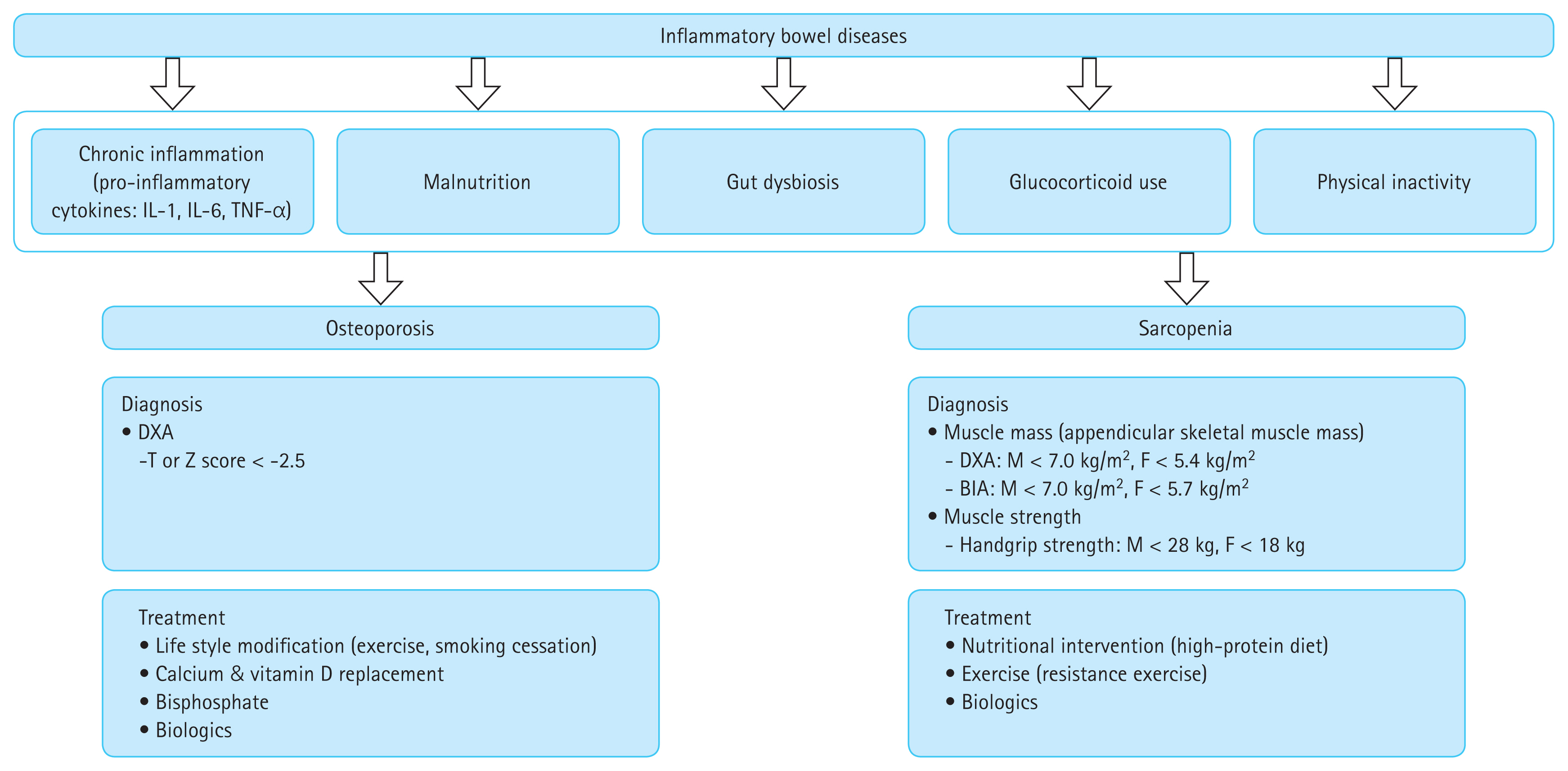
Overview of the pathophysiologies, diagnostic methods, and treatment strategies for metabolic musculoskeletal complications, including osteoporosis and sarcopenia. DXA, dual-energy X-ray absorptiometry; BIA, bioelectrical impedance analysis.
MS MANIFESTATIONS IN IBD
SpA
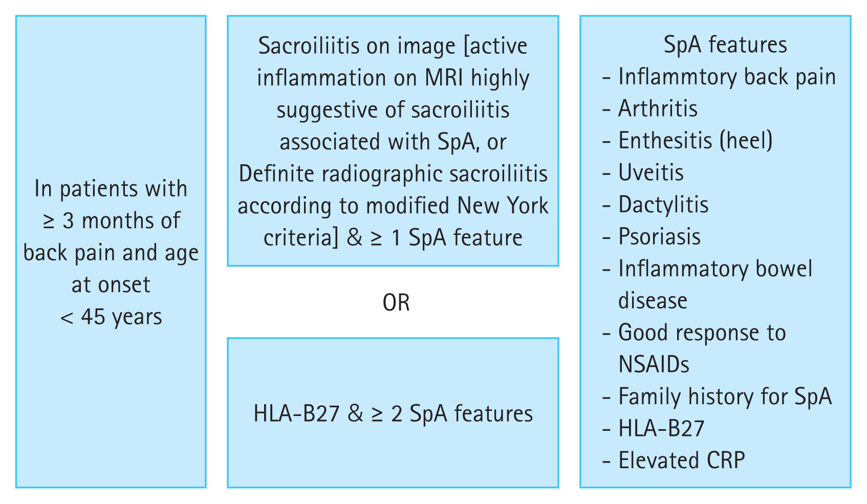
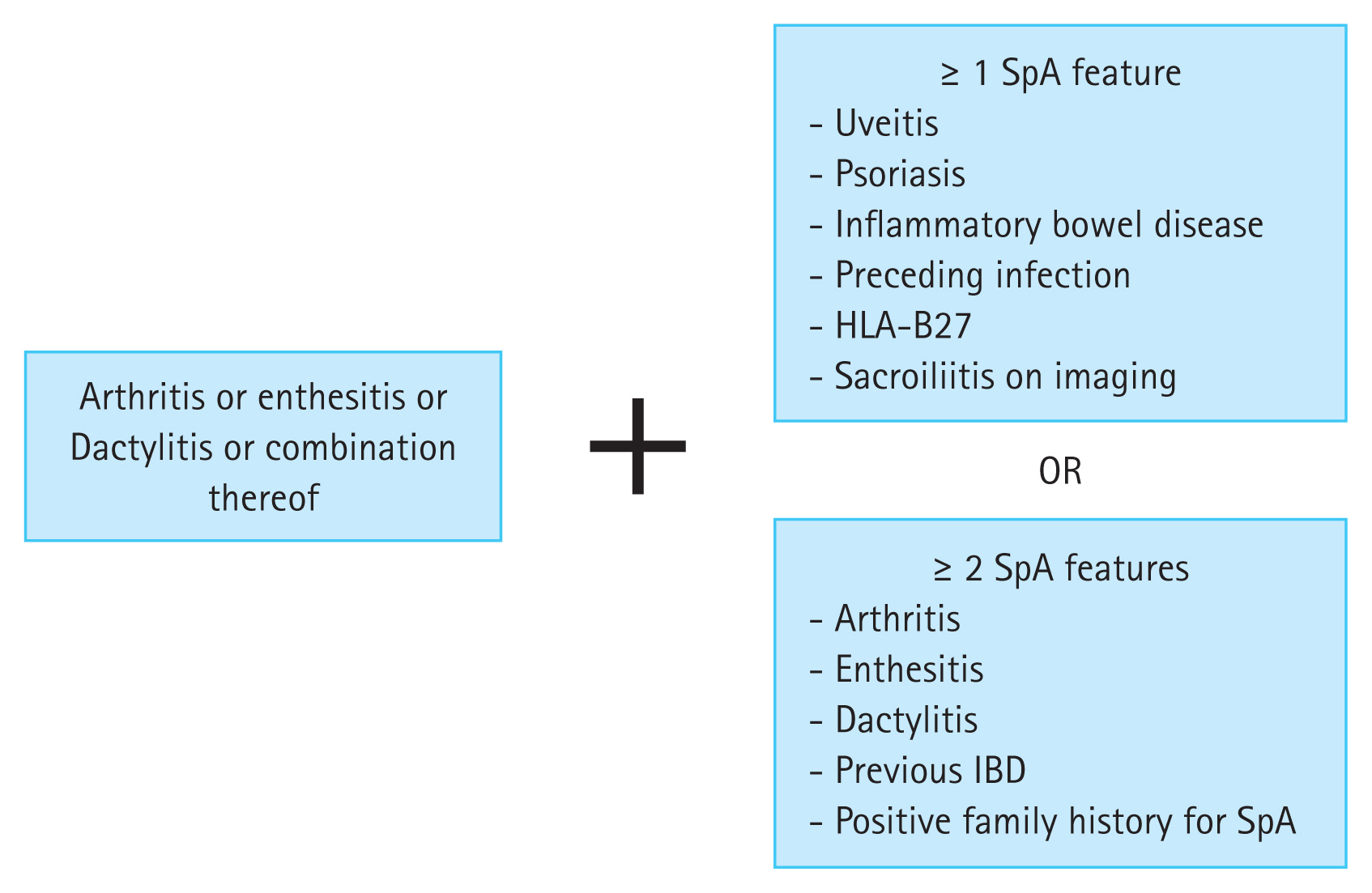
Chronic immune-mediated inflammation in IBD can affect peripheral joints, the axial skeleton, or both, a condition referred to as SpA. According to the classification criteria established by the Assessment of Spondyloarthritis International Society, SpA is broadly divided into axial and peripheral SpA based on symptom presentation (Fig. 2, 3) [75,76]. Axial SpA is characterized by symptoms such as back pain or morning stiffness of the spine, age < 45 years, sacroiliitis as determined via imaging, and human leukocyte antigen B27 (HLA-B27) positivity, whereas peripheral SpA involves the joints of the upper or lower limbs with sacroiliitis or other non-articular manifestations, such as psoriasis and uveitis [76,77].
The overlap between the extraintestinal manifestations of IBD and the features of SpA highlights a close relationship between the two conditions, emphasizing the importance of considering their potential coexistence. A systematic review reported that peripheral arthritis was the most common SpA presentation in patients with IBD, with a prevalence of 13%, followed by sacroiliitis at 10% and ankylosing spondylitis (AS) at 3% [78]. Although SpA is typically diagnosed after the onset of IBD, it can also precede the diagnosis. Peripheral arthritis occurs before IBD onset in 19.7% of cases, while axial SpA or AS occurs in 39.1% [79].
The pathophysiology of SpA involves a complex interplay of genetic, environmental, and immunological factors, although the precise mechanisms remain incompletely understood. A disrupted gut barrier is considered a primary process in SpA, particularly in individuals with genetic factors such as HLA-B27 [80]. HLA-B27 plays a central role in gut and joint inflammation. Individuals with this genetic marker have altered intestinal microbiota and subclinical gut inflammation, which are hallmarks of both axial SpA and AS [81]. An aberrant immune response to gut-derived antigens can trigger inflammation, leading to the upregulation of proinflammatory cytokines such as IL-1β, TNF-α, IL-23, and IL-17 [82].
This gut-induced inflammation may extend to MS sites, contributing to SpA through mechanisms such as molecular mimicry and immune cell trafficking [80,83]. Molecular mimicry occurs when an immune response to gut-derived antigens, such as those from intestinal microbiota, cross-reacts with normal host proteins in the synovial membrane, resulting in joint inflammation [83]. In addition, immune cell trafficking from the gut to skeletal sites is thought to play an important role in SpA development [80]. Integrins, such as alpha4-beta7 integrin, guide lymphocytes to gut or joint tissues [84]. Supporting this concept, studies have identified mucosa-associated invariant T cells in the synovial fluid of patients with AS, with integrin-expressing T cells being particularly abundant in their joints [84,85].
Genome-wide association studies further support the genetic link between SpA and IBD. Several genetic risk loci overlap between these conditions. For instance, NOD2/ CARD15, a gene associated with CD, has also been linked to sacroiliitis [86]. In addition, the Arg381Gln variant of IL23R, which is protective against CD, has been shown to reduce the risk of AS [87].
Axial SpA
Axial SpA refers to joint disease predominantly affecting the central axis of the body, which may occur with or without peripheral involvement (e.g., oligoarthritis, dactylitis, and enthesitis) or non-articular involvement (e.g., uveitis and psoriasis). In rheumatology, axial SpA is classified into non-radiographic axial SpA, representing an early stage of the disease, and radiographic axial SpA, also known as AS, both of which are considered part of the same disease specturm [88]. Axial manifestations in IBD have been studied under various definitions, including AS, inflammatory back pain, and isolated sacroiliitis.
AS (radiographic axial SpA)
AS has been reported in 10% of patients with IBD, a prevalence 20 times higher than in the general population. Most patients with AS test positive for HLA-B27 and present with both clinical symptoms of inflammatory back pain and radiographic evidence of sacroiliitis [89]. The diagnosis of AS is based on the 1984 Modified New York Criteria, which require at least one clinical symptom, such as inflammatory back pain, limited spinal mobility in the anteroposterior or lateral directions, or reduced chest expansion during deep breathing, along with radiographic evidence of sacroiliitis as viewed in plain X-ray [75]. In advanced cases, the characteristic “bamboo spine” appearance may be observed via X-ray.
For early diagnosis, MRI is a valuable tool because structural abnormalities may not be visible in X-rays during the early stages of the disease. MRI can detect characteristic signs of inflammation, such as bone marrow edema in the sacroiliac joints. Early diagnosis and timely treatment are essential for AS because if left untreated, the condition carries a poor prognosis due to progressive joint damage and deformities.
In patients with IBD who report symptoms of inflammatory back pain, HLA-B27 testing and sacroiliac joint X-rays should be performed. If sacroiliitis is suspected, prompt referral to a rheumatologist for multidisciplinary care is strongly recommended.
Inflammatory back pain
Inflammatory back pain is a distinct type of back pain that occurs without radiologic abnormalities and constitutes a significant proportion of axial manifestations in IBD, with a prevalence ranging from 5.2% to 42% [90]. Its characteristic features include morning stiffness, improvement of pain with physical activity, and worsening pain with rest, features that differentiate it from degenerative conditions such as herniated discs or spinal stenosis. Inflammatory back pain typically occurs in individuals under 40 years of age, has an insidious onset, and persists for more than 3 months.
This condition is considered an early presentation of axial SpA and may progress to AS over time [80]. Nonsteroidal anti-inflammatory drugs (NSAIDs) are generally effective for managing it.
Isolated sacroiliitis
Isolated sacroiliitis is diagnosed when radiographic examinations reveal sacroiliitis, characterized by erosion, sclerosis, narrowing, or widening of the sacroiliac joints, in the absence of clinical symptoms. It is the most common form of axial SpA, with a prevalence ranging from 16% to 46% [78]. However, progression to AS is rare [91]. HLA-B27 testing can aid diagnosis because isolated sacroiliitis is often HLA-B27-negative, unlike AS, in which more than 90% of patients test positive for HLA-B27 [82]. Because isolated sacroiliitis lacks clinical symptoms, treatment is typically unnecessary. However, regular monitoring for disease progression is recommended.
Treatment of axial SpA in patients with IBD
Non-pharmacological treatments, including physical therapy, rehabilitation, and exercise (e.g., swimming, stretching, and deep breathing exercises), can improve spinal mobility, alleviate symptoms, and prevent disability. First-line pharmacological treatment involves NSAIDs, which are effective for relieving symptoms and preventing joint deformities [92]. However, the adverse effects of NSAIDs on the GI tract have limited their use in patients with IBD. There is ongoing debate regarding whether NSAIDs exacerbate IBD. Recent meta-analyses suggest that they may increase the risk of exacerbation in CD but not in UC, although this effect was observed only in studies with a low risk of bias [93]. Short-term use of selective cyclooxygenase-2 (COX-2) inhibitors has not been associated with IBD exacerbation [94,95]. A meta-analysis reported that COX-2 inhibitor-associated IBD aggravation occurred in 14% of patients, primarily within the first few days to weeks of treatment [96]. Based on these findings, recent ECCO guidelines recommend case-by-case use of NSAIDs for SpA treatment in IBD, with selective COX-2 inhibitors allowed for short-term use [5].
For peripheral arthritis associated with axial SpA, sulfasalazine and methotrexate may be considered for managing peripheral symptoms such as arthritis and enthesitis [97]. However, neither sulfasalazine nor methotrexate is effective for axial SpA. Short-term, high-dose systemic glucocorticoids have shown modest efficacy for axial SpA but long-term systemic glucocorticoid use is not recommended because of potential side effects [92].
When NSAIDs are ineffective or symptoms are severe, biologic therapies may be considered. Several open-label studies have demonstrated the efficacy of anti-TNF agents for managing axial SpA [98–100]. However, there are limited studies on ustekinumab and vedolizumab for axial SpA, and the results have been inconsistent. One open-label study showed a clinical response in 43% of patients treated with ustekinumab, while a retrospective study reported worsening arthropathy in 22.5% of patients [101,102]. For vedolizumab, most studies have reported worsening of joint disease [102,103]. Therefore, ECCO guidelines recommend anti-TNF agents as the preferred biologic therapy for axial SpA in IBD, whereas ustekinumab and vedolizumab are not recommended.
Given the demonstrated efficacy of JAK inhibitors, such as tofacitinib and upadacitinib, in AS, these agents may be considered for treating axial SpA in patients with IBD [104,105].
Peripheral SpA
Peripheral SpA includes type 1 and type 2 peripheral arthritis, enthesitis, and dactylitis. Peripheral SpA is observed approximately twice as often in CD (10–20%) as in UC (5–14%) [6].
Type I peripheral arthritis
Type I peripheral arthritis is characterized by asymmetric oligoarthritis, typically involving five or fewer joints. It primarily affects the large joints of the lower extremities, such as the knees or ankles, as well as the hips, wrists, elbows, and shoulders. This type of arthritis affects approximately 3.6% to 6.0% of patients with IBD, often presenting early in the disease course and frequently associated with IBD flares. Joint damage, such as erosion or deformity, is rare, and the prognosis is generally favorable.
The diagnosis of type I peripheral arthritis is primarily clinical, based on physical examination findings such as tenderness, warmth, erythema, or swelling of the affected joints. While there are no definitive laboratory tests for this condition, the negativity of rheumatoid factor and anti-cyclic citrullinate peptide antibodies can support the diagnosis; however, a positive result does not exclude it [106]. In cases of monoarthritis, joint aspiration to assess the white blood cell count and synovial fluid culture is crucial to rule out infectious, traumatic, or crystal-induced arthritis [89]. This step is particularly important in patients receiving immunosuppressive therapy, as septic arthritis with atypical presentations should be considered.
Type II peripheral arthritis
Type II peripheral arthritis is characterized by polyarthritis involving more than five joints and typically affects the small joints of the upper extremities, such as the hands and wrists, in a symmetrical pattern [82]. Unlike type I, type II rarely precedes the diagnosis of IBD and tends to persist for months with repeated recurrences. In addition, it is largely independent of IBD activity and may persist even during periods of remission [107].
Joint erosions and damage may be observed in radiographic examinations, and the condition tends to follow a chronic course with a poorer prognosis than type I. Diagnosis relies on physical examination to confirm the presence of arthritis. Blood tests, including rheumatoid factor, antinuclear antibody, and anti-cyclic citrullinate peptide antibody tests, may help differentiate it from other autoimmune diseases, such as rheumatoid arthritis [82].
Enthesitis
Enthesitis refers to inflammation at the site where tendons attach to bone, commonly presenting as painful swelling at locations such as the Achilles tendon insertion on the heel, the plantar fascia insertion on the calcaneus, or the patellar ligament attachment to the kneecap [89]. It occurs in approximately 7% to 50% of patients with IBD and is a frequent appendicular manifestation in those with SpA. Chronic enthesitis can lead to structural changes in the bone, including osteopenia, abnormal new bone formation, irregularities, erosions of the bone cortex, and functional impairment [108].
Diagnosis is primarily clinical, based on tenderness at the enthesis during physical examination. Imaging is not always necessary in typical cases, but radiography may reveal erosions, spur formation, and ossification of the enthesis in advanced stages [108]. MS ultrasound is widely used for initial diagnosis and monitoring treatment efficacy, with characteristic findings including tendon edema, peritendinitis, increased Doppler signal, entheseal erosions, and calcifications in severe cases [109].
Dactylitis
Dactylitis, commonly referred to as a “sausage digit,” is characterized by inflammation of entire digits, resulting in significant pain and swelling in the affected fingers or toes. It affects up to 2% to 4% of patients with IBD and is associated with SpA [110]. Diagnosis is primarily clinical, based on symptoms and physical examination findings. However, MS ultrasound and MRI can help confirming the diagnosis. MS ultrasound may reveal features such as flexor tendon tenosynovitis and joint synovitis, which are characteristic of dactylitis [111].
Treatment of peripheral SpA in patients with IBD
Type I peripheral arthritis is generally self-limiting, with joint symptoms typically improving within 8 to 10 weeks when IBD is appropriately controlled [112]. By contrast, type II peripheral SpA is a chronic condition with adverse outcomes that require active treatment. For symptom control, acetaminophen, NSAIDs, or short-term COX-2 inhibitors may be used. In cases requiring additional management, short-term low-dose oral glucocorticoids may be considered for peripheral arthritis [80]. If infectious arthritis is ruled out, intra-articular steroid injections can provide symptomatic relief.
When acetaminophen, NSAIDs, or short-term COX-2 inhibitors are ineffective, sulfasalazine, commonly used for UC, can also be employed for peripheral arthritis. The typical starting dose is 500 mg twice daily for 2 to 3 weeks, with a gradual increase to a maximum of 3,000 mg per day [89]. If sulfasalazine proves ineffective, oral methotrexate at a dose of 7.5 mg once weekly, accompanied by folic acid, may be considered [113]. However, azathioprine has no role in treating peripheral SpA [80].
In terms of biologic therapy, while no RCTs specifically address biologics for non-axial SpA, studies have demonstrated that anti-TNF-α agents effectively reduce arthralgia and arthritis in patients with IBD [114,115]. In a post hoc analysis of adalimumab, improvements in arthritis and arthralgia compared to placebo were observed in patients with CD, with male sex and moderate disease activity identified as predictors of response [116]. However, a post hoc analysis of ustekinumab showed no beneficial effects for managing arthralgia or arthritis in CD, although a systematic review of nine studies have reported its efficacy in treating psoriatic arthralgia and arthritis [117,118].
Vedolizumab has shown inconsistent results for peripheral arthropathies. A post hoc analysis of the GEMINI study reported improvements in the severity of existing peripheral arthropathy and reduced new joint symptoms, whereas a multicenter retrospective study noted the development of new arthralgia symptoms during vedolizumab treatment [102,119]. Consequently, ECCO guidelines recommend anti-TNF-α agents for treating non-axial SpA in IBD. In addition, methotrexate, sulfasalazine, and ustekinumab can be considered.
Tofacitinib, a JAK inhibitor approved for psoriatic arthritis, showed some benefit for IBD-associated peripheral arthritis in a post hoc analysis of the OCTAVE studies and may be suggested for treating peripheral SpA. However, definitive evidence supporting its use for joint disease in IBD remains limited [8,120].
Table 1 summarizes the treatment of axial and peripheral SpA.

Treatment approaches for axial and peripheral spondyloarthritis
Fibromyalgia
Fibromyalgia is characterized by generalized pain, tenderness, fatigue, and cognitive difficulties persisting for more than 3 months. While the association between fibromyalgia and IBD is debated, some studies have reported a higher prevalence in patients with IBD, particularly those with CD, with an overall prevalence of 3.0% to 3.7%. This suggests a lower pain threshold in patients with IBD compared to healthy individuals [121,122].
The American College of Rheumatology’s 2010 diagnostic criteria, which include the Widespread Pain Index (WPI) and the Symptom Severity Scale (SSS), are used for diagnosis [123]. A diagnosis is made if WPI ≥ 7 and SSS ≥ 5, or if the WPI is between 3 and 6 with SSS ≥ 9.
Treatment for fibromyalgia in patients with IBD is similar to that in the general population. Tricyclic antidepressants, such as amitriptyline or nortriptyline, are considered first-line treatments [82]. More recently, medications such as the anticonvulsant pregabalin and serotonin-norepinephrine reuptake inhibitors, including duloxetine and milnacipran, have shown effectiveness. However, these medications can cause side effects, including dry mouth, dizziness, insomnia, and depression due to their anticholinergic effect; thus, caution is required.
If these medications are ineffective, consulting a rheumatologist is advisable because fibromyalgia can be challenging to manage, even with appropriate treatment.
CONCLUSIONS
MS manifestations and complications, such as osteoporosis, sarcopenia, and axial and peripheral SpA, are frequently observed in patients with IBD and contribute significantly to morbidity and reduced quality of life. Shared pathophysiological mechanisms, including chronic inflammation, glucocorticoid use, malnutrition, and gut dysbiosis, highlight the need for an integrated approach to managing these conditions. Osteoporosis and sarcopenia, often coexisting as osteosarcopenia, heighten the risk of fractures, diminished physical performance, and overall poor outcomes. Comprehensive management strategies incorporating lifestyle modifications, calcium, and vitamin D supplementation, resistance exercise, and pharmacologic therapies are essential to mitigate their impact. For spondyloarthropathies, early recognition and appropriate treatment with NSAIDs and biologics are critical to preventing long-term joint damage and disability.
Looking ahead, future research should prioritize personalized treatment approaches based on individual risk factors, such as age, disease activity, and genetic predisposition. In addition, further studies on the gut–muscle–bone axis and the role of gut microbiota in MS manifestations and complications could uncover new therapeutic opportunities. Multidisciplinary care, involving collaboration among gastroenterologists, rheumatologists, and endocrinologists, remains essential for improving outcomes and quality of life in patients with IBD and MS involvement.
Notes
CRedit authorship contributions
Young Joo Yang: conceptualization, methodology, data curation, formal analysis, writing - original draft; Seong Ran Jeon: conceptualization, methodology, data curation, formal analysis, writing - review & editing, supervision, funding acquisition
Conflicts of interest
Jeon SR was supported by a grant from Soonchunhyang University Research Fund. Yang YJ has no conflicts to declare.
Funding
This work was supported by the Soonchunhyang University Research Fund.