Ectopic Thyroid Nodule in Thyroglossal Duct
Article information
A 52-year-old woman presented with a small midline anterior neck mass and a 3-kg-weight loss over the course of one month. The patient was diagnosed with Graves disease based on increased free T4 levels (3.30 ng/dL), suppressed thyroid stimulating hormone (TSH) levels (<0.001 IU/mL), and a high titer of TSH receptor antibodies (32.6 IU/L). Scintigraphy with Tc-99m pertechnetate (Fig. 1) also revealed a diffusely enlarged thyroid gland with homogenous uptake, suggesting diffuse toxic goiter. Another uptake focus was noted apart from the pyramidal lobe, raising the possibility of ectopic thyroid tissue. Sonography of the thyroid gland (Fig. 2) showed a 5.1×10.1×16.1 mm-sized isoechoic solid nodule within the thyroglossal duct, which would have enlarged as a result of hyperthyroidism. The cytology specimen of this nodule, which was obtained by fine needle aspiration, showed benign follicular cells. The patient is now being treated with 20 mg of methimazole.

Scintigraphy with Tc-99m pertechnetate revealed a diffusely enlarged thyroid gland with homogenous uptake, suggesting diffuse toxic goiter. Uptake of the ectopic thyroid tissue (arrow) is seen apart from the pyramidal lobe (arrowhead).
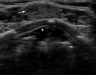
Sonography of the thyroid gland showed a 5.1×10.1×16.1 mm-sized ovoid to round, isoechoic solid nodule (arrow) within the thyroglossal duct in front of the thyroid cartilage (arrowhead).
Thyroglossal duct remnants are the most common midline neck masses found during childhood, representing more than 75% of such masses [1]. Approximately 7% of the adult population still has these remnants [2]. The presence of a solid mass along the thyroglossal duct cyst should raise the suspicion of ectopic thyroid tissue, as it is estimated that 35-70% may contain thyroid tissue in their wall [3]. Carcinoma arising from thyroglossal duct cysts is rare (only 1% of thyroglossal duct cases) and approximately 85-92% of all thyroglossal duct cyst carcinomas are papillary carcinomas [4]. Although they are characterized by relatively non-aggressive behavior and rare lymphatic spread [5], the possibility of occult malignancy should be evaluated in such cases.
Notes
No potential conflict of interest relevant to this article was reported.