N-terminal pro-B-type natriuretic peptide as a marker of disease severity in patients with pericardial effusions
Article information
Abstract
Backgraound/Aims
N-terminal pro-B-type natriuretic peptide (NT-proBNP) has recently been introduced as a useful marker in diagnosing underlying disease in patients with dyspnea and for determining the prognosis of patients with heart failure. The purpose of this study was to evaluate the value of the NT-proBNP as a marker of disease severity in patients with pericardial effusions.
Methods
We enrolled 69 consecutive patients who showed moderate or large pericardial effusion with preserved left ventricular (LV) systolic function; 42 patients finally participated in the study, and 13 (31.0%) of them showed cardiac tamponade. We analyzed the etiologies, the clinical and echocardiographic variables, and the serum NT-proBNP levels in these patients.
Results
The mean NT-proBNP level was 751±1002 ng/L (range 5 to 5289), and the median level was 385 ng/L (interquartile range 152 to 844). The NT-proBNP levels were higher in those patients with jugular venous distension (p=0.002), pulsus paradoxus (p=0.016), heart rate ≥100/min (p=0.006), cardiac tamponade (p=0.001), large pericardial effusion (p=0.029), exaggerated respiratory variation of the transmitral inflow (p=0.006), or plethora of the inferior vena cava (p=0.01). The NT-proBNP levels showed significant correlation with heart rate (r=0.517, p<0.001) and the diameter of the inferior vena cava (r=0.329, p=0.03).
Conclusion
NT-proBNP may be useful as a marker of disease severity in patients suffering from pericardial effusion, but further prospective studies with more patients will be needed.
INTRODUCTION
Pericardial effusion produces a continuum of pressure increases in the pericardium and therefore leads to a wide spectrum of hemodynamic and clinical manifestations1). The actual effect of accumulating pericardial fluid on raising the intrapericardial pressure is determined by both the rate of fluid accumulation relative to pericardial stretch and by the effectiveness of compensatory mechanisms2). Accordingly, once the total intrapericardial volume has caused the pericardium to reach the noncompliant region of its pressure-volume relation, critical tamponade can rapidly develop3).
To establish the severity of disease in patients with pericardial effusion, especially in those suffering from cardiac tamponade, physicians need to consider the symptomatic manifestations, physical examination signs, echocardiographic features, and hemodynamic characteristics. This process is complex and difficult, and a simple diagnostic supplement, such as a biomarker, is helpful.
B-type natriuretic peptide (BNP) is a 32-amino acid peptide hormone that is primarily released from the cardiac ventricles. It is synthesized as an inactive 108-amino acid pro-hormone pro-BNP, which is split into the active hormone BNP and the inactive N-terminal fragment (NT-proBNP), a 76-amino acid peptide. Cardiac myocytes constitute the major source of BNP-related peptides, and the main stimulus for synthesis and secretion is myocyte stretch. BNP has a number of systemic effects, including diuresis, vasodilatation, and inhibition of the renin-angiotensin-aldosterone system and the sympathetic nervous system4, 5). The rapid bedside assay for BNP and NT-proBNP has previously been shown to be useful for ruling out heart failure6). The test is also useful for making the diagnosis of systolic7) or diastolic heart failure8). BNP and NT-proBNP are predictors of cardiovascular morbidity and mortality in patients suffering from stable coronary heart disease9), acute coronary syndrome10), or heart failure11).
However, for patients with pericardial effusions, the role of natriuretic peptides as markers for disease severity, progression, or prognosis is unknown. Therefore, we undertook the present study to evaluate the hypothesis that NT-proBNP could be a useful marker for assessing disease severity in patients with pericardial effusions.
MATERIALS AND METHODS
Subjects
Between October 2004 and September 2006, we prospectively enrolled 69 consecutive patients with new onset moderate or large pericardial effusions and preserved LV function (ejection fraction ≥45%) on echocardiography, all of whom were admitted to our hospital. Moderate pericardial effusion was considered to be present when the anterior plus posterior echo-free spaces were 1 to 2 cm, and large effusion was considered when this figure was more than 2 cm. Patients with postoperative pericardial effusions, serum creatinine level more than 1.5 mg/dL, LV ejection fraction less than 45%, known coronary or valvular heart disease, atrial fibrillation, or severe circulatory congestion were excluded. Hence, 43 patients were finally selected for study participation. All patients underwent echocardiographic and Doppler examinations, and blood samples for determining the serum creatinine and NT-proBNP levels were collected on the same day.
Clinical Data
Age, gender, weight, height, body mass index, NYHA classification, blood pressure, presence or absence of pulsus paradoxus (a reduction in systolic blood pressure greater than 10 mmHg during inspiration), heart rate, respiratory rate, and electrocardiographic findings were all recorded. Characteristic clinical findings2) (dyspnea, pulsus paradoxus, jugular venous distension, tachycardia and hypotension) were also recorded. In our study, "overt clinical tamponade" was considered present when heart rate was ≥100/min, and there was documented evidence of systolic blood pressure less than 90 mmHg. Cardiac tamponade was confirmed by two- dimensional (2D) and Doppler echocardiographic findings; this "echocardiographic tamponade" was defined as right atrial (RA) or right ventricular (RV) collapse, inferior vena cava (IVC) plethora without inspiratory collapse, and exaggerated respiratory variation of mitral early filling velocity.
Etiologic investigation was done according to the following protocol suggested by Sagrista-Sauleda et al.12) The initial evaluation included basic laboratory studies, chest radiograph, physical examination, echocardiography, evaluation for tuberculosis, measurement of serum antinuclear antibodies and thyroid hormones, and other investigations that were warranted by the individual clinical findings. Advanced evaluation included pericardiocentesis for those patients with cardiac tamponade, a suspicion of purulent pericarditis, or a chronic large pericardial effusion. In addition to routine cytological and biochemical studies of the pericardial fluid, adenosine deaminase activity was measured, and the results of polymerase chain reaction (PCR) for tubercle bacilli were recorded. Subxiphoid pericardial biopsy with histologic examination and culture of the biopsy specimen were not routinely performed in this study.
Echocardiographic Study
All echocardiographic studies were carried out using a standard protocol on a commercially available system (Vivid 7, GE Medical Systems, Horten, Norway). Echocardiograms were stored digitally and analyzed offline by two cardiologists who had no knowledge of the NT-proBNP data. M-mode, 2D, and Doppler recordings were carried out in all patients whose clinical status did not make the measurements impossible. All measurements were recorded through the whole respiratory cycle and averaged over five cardiac cycles. The dimensions of the cardiac walls and chambers were measured according to the American Society of Echocardiography guidelines13). The interventricular septal thickness (IVST) at end diastole, LV posterior wall thickness (PWT) at end diastole, and LV internal dimension at end diastole and end systole (LVIDd and LVIDs, respectively) were measured via 2D echocardiographically guided M-mode examination. The left atrial (LA) volume was measured by the length-diameter prolate ellipsoid method at end systole. On Doppler echocardiographic examination, the transmitral early filling velocity (E), late filling velocity (A), deceleration time (DT) of E flow, E/A ratio, early velocity of the septal annulus (E'), and E/E' were also measured. For estimating RV systolic pressures non-invasively, we measured the maximal velocity in the tricuspid regurgitant jet (TRVmax) and obtained RV systolic pressures by the following equation:
RV systolic pressure = 4(TRVmax)2 + RA pressure
RA pressure was estimated based on evaluation of IVC diameter during respiration. If IVC diameter was small (<1.5 cm) and collapsed by at least 50% with respiration, then estimated RA pressure was 5 mmHg. If IVC diameter was normal (1.5-2.5 cm), estimated RA pressure was 10 mmHg or 15 mmHg according to the presence or the absence of IVC collapse with respiration, respectively. If the IVC was dilated (>2.5 cm), then estimated RA pressure was 20 mmHg.
Morphologic features suggestive of tamponade physiology, as confirmed by 2D echocardiography, included RA compression during late diastole14), RV collapse during early diastole15), and IVC plethora with a lack of inspiratory collapse16). Doppler hemodynamic findings suggestive of tamponade physiology included exaggerated respiratory variation (difference >25%) of the mitral E velocity during inspiration and expiration17, 18), accompanied by an increase in hepatic venous diastolic flow reversal on expiration18).
Measurement of Serum NT-proBNP Levels
Peripheral venous blood samples were carefully obtained from the antecubital vein and then transferred into standard sampling tubes. NT-proBNP was measured by employing the electrochemiluminescence principle (Elecsys® 2010/Molecular analytic E710, Roche Diagnostics, Indianapolis, Ind., USA) using sandwich immunoassay with two polyclonal antibodies.
Statistical analysis
SPSS Inc. for Windows Standard version 12.0 was used for statistical analysis. The data are expressed as means±SDs (range) for the normally distributed variables or as medians and interquartile ranges for the asymmetrically distributed variables. The quantitative normally distributed data was compared using the Student's t-test. The chi-square test was used for non-continuous variables. Pearson's correlation coefficient was used to assess correlations between the NT-proBNP levels and the normally distributed clinical and echocardiographic variables. Natural logarithmic transformation (ln) was used to adjust for the asymmetrically distributed variables, including the NT-proBNP levels. Multivariate regression analysis was performed to establish the independent clinical and echocardiographic variables that determined serum NT-proBNP levels. Logistic regression was performed to assess whether NT-proBNP would add diagnostic information over clinical parameters. A p value of < 0.05 was considered statistically significant.
RESULTS
Baseline patient characteristics
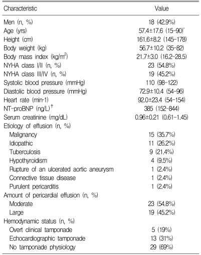
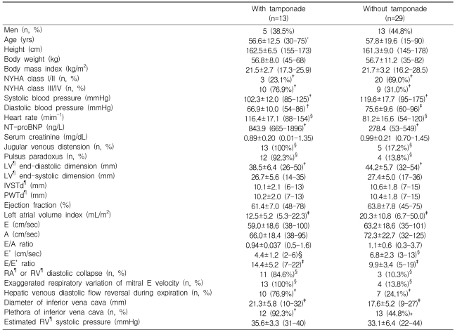
The baseline clinical and echocardiographic characteristics are listed in Table 1. In 11 (26.2%) of 42 patients, the etiology of the pericardial effusion was unknown. The three most frequent identifiable causes were: malignancy in 15 (35.7%), tuberculosis in 9 (21.4%), and hypothyroidism in 4 (9.5%). Three (7.2%) patients had other unusual causes: rupture of an ulcerated aortic aneurysm, connective tissue disease, and purulent pericarditis complicated by mediastinal empyema, respectively. Effusion was moderate in 23 patients and large in 19 patients. On the basis of clinical and echocardiographic findings, 5 patient had overt clinical tamponade (19%), 13 patients (31.0%) had echocardiographic cardiac tamponade, and 29 (69.0%) patients had no tamponade physiology (Table 2). All patients with cardiac tamponade underwent pericardiocentesis and exhibited subsequent clinical and hemodynamic improvement. Diagnostic pericardiocentesis was carried out in 13 patients who were without cardiac tamponade. NYHA class III/IV, jugular venous distension, pulsus paradoxus, RA or RV collapse, exaggerated respiratory variation of mitral E velocity, hepatic venous diastolic flow reversal during expiration, and IVC plethora were all more common in patients with cardiac tamponade (Table 2). In patients with cardiac tamponade, the values of systolic and diastolic blood pressure, LV end-diastolic dimension, LA volume index, and E' velocity were lower than in patients without tamponade; however, the values of heart rate, NT-proBNP levels, E/E' ratio, and IVC diameter were higher (Table 2).
Relationship between NT-proBNP and clinical features
The mean ln NT-proBNP level was 2.6±0.59 ng/L (mean 751±1002 [range 5 to 5,289], median 385 [interquartile range 152 to 844]). Higher ln NT-proBNP levels were observed in those patients with jugular venous distension (2.9±0.50 vs 2.3±0.54 ng/L, respectively, p=0.002), and in those patients with pulsus paradoxus (2.8±0.41 vs 2.4±0.62 ng/L, respectively, p=0.016) (Figures 1B and 1C). The ln NT-proBNP levels were higher in patients with heart rates ≥100/min, compared to those patients with heart rates <100/min (2.9±0.42 vs 2.4±0.59 ng/L, respectively, p=0.006) (Figure 1A). Furthermore, higher ln NT-proBNP levels were observed in patients with cardiac tamponade than in patients without this finding (3.0±0.34 vs 2.4±0.58 ng/L, respectively, p=0.001) (Figure 1D). NYHA class III or IV patients showed a trend of elevated ln NT-proBNP levels compared with NYHA class I or II patients, but this was not statistically significant (2.7±0.51 vs 2.4 0.61 ng/L, respectively, p=0.074). There was positive correlation between ln NT-proBNP level and heart rate (r=0.517, p<0.001) (Figure 2A). There was no relation between ln NT-proBNP level and either age, gender, or systolic or diastolic blood pressure.

Distribution of ln NT proBNP in patients with pericardial effusion, according to heart rate (A) and presence or absence of jugular venous distension, pulsus paradoxus, and cardiac tamponade (B, C and D). The plots display the median, and 25th and 75th percentiles (lower and upper limits of the boxes), along with error bars. JVD, jugular venous distension; PP, pulsus paradoxus; Tamp, cardiac tamponade.
Relationship between NT-proBNP and echocardiographic findings
Higher NT-proBNP levels were observed in patients with large pericardial effusions than in patients with moderate pericardial effusions (2.8±0.47 vs 2.4±0.61 ng/L, respectively, p=0.029). The ln NT-proBNP levels were higher in patients with exaggerated respiratory variation of the transmitral inflow, compared to patients without this finding (2.9±0.40 vs 2.4±0.61 ng/L, respectively, p=0.006). The ln NT-proBNP levels were higher in patients with IVC plethora, as compared to patients without this finding (2.7±0.51 vs 2.3±0.60 ng/L, respectively, p=0.01). The ln NT-proBNP levels were not significantly different between patients with an E/E' ratio ≥10 and patients with an E/E' ratio < 10. There was a positive correlation between the ln NT-proBNP level and IVC diameter (r=0.329, p=0.03) (Figure 2B). There was no significant correlation between the ln NT-proBNP level and either LV cavity dimensions, LV wall thickness, LA volume index, E velocity, A velocity, E/A ratio, E', E/E' ratio, or estimated RV systolic pressure.
Multivariable analysis
For multivariable analysis, we selected variables such as age, body mass index, systolic blood pressure, heart rate, end-diastolic dimension of LV, ejection fraction, LA volume index, E', E/E' ratio, and IVC diameter. On multivariable analysis, the ln NT-proBNP level was only independently related to heart rate (p=0.04), with an overall r2=0.40.
Increased NT-proBNP as a diagnostic marker of cardiac tamponade
The receiver operating characteristic (ROC) analysis for NT-proBNP as a diagnostic marker of cardiac tamponade showed that the area under the curve was 0.83 (95% confidence interval 0.70-0.96) (p=0.001). A cut-off NT-proBNP value of 436 ng/L had a specificity of 76% and a sensitivity of 92% in diagnosing cardiac tamponade (Figure 3). Multivariate logistic regression models were used to assess the diagnostic value of NT-proBNP, when combined with clinical parameters (e.g. systolic blood pressure and heart rate). We found that NT-proBNP added significant diagnostic information to the clinical parameters when diagnosing cardiac tamponade (Figure 4).

Receiver-operating characteristic curve analysis of ln NT-proBNP level, according to cardiac tamponade. Area under the curve was 0.83 (95% confidence interval [CI] 0.70-0.96), and optimal cut off value for prediction of the presence of cardiac tamponade was 436 ng/L.
Intraobserver and interobserver variability
The mean of differences between two measurements made by the same examiner were 0.17±6.59 cm/s (from -9.2 to 16.8 cm/s) for E, -3.08±2.10 cm/s (from -4.8 to 0.20 cm/s) for A, -0.045±0.039 (from -0.06 to 0.03) for E/A ratio, -0.075±0.413 cm/s (from -0.80 to 0.60 cm/s) for E', -0.666±1.129 (from -2.30 to 0.86) for E/E' ratio, and -1.70±7.18 mL/m2 (from -13.0 to 7.0 mL/m2) for LA volume index. Interobserver values for E, A, E/A ratio, E', E/E' ratio and LA volume index from two independent observers were in good agreement (r=0.960 for E, r=0.992 for A, r=0.983 for E/A ratio, r=0.964 for E', r=0.980 for E/E' ratio, and r=0.976 for LA volume index). However, the agreement for the DT was less robust (r=0.597), and the value of this parameter was excluded from the analysis.
DISCUSSION
We performed a cross-sectional study to investigate the role of NT-proBNP in assessing disease severity in patients with pericardial effusions. Our study showed that the NT-proBNP serum levels were increased in patients with jugular venous distension, pulsus paradoxus, heart rate ≥100/min, exaggerated respiratory variation of the transmitral inflow, IVC plethora, and cardiac tamponade. The NT-proBNP levels showed positive correlation with the heart rate and IVC diameter. On ROC curve analysis, a cut-off NT-proBNP value of 436 ng/L showed good specificity and sensitivity in diagnosing cardiac tamponade. We also found that NT-proBNP added significant diagnostic information to the clinical parameters
There are few reports concerning natriuretic peptide assay in pericardial effusion patients. In the study by Spodick et al,19) the levels of atrial natriuretic peptide did not increase because the compressed myocardium could not stretch. Recently, Fernandes et al.20) reported that NT-proBNP was increased in pericardial diseases and was associated with diastolic dysfunction. However, this study did not show the relation between NT-proBNP levels and pericardial effusion severity.
Effect of compensatory mechanisms
Patient with severe pericardial effusion have more severely compromised hemodynamics. In patients suffering from critical tamponade, cardiac compensatory mechanisms such as increased heart rate principally induced by beta-adrenergic activity, peripheral vasoconstriction, increased ejection fraction, and blood volume expansion are activated to maintain cardiac output and blood pressure2). Any additional compensation provided by neurohormonal stimulation is similar to what occurs during heart failure21).
In our study, NT-proBNP levels were related to the presence of jugular venous distension and IVC plethora. Jugular venous distension and IVC plethora result from compensatory vasoconstriction and fluid retention and subsequently increased blood volume and venous pressure via activation of the renin-angiotensin system2, 3, 16). The NT-proBNP levels were also correlated with heart rate, which increases with compensatory beta-adrenergic stimulation. Hanford et al.22) reported that catecholamine infusion in rats is associated with increased BNP levels. Furthermore, angiotensin II effects a decrease in BNP degradation23), and together with endothelin, stimulates BNP gene expression24). Vinch et al.25) suggested that the paradoxical increase in BNP levels seen after intravenous conscious sedation may be the result of counter-regulatory hormone release caused by an acute decrease in blood pressure.
Effect of hemodynamic changes and reduced coronary flow
In the current study, NT-proBNP levels were increased in patients with pulsus paradoxus and with exaggerated respiratory variations of transmitral inflow related to increased pericardial pressure and subsequent accentuated interdependence of the cardiac chambers2, 3, 26, 27). In those patients displaying pulsus paradoxus and exaggerated respiratory change of transmitral inflow, the stroke volume and cardiac output are usually decreased, and this leads to absolute or relative hypotension. The decrease in blood pressure may induce a compensatory increase in counter-regulatory hormones, as mentioned above, and reduce coronary flow. It is well known that elevated BNP and NT-proBNP levels may be the result of cardiac ischemia via reduced coronary flow28). However, although coronary blood flow is reduced in tamponade, there is no ischemic component because the coronary flow remains in proportion to the reduced work of the heart29). Therefore, reduced coronary flow may not be the main impetus behind the elevated NT-proBNP levels observed in the present study.
Effect of diastolic function
Once the total intrapericardial volume has caused the pericardium to reach the noncompliant region of its pressure-volume relation, even small increases in the intrapericardial volume sharply increase the intrapericardial pressure1). Therefore, as the ventricles begin to relax at the end of systole, they are initially constrained by the elevated intrapericardial pressure, resulting in a pattern of parallel upward displacement of the diastolic pressure-volume curve30). Obtaining only echocardiographic evidence of increased diastolic filling pressures is insufficient. Chamber dimensions obtained in a single echocardiography exam cannot accurately predict the presence or severity of cardiac tamponade1, 31).
In the current study, there was no significant correlation between the NT-proBNP levels and either the LV cavity dimensions or the LA volume index. One possible explanation for this is that, although the diastolic filling pressures were increased, total cardiac volume was limited by the pericardial effusion, and the myocytes could not stretch. Hence, the elevated NT-proBNP levels in the study patients may not be related to chamber dimensions.
In the current study, there was no significant correlation between the NT-proBNP levels and Doppler parameters, such as E velocity, A velocity, E/A ratio, E', and E/E' ratio. There are several reasons why the NT-proBNP levels were not related with the Doppler parameters of diastolic function. First, the E/E' ratios of many patients (49%) in our study ranged between 8 and 15, but only 8 patients (19%) had E/E' values > 15. The range of 8 to 15 is considered to be a "gray zone" in the estimation of LV filling pressures, and therefore other parameters are required for an accurate prediction of filling pressure32, 33). Second, as mentioned in previous studies, BNP showed weak correlation with LV end-diastolic pressure and low sensitivity for the detection of elevated LV end-diastolic pressure in patients with normal systolic function34). Therefore, we believe that the effect of compensatory mechanisms on the production of NT-proBNP, as mentioned above, was robust enough to mask the effect of diastolic dysfunction on the production of the NT-proBNP in patients with pericardial effusion.
Limitations of the study
There are several limitations to this study. First, the number of patients in our study was small. Second, the patient group was heterogeneous; the etiology of pericardial effusion was diverse, and the duration of pericardial effusion was uncertain, so the influence of these variables on NT-proBNP levels was unclear. Third, we could not check NT-proBNP levels after pericardiocentesis, so we lack comparison between NT-proBNP levels before and after pericardiocentesis. Fourth, our study lacked invasive measurement of hemodynamics. There were also some limitations in the measurement of echocardiographic parameters in patients with unstable hemodynamics. In the current study, the interobserver values for the DT from two independent observers were not in good agreement (r=0.597), perhaps because of rapid heart rate and the summation of E and A velocity, especially in patients with cardiac tamponade. However, interobserver values for E, A, E/A ratio, E', E/E' ratio and LA volume index were in good agreement. Therefore, we do not believe that interobserver variability of the measurements affected the results significantly.
Conclusion and clinical implications
Abnormally elevated NT-proBNP levels are a special feature of patients with pericardial effusions. The mechanism behind the increased NT-proBNP levels is not entirely clear, but NT-proBNP levels may be the result of counter-regulatory hormone release induced by compensatory activation of the adrenergic and renin-angiotensin systems. However, the relation between NT-proBNP levels and diastolic dysfunction could not be clearly assessed and would be of interest in future studies.
We believe that NT-proBNP measurement has potential as a simple, relatively cheap method for assessing disease severity in patients with pericardial effusions. Furthermore, it may be used in the diagnosis of cardiac tamponade. Our findings should be further assessed in prospective studies with more data that could address the role of NT-proBNP in disease progression and more precisely determine its role as a prognostic marker in patients suffering from pericardial effusion.


