Isolated right ventricular infarction during percutaneous coronary intervention
Article information

A 79-year-old male with hypertension was admitted with chest pain. An electrocardiogram (ECG) was obtained (Fig. 1A). The troponin I level was increased to 0.11 ng/mL (reference range, 0–0.04 ng/mL). Coronary angiography revealed an irregular stenosis of 80% in the middle portion of the right coronary artery (RCA) (Fig. 1B). The left coronary system exhibited no significant lesions. Balloon angioplasty and coronary stenting were performed over the middle RCA. After coronary stenting, the right ventricular (RV) branches were jailed (Fig. 1C), and he complained of recurrent chest pain.
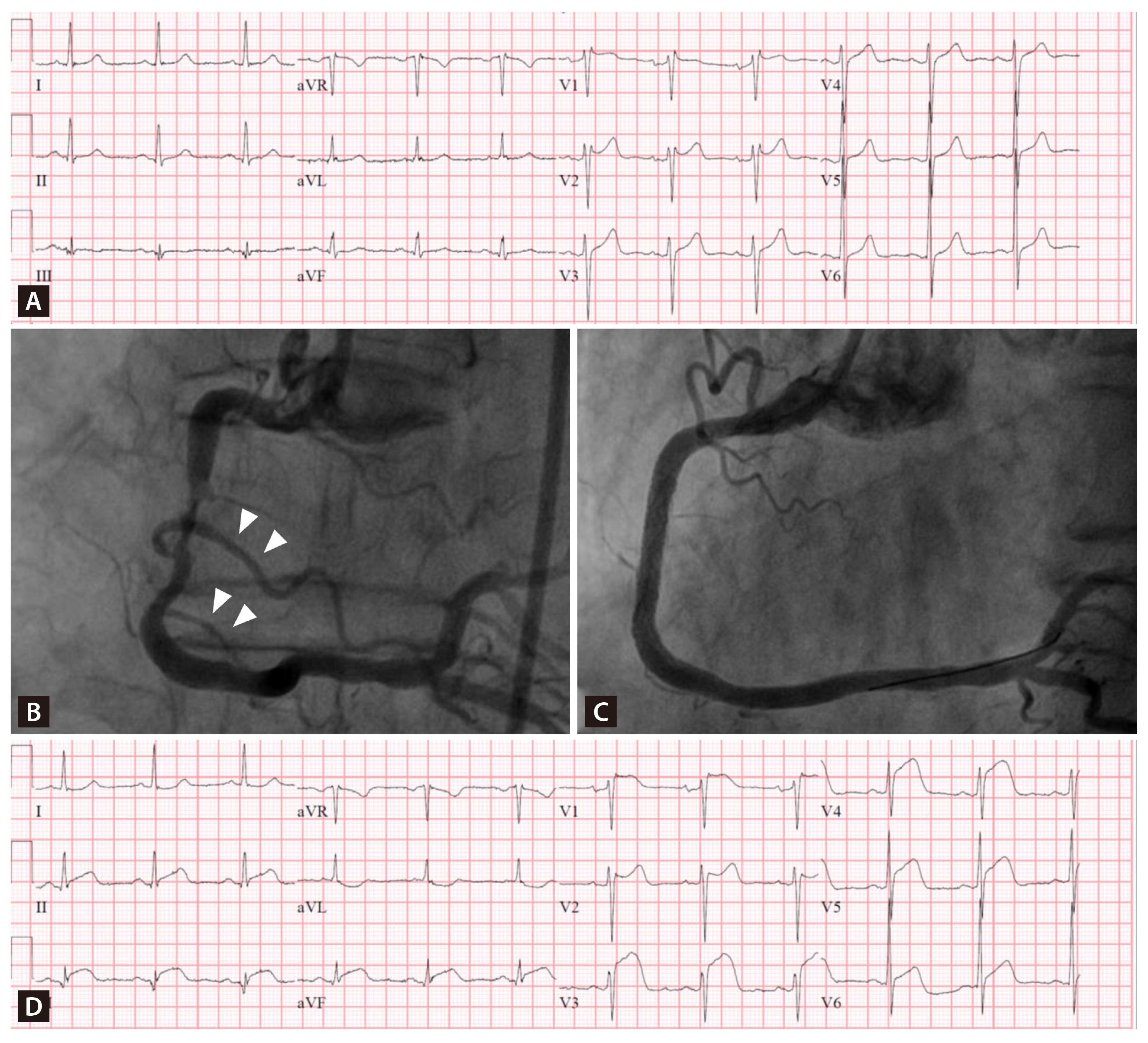
Electrocardiogram and Right coronary angiogram findings. (A) Initial electrocardiogram (ECG). (B) Coronary angiography revealed an irregular stenosis of up to 80% in the middle portion of the right coronary artery (RCA). The right ventricular branches are intact (arrowheads). (C) After coronary stenting of the middle RCA, the right ventricular branches of the RCA were jailed. (D) The ECG after stenting shows newly developed marked ST elevation in leads V1–V5, and slight ST elevation in the inferior leads (more in lead III than lead II).
A repeat ECG (Fig. 1D) showed newly developed marked ST elevation (STE) in leads V1–V5, and slight STE in the inferior leads (more in lead III than lead II). A repeat left coronary angiogram revealed no occlusion. Attempted revascularization of the RV branches failed, but his chest pain somewhat improved after an intracoronary infusion of nicorandil. The post-procedural echocardiogram revealed RV systolic dysfunction with a wall motion abnormality of the RV free wall, which was compatible with an RV myocardial infarction (MI, RVMI). The post-procedural troponin I level maximally increased to 7.7 ng/mL. His symptom and STE gradually improved, and he was discharged 4 days later.
Isolated STE due to RVMI is uncommon in clinical practice. Since RVMI usually occurs with left ventricular inferior infarction, the electrical current of injury from the left ventricle dominates the RV electrical forces. The typical ECG changes in this setting are STE in the inferior leads with concomitant ST depression in the precordial leads [1]. However, the ECG presentation of an isolated RVMI can mimic an anterior STEMI because of the anterior location of the RV. The absence of Q-wave development in the anterior precordial leads may help the diagnosis of RVMI [2], as was observed in current case.
Notes
CRedit authorship contributions
Chan-Hee Lee: conceptualization, data curation, writing - original draft; Young-Sang Jeong: investigation, data curation, writing - original draft, visualization; Melvin M. Scheinman: writing - review & editing, supervision
Conflicts of interest
The authors disclose no conflicts.
Funding
None