Femoral sheath fracture and migration during temporary pacemaker removal: Successful bail-out using a balloon retrieval technique
Article information

A 93-year-old female presented with syncope and was diagnosed with an inferior wall ST-segment elevation myocardial infarction complicated by shock and complete atrioventricular block. Emergent temporary pacemaker (TPM) insertion, veno-arterial extracorporeal membrane oxygenation, and percutaneous coronary intervention (PCI) were performed.
The patient recovered uneventfully, and several days later underwent additional PCI for a non-culprit left main lesion. During removal of the right ventricular TPM, sheath fracture with migration occurred (Fig. 1). The patient was markedly obese (body mass index 37.8 kg/m2), and the relatively vertical course of the sheath in the right common femoral vein likely predisposed it to kinking and fracture.
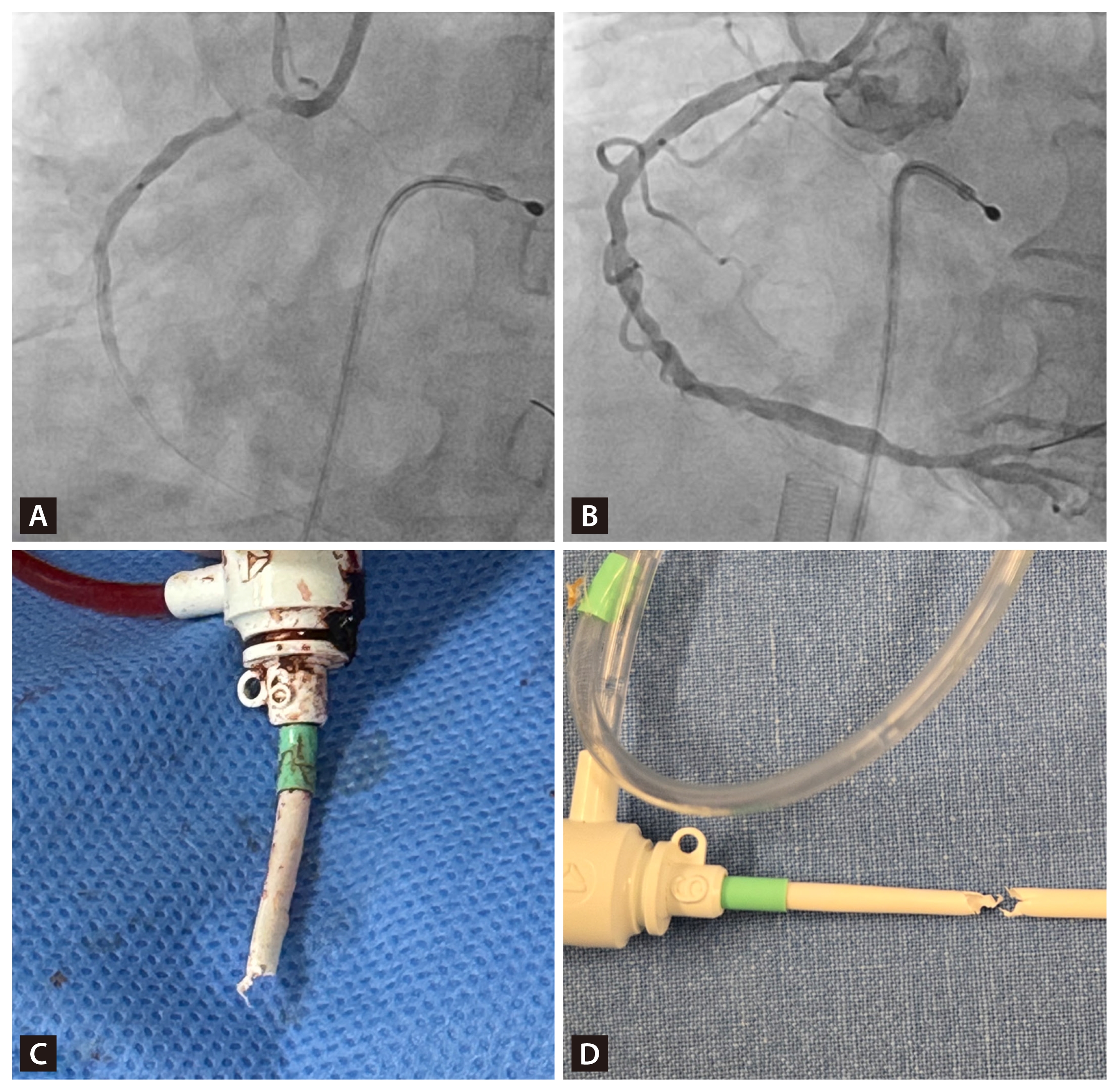
Coronary angiography and gross images of fractured sheath. (A) Coronary angiography showing a culprit lesion in the proximal right coronary artery. Because of complete atrioventricular block and severe bradycardia, a temporary pacemaker was inserted first. (B) Veno-arterial extracorporeal membrane oxygenation was applied, followed by successful stent implantation without catastrophic events. (C) Intraoperative image showing the proximal portion of the fractured sheath. (D) Entire sheath with a visible fracture line along the shaft, confirming complete disruption.
Fortunately, fluoroscopy showed that the fractured sheath had not migrated far, remaining close to the adjacent femoral artery sheath. For bail-out, a repeat puncture was performed distal to the original sheath site and a 9 French sheath was inserted. A 0.014″ guidewire was advanced, followed by positioning of a 2.5 mm balloon within the lumen of the floating remnant. Inflation of the balloon allowed successful retrieval of the fragment into the 9 French sheath. To facilitate secure capture and retrieval of the irregularly torn sheath remnant, the distal end of the 9-French sheath was partially cut with scissors to widen and modify the tip (Fig. 2).
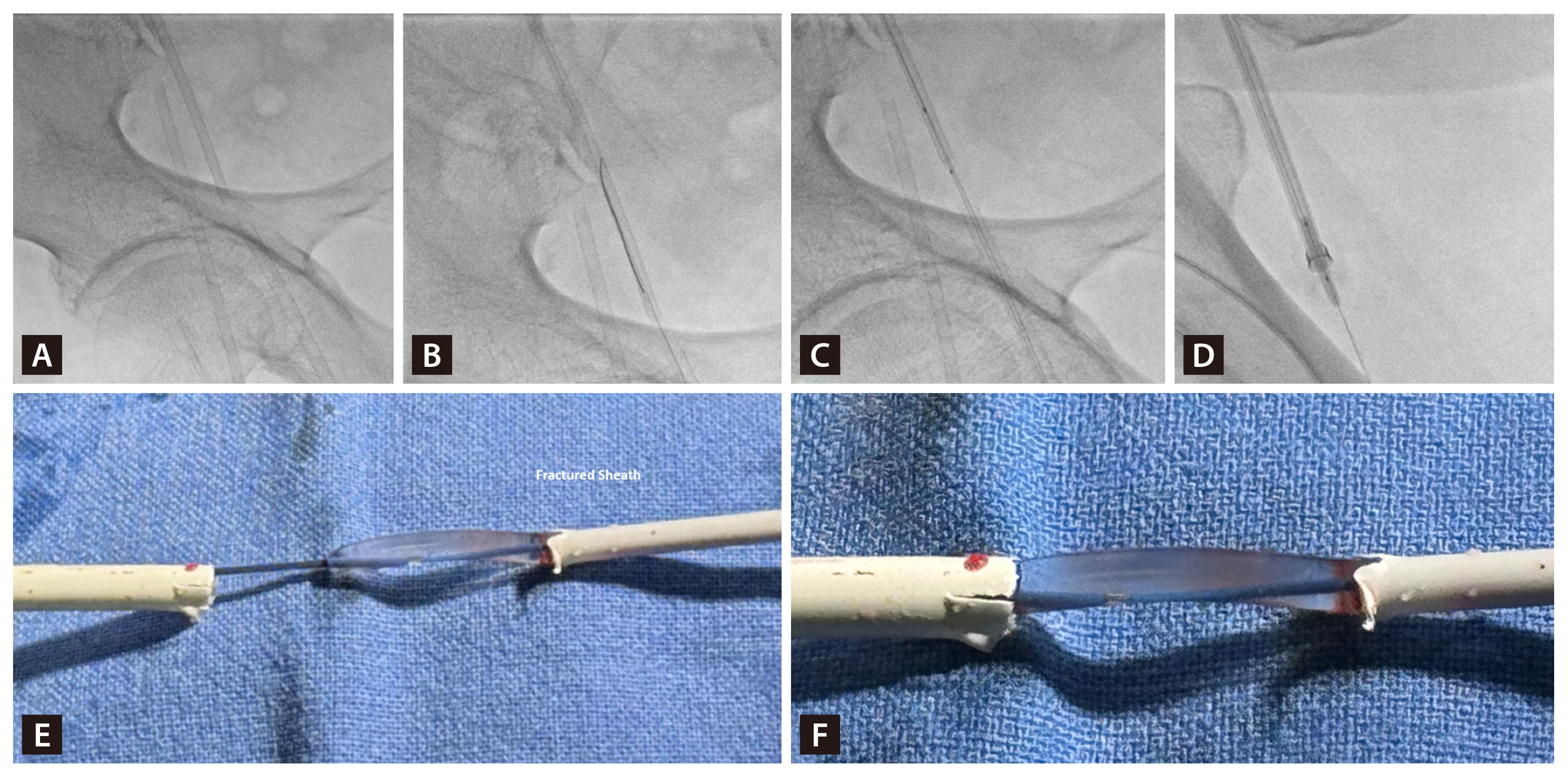
Fluoroscopic images and intaroperative images of balloon retrieval of fractured sheath. The right common femoral artery sheath was left in place throughout the procedure. (A) Repeat puncture of the right superficial femoral vein distal to the previous lock sheath site, followed by insertion of a 9 French sheath. (B) Advancement of a 0.014″ guidewire into the true lumen of the remnant sheath. (C) Placement of a 2.5 × 15 mm non-compliant balloon into the true lumen over the wire. (D) Balloon inflation and successful retrieval of the remnant sheath through the distal 9 French sheath. (E) Balloon inflated within the lumen of the fractured sheath remnant, securing the fragment. (F) The fractured sheath being drawn into the 9 French sheath by balloon traction, enabling successful retrieval.
Foreign bodies within the venous system, if lodged in the pulmonary artery, right atrium, or right ventricle, may rarely cause ventricular arrhythmia or thrombus formation; thus, removal is mandatory [1–3]. The most common etiology is catheter fracture, usually managed by snaring, with percutaneous retrieval success rates nearly 100% [1–3]. In this case, although snare technique was possible, the relatively large 6 French sheath fragment prompted the use of balloon traction to minimize vascular injury.
Informed consent was obtained from patients.
Notes
CRedit authorship contributions
Daeung Ohn: conceptualization, investigation, writing - original draft, writing - review & editing, visualization; Yeon-Jik Choi: writing - review & editing, project administration; Junghoon Lee: writing - review & editing, supervision; Suk-Min Seo: writing - original draft, writing - review & editing, supervision, project administration
Conflicts of interest
The authors disclose no conflicts.
Funding
None