Clinical features of moderate-to-severe antibiotic allergy in Korea: analysis from the drug allergy safety information sharing system database
Article information

Abstract
Background/Aims
Antibiotics are major causes of drug allergy. Sharing standardized information on culprit agents is essential to prevent recurrent drug allergy. In 2024, the Ministry of Health and Welfare established the Drug Allergy Safety Information Sharing System (DASI), integrated with the Personal Health Record (PHR). We analyzed the epidemiology, culprit antibiotics, clinical manifestations, and diagnostic practices of antibiotic allergy in Korea using DASI data.
Methods
We retrospectively reviewed 807 cases of moderate-to-severe antibiotic allergy from eight tertiary hospitals. Clinical features were assessed according to reaction type, severity, MedDRA system organ class, culprit antibiotics, and diagnostic methods.
Results
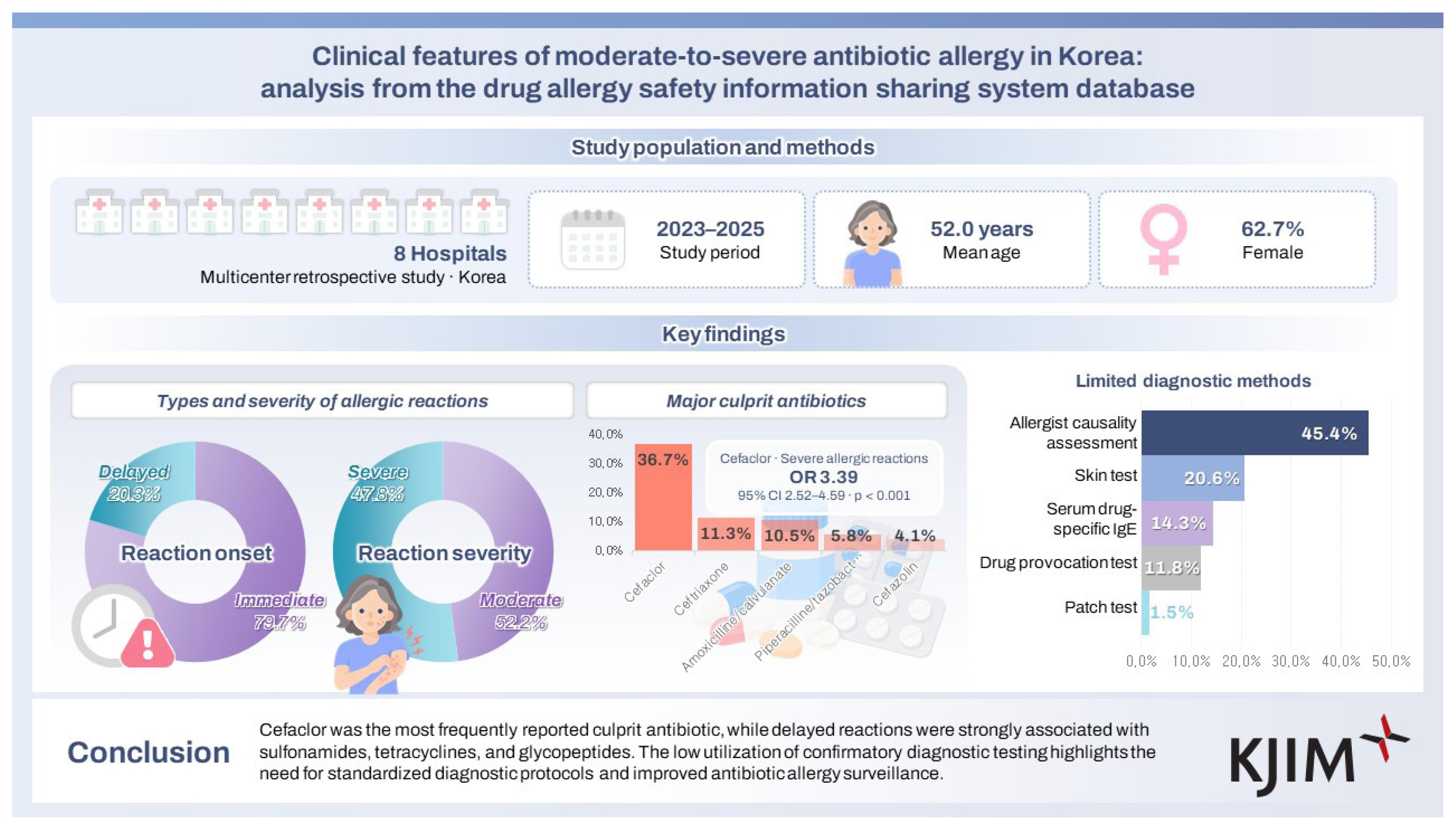
Of 807 cases, the most common manifestations were cutaneous (77.1%), immune (35.2%), and respiratory (23.7%) symptoms. Most cases were caused by β-lactams, particularly second-generation cephalosporins (43.1%). Regarding single agent, cefaclor was the leading culprit (36.7%) and showed significant associations with immediate (odds ratio [OR] 13.88, 95% confidence interval [CI] 7.37–29.75) and severe reactions (OR 3.39, 95% CI 2.52–4.59). Immediate reactions frequently involved multiple organs, while 95.7% of delayed reactions involved the skin. β-lactams were the leading culprits of both immediate and delayed reactions, while sulfamethoxazole/trimethoprim, tetracyclines, and glycopeptides were identified as the major culprits of delayed reactions. Diagnosis relied mainly on clinical assessment (45.4%), while confirmatory drug provocation testing was performed in only 11.8%.
Conclusions
Immediate antibiotic allergy in Korea was mainly caused by β-lactams, with cefaclor identified as the leading culprit for severe reactions. Delayed reactions were characterized by a relatively greater contribution of non-β-lactam antibiotics than immediate reactions.
INTRODUCTION
Antibiotics are among the most widely used medications in clinical practice, yet they remain a leading cause of adverse drug reactions (ADRs) [1]. The clinical burden of antibiotic-related ADRs in Korea is particularly concerning given the nation’s high antibiotic consumption rate. According to recent data, antibiotic use in Korea increased by 29.6% between 2016 and 2023, substantially exceeding the global average of 10.9% [2].
According to previous reports, antibiotics are the culprit of 22.3% to 40.9% of total ADRs, and a substantial proportion of these are attributed to antibiotic allergy caused by hypersensitivity reactions [3,4]. However, the number of true antibiotic allergy cases can be remarkably lower than reported, according to recent studies. For example, approximately 10% of patients report a history of penicillin allergy, yet fewer than 1% are confirmed as truly allergic upon diagnostic testing [5]. Based on these results, there has been growing international interest in antibiotic allergy delabeling and the importance of surveillance, but there remains a lack of sufficient data on antibiotic allergy in Korea.
Major contributors to the overdiagnosis of antibiotic allergy include the lack of standardized diagnostic protocols due to limited use of traditional allergy testing methods (skin test, patch test, drug provocation test), inadequate documentation of drug allergy, and inadequate sharing of information across clinical settings [6,7].
To overcome these limitations, the Ministry of Health and Welfare launched a pilot project named ‘Patient-centered Drug Allergy Information Sharing System’ in 2024. This project enables patients to access their drug allergy information as needed and to share it with healthcare providers when necessary, by granting access to the standardized medical records via Personal Health Record (PHR) platform. Eight hospitals in Korea (Seoul National University Hospital, Severance Hospital, Seoul St. Mary’s Hospital, Seoul National University Bundang Hospital, Chungbuk National University Hospital, Dankook University Hospital, Kyungpook National University Hospital, and Jeonbuk National University Hospital) participated in this project and contributed to the formation of Drug Allergy Safety Information Sharing System (DASI), which enabled patients and healthcare providers to systematically explore patients’ drug allergy history.
The main objective of this study is to identify major culprits, clinical manifestations, and current diagnostic practices of antibiotic allergy using cases collected in DASI.
METHODS
Study subjects
This retrospective study included cases of antibiotic allergy from eight hospitals which participated in the development of DASI, which was established as part of the Patient-centered Drug Allergy Information Sharing System pilot project in 2024.
The inclusion criteria were as follows: 1) cases in which an antibiotic was identified as the culprit drug; 2) cases classified as ‘probable’ or higher according to the World Health Organization-Uppsala Monitoring Centre (WHO-UMC) Causality Assessment; and 3) cases categorized as moderate or severe based on the Common Terminology Criteria for Adverse Events (CTCAE) version 5.0.
The exclusion criteria were as follows: 1) cases determined to represent Type A ADRs which are predictable and dose-dependent; 2) cases classified as ‘possible’ or lower according to the WHO-UMC Causality Assessment; and 3) cases in which the severity was graded as mild based on the CTCAE version 5.0.
Analyzed variables
We analyzed the clinical characteristics of antibiotic allergy in cases selected. Analyzed variables were as follows: 1) demographic characteristics (age, sex); 2) culprit antibiotic classes and frequencies of each class; 3) type of hypersensitivity reaction: immediate (≤ 1 hour) vs. delayed (> 1 hour); 4) primary clinical manifestations; 5) severity; 6) diagnostic methods (causality assessment by allergists, skin test, serum drug-specific IgE, drug provocation test, and patch test); and 7) causality level assessed (‘probable’ or ‘certain’).
Classifications of clinical manifestations were determined based on the System Organ Class (SOC) framework of the Medical Dictionary for Regulatory Activities (MedDRA), version 28.0. Symptoms were classified into the following groups: skin and subcutaneous tissue disorders (“Cutaneous”); vascular disorders (“Vascular”); respiratory, thoracic, and mediastinal disorders (“Respiratory”); gastrointestinal disorders (“Gastrointestinal”); nervous system disorders (“Nervous”); immune system disorders (“Immune”); blood and lymphatic system disorders (“Hematologic”); and other symptoms.
Causality and severity assessments were conducted using the WHO-UMC system and CTCAE version 5.0, respectively, as described above. Using the CTCAE 5.0 keyword definition for “Allergic reaction” as a baseline evaluation tool, severity grades were classified as follows: Grade 1 = mild, Grade 2 = moderate, and Grade ≥ 3 = severe. All cases of anaphylaxis were categorized as severe, as they are uniformly graded ≥ 3 according to CTCAE 5.0. Similarly, severe cutaneous adverse reactions (SCAR) cases, comprised of drug reaction with eosinophilia and systemic symptoms (DRESS), toxic epidermal necrolysis (TEN), Stevens–Johnson syndrome (SJS), and acute generalized exanthematous pustulosis (AGEP) were also classified as severe. For symptoms not explicitly listed as CTCAE 5.0 keywords, severity was determined based on the closest corresponding term within the CTCAE 5.0 framework.
Diagnostic methods were categorized as follows: cases diagnosed based on clinical history and the decision of an allergist were described as ‘causality assessment by allergists.’ WHO-UMC criteria were applied for clinical diagnosis in this study, and their components (temporal relationships, previous knowledge of the suspected drug, dechallenge and rechallenge events, and the possibility of an alternative diagnosis) were evaluated. Other cases were classified according to the specific diagnostic tests performed.
Statistical analysis
Statistical analyses were performed using the collected data. R 4.4.3 (R Foundation for Statistical Computing, Vienna, Austria) was used for the statistical analyses and the preparation of tables and figures. Continuous variables were presented as mean ± standard deviation, and categorical variables were summarized as counts and percentages (n [%]). Student’s t-test was used for the analysis of continuous variables, whereas Chi-square test or Fisher’s exact test was used for the analysis of categorical variables. Statistical significance was defined as p < 0.05. The association between antibiotic class and reaction type (immediate vs. delayed) and between antibiotic type and reaction severity were evaluated using univariate logistic regression, and the results were expressed as odds ratios (ORs) with 95% confidence intervals (CIs).
Institutional Review Board approval
This study was approved by the Institutional Review Board of Seoul National University Hospital (IRB No. H-2512-063-170). The requirement for informed consent was waived because of the retrospective design and use of de-identified data.
RESULTS
Overview of cases
A total of 807 antibiotic allergy cases collected from eight hospitals in Korea between 2023 and 2025 were included in this study. Seoul National University Hospital reported the largest number of 231 cases (28.6%), followed by Kyungpook National University Hospital with 121 cases (15.0%) and Severance Hospital with 116 cases (14.4%).
Among the 807 cases, 301 (37.3%) occurred in men and 506 (62.7%) in women. The mean age was 52.0 ± 18.9 years, with individuals in their seventh decade constituting the largest age group. When classified by the onset of allergic reaction, 643 cases (79.7%) were immediate and 164 cases (20.3%) were delayed. When categorized by severity, 421 cases (52.2%) were moderate and 386 cases (47.8%) were severe (Table 1).

Baseline demographics and general characteristics of 807 cases
Using the MedDRA 28.0 SOC classification, cutaneous symptoms were the most frequently reported symptoms (622 cases, 77.1%), followed by immune (284 cases, 35.2%), respiratory (191 cases, 23.7%), nervous (86 cases, 10.7%), vascular (82 cases, 10.2%), and gastrointestinal symptoms (72 cases, 8.9%). In immediate reactions, cutaneous (72.3%), immune (42.3%), and respiratory symptoms (29.2%) were common and multiple organ systems were frequently involved in a single case. In comparison, cutaneous symptoms were predominant (95.7%) and were typically the only system involved in delayed reactions (Fig. 1).
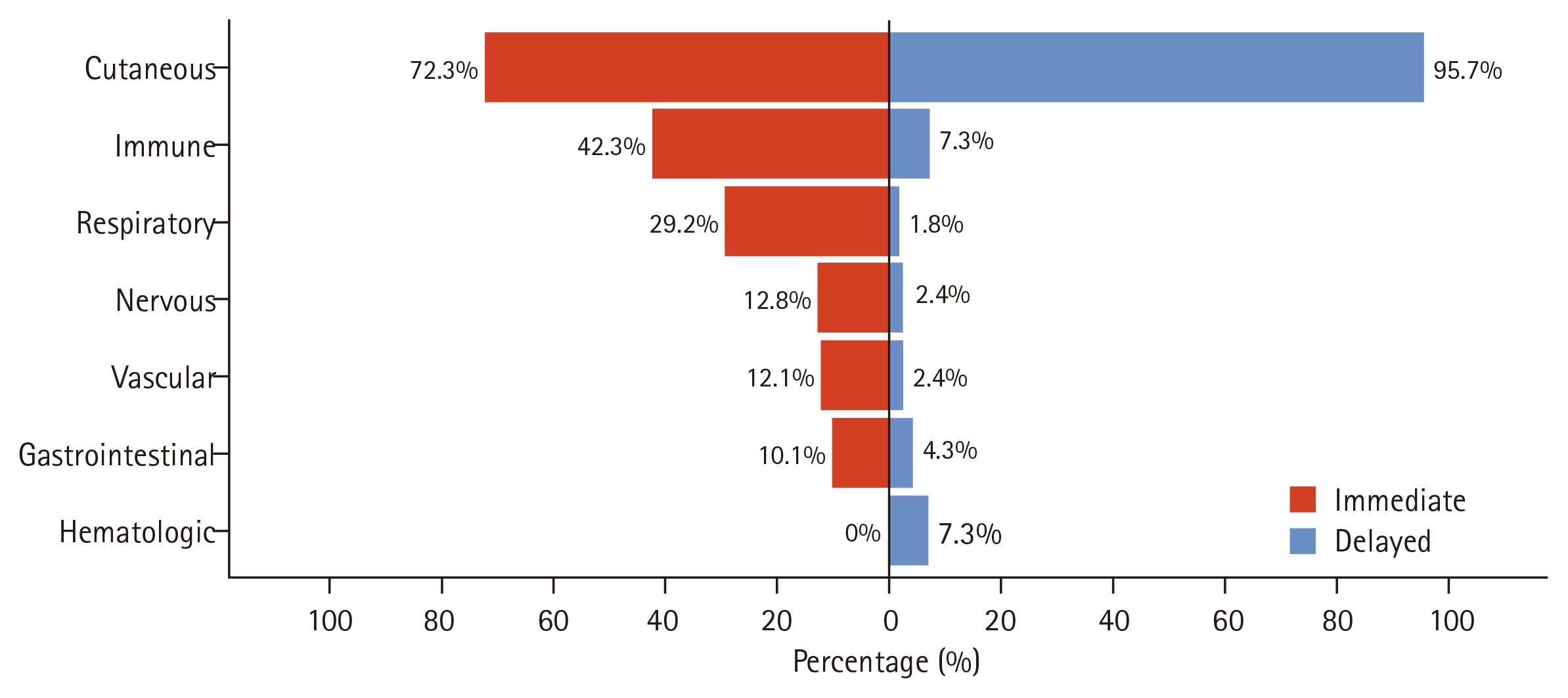
Proportion of clinical symptoms in immediate (n = 643) vs. delayed (n = 164) antibiotic allergic reactions. Clinical manifestations were categorized according to MedDRA system organ classes. Immediate reactions most frequently involved skin/subcutaneous tissue (72.3%), immune (42.3%), and respiratory (29.2%) disorders, often affecting multiple organ systems, whereas delayed reactions were predominantly cutaneous (95.7%) with minimal systemic involvement.
Among 386 severe cases, immediate reactions predominated (n = 336, 87.0%), with anaphylaxis constituting 92.3% (n = 310). The remaining 26 immediate severe cases included isolated syncope (n = 14) and severe cutaneous reactions not meeting anaphylaxis criteria (n = 12). Delayed severe reactions (n = 50, 13.0%) were primarily SCAR (n = 26): SJS (n = 12) and TEN (n = 2), DRESS (n = 10), and AGEP (n = 2). Sulfamethoxazole/trimethoprim (n = 9, 34.6%) and vancomycin (n = 7, 26.9%) were the predominant culprits in SCAR cases. The remaining 24 delayed severe cases included severe, isolated cutaneous reactions (n = 16) and reactions with systemic and cutaneous symptoms not meeting the criteria for SCAR (n = 8).
Culprit antibiotics and their correlation with reaction severity and onset
In this study, β-lactam antibiotics were identified as culprit for 86.5% of all cases, with second-generation cephalosporins being the most common antibiotics class with 348 cases (43.1%). Penicillins came next with 137 cases (17.0%), third-generation cephalosporins with 136 cases (16.9%), and first-generation cephalosporins with 59 cases (7.3%) (Table 2). Fluoroquinolones were the most common culprit (5.3%) among non-β-lactam antibiotics, followed by glycopeptides (3.2%), sulfamethoxazole/trimethoprim (2.1%), and tetracyclines (1.2%).

Classes of culprit agents of antibiotic allergy
Among individual agents, cefaclor showed the highest frequency of reported cases, accounting for 296 cases (36.7%), followed by ceftriaxone (11.3%), amoxicillin/clavulanate (10.5%), piperacillin/tazobactam (5.8%), and cefazolin (4.1%) (Table 3). It is worth noting that these analysis results represent the absolute frequency of reported events within the database, which are not adjusted for the total prescription volume in clinical practice.

Culprit agents of antibiotic allergy
Among 643 immediate reactions, β-lactams were identified as the culprit in 584 cases (90.8%). In contrast, the proportion of β-lactams causing delayed reactions was relatively lower, with 114 out of 164 cases (69.5%) attributed to β-lactams. By antibiotic class, second-generation cephalosporins were significantly more likely to be associated with immediate reaction (OR 5.92, 95% CI 3.81–9.59). Cefaclor, the most frequently implicated second-generation cephalosporin in this study, was strongly associated with immediate reactions (OR 13.88, 95% CI 7.37–29.75). This association likely reflects the predominant pattern of reported cefaclor-related reactions in this cohort. In contrast, sulfonamides (OR 0.01, 95% CI 0.001–0.07), tetracyclines (OR 0.16, 95% CI 0.04–0.58), and glycopeptides (OR 0.24, 95% CI 0.11–0.53) showed significant associations with delayed reactions (Fig. 2A).
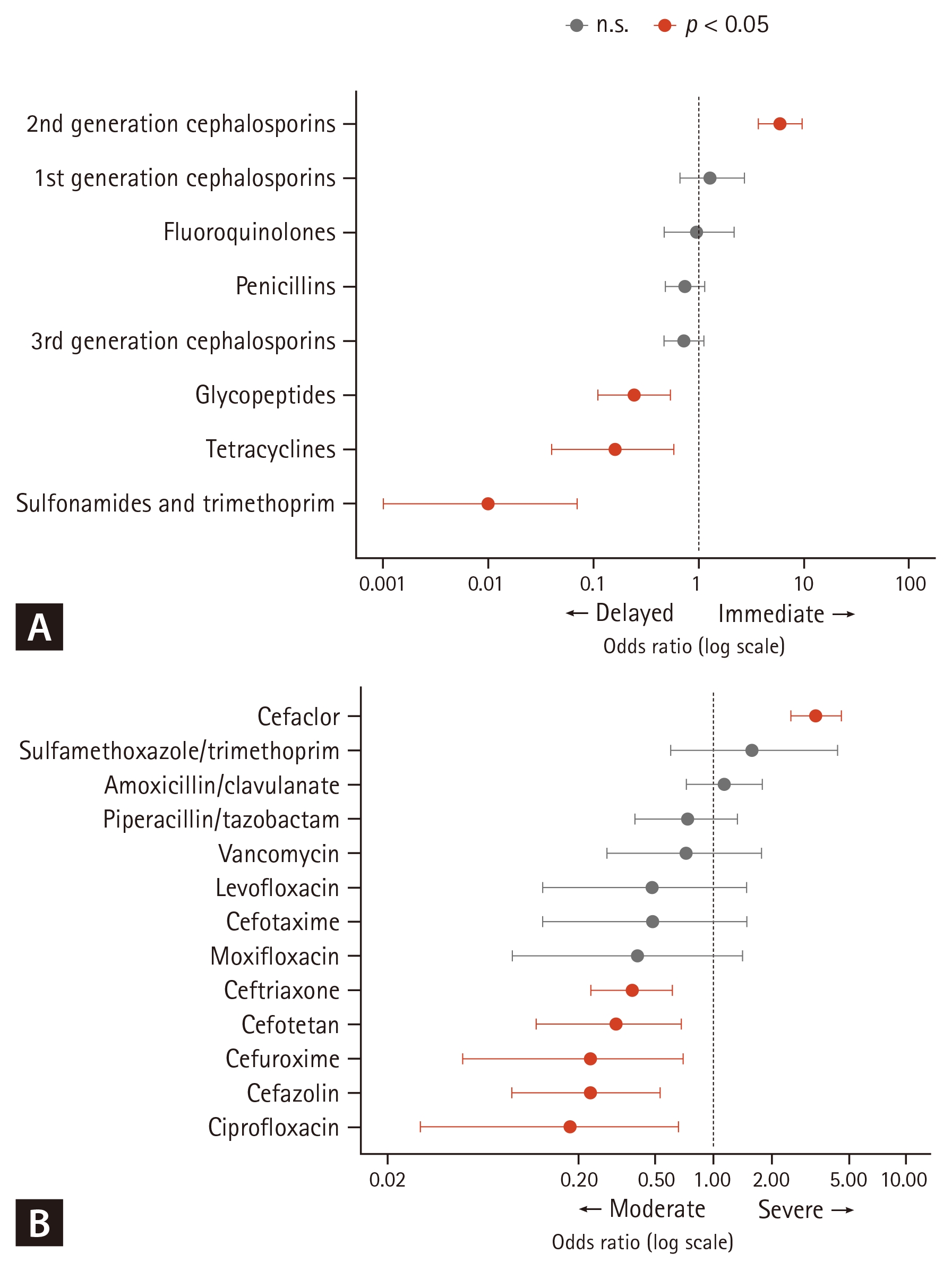
Associations with reaction characteristics and antibiotics. (A) Delayed vs. immediate reactions: ORs with 95% CIs for immediate reactions compared with delayed reactions. ORs > 1.0 indicate increased association with immediate reactions; ORs < 1.0 indicate association with delayed reactions. (B) Severe vs. moderate reactions: ORs for severe reactions compared with moderate reactions. Reference group: all other antibiotics combined. OR, odds ratio; CI, confidence interval.
Culprit antibiotics with ≥ 10 reported cases were included in univariate logistic regression models to evaluate associations with reaction severity. Cefaclor showed higher odds of severe reactions compared with other reported antibiotics (OR 3.39, 95% CI 2.52–4.59, Table 4, Supplementary Table 1). As exposure-adjusted data were not available, this finding should be interpreted as reflecting the relative frequency of severe reactions among reported cases rather than an inherently higher allergenic risk.

Univariate logistic regression analysis of allergic reaction severity, based on culprit antibiotics
Diagnostic practices
The most common diagnostic method in 807 cases was causality assessment by allergists, accounting for 366 cases (45.4%). This was followed by skin test (20.6%), serum drug-specific IgE test (14.3%), provocation test (11.8%), and patch test (1.5%). Among immediate reactions, skin test, serum drug-specific IgE test, and provocation test were used in 25.2%, 17.7%, and 12.9% of cases, respectively. In contrast, causality assessment by allergists was predominantly used in 69.5% of delayed allergic reactions, followed by patch test (7.3%) and provocation test (7.3%, Supplementary Table 2).
DISCUSSION
This multicenter study analyzed 807 cases of antibiotic allergy collected from eight tertiary hospitals in Korea. Approximately 80% of cases were presented as immediate reactions, and nearly half of all cases were classified as severe allergic reactions. Oral second-generation cephalosporins, particularly cefaclor, were identified as the major culprit, while sulfamethoxazole/trimethoprim, tetracyclines, and glycopeptides demonstrated higher odds of causing delayed hypersensitivity reactions. In the diagnosis of delayed reactions, the role of diagnostic testing was limited, as most cases were identified by the causality assessment of allergists rather than confirmatory tests.
Antibiotics remain among the most widely used medications worldwide, and recent reports indicate that antibiotic consumption increased by 10.9% globally and by 29.6% in Korea in 2023 compared with 2016 [2]. Given the substantial proportion of ADRs attributable to antibiotics, this increase in antibiotic consumption further underscores the clinical importance of antibiotic allergy.
Nevertheless, domestic research on overall trends in antibiotic allergy has largely been limited to single-center analyses. In a study analyzing antibiotic allergy cases in 2015 at a Korean tertiary hospital, penicillins and quinolones were reported as the most common culprit antibiotic classes [8]. Meanwhile, multicenter, systematic analyses in Korea have largely been limited to studies focusing specifically on anaphylaxis or SCARs [9–11]. For example, an analysis of cephalosporin-induced anaphylaxis conducted across 12 general hospitals in Korea between 2013 and 2015 reported that ceftizoxime and cephalosporins sharing the same R1 side chain were the most frequent culprits, which differs from the findings of our study [9]. In a study analyzing cases of anaphylaxis caused by intravenous β-lactam antibiotics across nine hospitals in Korea, cephalosporins accounted for 77% of all cases, with ceftriaxone being the most common culprit antibiotic (23%) [10]. Lastly, an analysis of SCAR cases reported between 2010 and 2015 through the Korean SCAR Registry, which involved 28 tertiary hospitals nationwide, showed that cephalosporins were the most common culprit drug among antibiotic-related SCAR cases [11]. Different from the prior Korean SCAR registry data showing β-lactams as leading culprits, our DASI-based analysis highlights that non-β-lactam agents, especially sulfamethoxazole/trimethoprim and vancomycin, also make substantial contributions to delayed severe reactions.
Our study collected moderate and severe antibiotic allergy cases from eight tertiary hospitals in Korea and analyzed not only SCAR and anaphylaxis but also a broader spectrum of allergic reactions. In doing so, this study addresses a key limitation of previous domestic research by providing a multicenter analysis of overall characteristics of antibiotic allergy, rather than focusing solely on SCAR or anaphylaxis. Furthermore, this study offers a novel perspective by examining diagnostic approaches for antibiotic allergy and elucidating how immediate and delayed allergic reactions are currently diagnosed in real-world clinical practice.
Previous literature on antibiotic allergy and anaphylaxis indicates that the distribution of culprit antibiotics varies considerably across countries. According to the Allergy Vigilance Network, which collected anaphylaxis data from French-speaking countries, amoxicillin was the most common culprit among 165 cases of antibiotic-induced anaphylaxis reported between 2002 and 2010, accounting for 97 cases, followed by cephalosporins with 41 cases [12]. In a 2003 study analyzing medical records from Hungary, Spain, Sweden, and India, the incidence of anaphylaxis caused by intravenous penicillin was the highest among all antibiotics, with a rate of 32 cases per 100,000 individuals [13]. An analysis of fatal anaphylaxis cases in the United States from 1999 to 2010 reported that, among 149 deaths attributed to antibiotic-induced anaphylaxis, penicillins were the most common culprits (35 cases), followed closely by cephalosporins (33 cases) [14]. In contrast, an analysis of self-reported antibiotic allergy based on medical records from San Diego County in 2007 showed that sulfonamides had the highest incidence of self-reported allergy, regardless of sex [15]. To summarize, penicillin was the most common culprit antibiotic class in Europe and the United States, although allergic reactions to cephalosporins and sulfonamides were also frequently reported in the United States. Our findings highlight a distinctive reporting pattern of moderate-to-severe antibiotic allergy in Korea, in which oral cefaclor was the most frequently reported culprit rather than penicillin. However, this trend may reflect widespread outpatient prescribing patterns and high exposure frequency rather than an elevated intrinsic allergenic risk of cefaclor. Given that cefaclor is one of the most prescribed oral antibiotics in the outpatient setting, repeated and frequent exposure to cefaclor may have contributed to exposure-driven sensitization [16,17]. In the absence of denominator data such as prescription volume or actual antibiotic consumption, this finding should be interpreted as the frequency of cefaclor implication among reported moderate-to-severe antibiotic allergy cases, rather than as evidence of a higher intrinsic allergenic risk. Therefore, the relative contribution of cefaclor’s inherent potential to induce hypersensitivity and its widespread outpatient use as a driver of sensitization warrants further investigation. Our dataset, drawn from eight geographically representative tertiary hospitals across Korea, provides reasonable nationwide representativeness. However, since this study was limited to moderate-to-severe cases and relied on reported cases rather than population-level surveillance, the incidence of antibiotic allergy based on actual antibiotic consumption in Korea could not be assessed.
According to a recent study in Korea, the sensitivity of the intradermal test and serum specific IgE assay in patients with immediate cefaclor hypersensitivity were reported to be 0.863 and 0.496, respectively. When the two tests were used in combination, the sensitivity increased to 0.908, while the specificity of both tests was reported to be 1.0 [18]. These findings suggest that incorporating both skin test and serum drug-specific IgE testing more proactively in patients with suspected cefaclor allergy may improve diagnostic accuracy.
Drug hypersensitivity reactions are classified into immediate and non-immediate types according to the onset of symptoms [19]. Immediate reactions typically occur within one hour after drug administration and are primarily mediated by IgE and commonly manifest as urticaria, angioedema, or anaphylaxis [20]. In contrast, non-immediate reactions occur several hours or days after drug exposure and can be further divided into accelerated and delayed reactions. Accelerated reactions typically appear between an hour and 24–48 hours after drug exposure and commonly present with mild cutaneous or respiratory symptoms. Clinically, they can be easily confused with either immediate or delayed reactions, and their pathophysiology may involve overlapping mechanisms of both types [19]. Delayed reactions generally occur several days after drug exposure and are primarily mediated by T-cell–driven immune responses. They may present with exanthematous eruptions and high fever, and in rare cases manifest as SCARs like DRESS or SJS/TEN [21].
In this study, immediate hypersensitivity reactions accounted for approximately 80% of all cases. The remaining cases were non-immediate reactions, all of which were classified as delayed reactions. However, this pattern may reflect reporting bias inherent to the current surveillance system. Although some studies have reported that non-immediate reactions are more common than immediate reactions [22,23], there are clinical factors that lead to the underestimation of non-immediate reactions. They often occur outside the clinical settings and are frequently omitted from medical records or adverse event reporting systems. They also occur later, are generally less severe, and are often more difficult to diagnose due to limitations in available diagnostic tools. The finding in our study that the patch test and other diagnostic tests were rarely used supports this limitation and underscores the absence of a standardized ADR reporting system.
Significant association between higher odds of delayed reactions and sulfonamides, tetracyclines, and glycopeptides shown in this study is consistent with previous reports indicating that these agents can induce T-cell–mediated immune responses such as DRESS and maculopapular eruptions [24,25]. However, this study has a limitation in that it did not incorporate confirmatory tests such as histopathology, lymphocyte transformation test, or cytokine assays.
Our study highlighted the result that oral antibiotics prescribed in the outpatient setting were identified as major culprits of antibiotic allergy, allowing us to examine antibiotic safety issues from the perspective of primary care—an aspect that differs from previous inpatient-based studies. In addition, the use of the DASI system enabled the collection of multicenter data, providing nationwide representativeness and further strengthening the significance of this study. By integrating the DASI system with the PHR platform, this study analyzed reliable, expert-validated drug allergy safety data collected from multiple institutions across the country. As such, it provides important evidence for the future development of a guideline for the diagnosis and management of antibiotic allergy in Korea.
However, this study has several limitations. First, because it was based on data from tertiary hospitals, it may not fully reflect the patterns observed in primary and secondary clinical settings. Although this tertiary hospital-based dataset may be subject to selection bias and may not represent the population-level epidemiology of antibiotic allergy, it should be interpreted in the context of the Korean healthcare system. In Korea, detailed evaluation of drug allergy is usually performed in tertiary referral hospitals, where most allergy specialists and specialized diagnostic resources are concentrated. Therefore, the present study primarily reflects clinically relevant, moderate-to-severe antibiotic allergy cases evaluated by allergy specialists in real-world practice. Second, most patients included in this study were adults, limiting the applicability of our findings to pediatric populations. Third, because many diagnoses relied on clinical assessment rather than standardized confirmatory testing, subjectivity in the evaluation of diagnostic criteria and the possibility of underreporting may remain. However, a higher degree of consistency was ensured, as all eight participating institutions operate as regional pharmacovigilance centers with specialized training in ADR causality assessment, and the overall system is supervised by allergy specialists. The consensus application of WHO-UMC criteria across these centers further strengthens the reliability of diagnoses. This is also a potential strength of our study, since confirmatory tests are often not feasible in real-world practice, particularly in moderate-to-severe cases. Fourth, accelerated reactions could not be reliably separated from immediate or delayed reactions and were analyzed together with delayed reactions because time-to-onset was often recorded in a simplified manner in routine practice. In addition, mild cases were not included, which may have prevented the results from capturing the full spectrum of antibiotic allergy. Nevertheless, given that this study focused on moderate-to-severe reactions that are directly linked to patient safety, its clinical relevance remains substantial.
Nonetheless, this study provides several meaningful insights. First, the development and broader implementation of standardized diagnostic systems are urgently needed for the prevention and management of antibiotic allergy in Korea. Second, the inspection of prescribing practices and the implementation of alert systems for frequently implicated oral antibiotics such as cefaclor should be considered. Indeed, a recent nationwide Health Insurance Review and Assessment Service (HIRA)-based study highlighted that cefaclor had the highest exposure counts among all analyzed anaphylaxis culprits, reinforcing that its high clinical frequency in Korea is heavily driven by widespread prescription volume rather than an exclusively higher intrinsic allergenic risk [26]. However, it remains to be elucidated whether the high frequency of cefaclor allergy stems primarily from its widespread prescription volume or its intrinsic risk as an allergen. Third, platforms for sharing information and current status of allergy such as DASI should be further expanded to establish a robust pharmacovigilance system that enables the continuous collection, analysis, and feedback of diverse allergic reactions occurring in clinical practice. Using this information, the goal would be to develop the policies for effective antibiotic allergy surveillance.
In conclusion, this multicenter study demonstrates distinctive patterns of moderate-to-severe antibiotic allergy in Korea, with cefaclor as the predominant culprit of immediate severe reactions.
KEY MESSAGE
1. This nationwide multicenter study reveals distinctive patterns of antibiotic allergy in Korea, with cefaclor identified as the most frequently reported culprit.
2. Delayed reactions were more strongly associated with sulfonamides, tetracyclines, and glycopeptides.
3. The low utilization of confirmatory diagnostic testing underscores an urgent need for standardized diagnostic protocols.
Notes
Acknowledgments
We extend our sincere gratitude to the information technology and medical informatics staff at each participating institution for their assistance with data collection, refinement, and quality management for this study: Tae Woo Kim, Moonsu Kang, Yeontak Jeong, Eunseok Cho, Minji Kim (Seoul National University Hospital), Jisun Kim, Yunju Lee (Kyungpook National University Hospital), Yuri Choi, Younghee Suh, Suheon Whang, Taegeon Jeong (Seoul St. Mary’s Hospital), Eunhye Kang, Young ah Kim, Jaehyeon Park, Jimin Seo, Younghee Lim (Division of Digital Health, Yonsei University Health System), Ga-Young Oh, Hyun-Joo Park, Mi-Seon Nam (Seoul National University Bundang Hospital), Minkyu Kim, SuSung Kim, Yusin Choi (Jeonbuk National University Hospital), Kyungman Jeong, Sunkyung Park, Kisuk Kim, Misun Yoon, Youngju Choi, Hyunju Kwon, Kichul Yu (Chungbuk National University Hospital), Junseong Lee (ezCaretech), Hyungki Kim (Seowoo Soft), Mijeong Shin, Yejeong Jeong, SeokJoo Lee (Dankook University Hospital), Jaeyong Shin (Korea IT consulting).
CRedit authorship contributions
Eunsik Bang: methodology, investigation, data curation, formal analysis, original draft, visualization; Min-Ho Kang: data curation, review & editing; Jee Hyun Kim: data curation, review & editing; Won Jong Song: data curation; Su Jeong Song: data curation; Eunyoung Sa: data curation; Jeong-Min Park: resources, review & editing; Ji-Hyang Lee: resources, review & editing; Jeong-Eun Yun: resources, review & editing; Jiung Jeong: resources, review & editing; Sujeong Kim: resources, review & editing; Hwa Young Lee: resources, review & editing; Kyung Hee Park: resources, review & editing; Min-Gyu Kang: resources, review & editing; Sang Min Lee: resources, review & editing; So Ri Kim: resources, review & editing; Sae-Hoon Kim: resources, review & editing; Hye-Ryun Kang: conceptualization, methodology, resources, review & editing, supervision, project administration, funding acquisition
Conflicts of Interest
The authors disclose no conflicts.
Funding
This research was supported by a grant from the Korea Health Information Service in 2024.