Association of age-adjusted skeletal muscle index and modest alcohol intake with hepatic steatosis and fibrosis in males
Article information

Abstract
Background/Aims
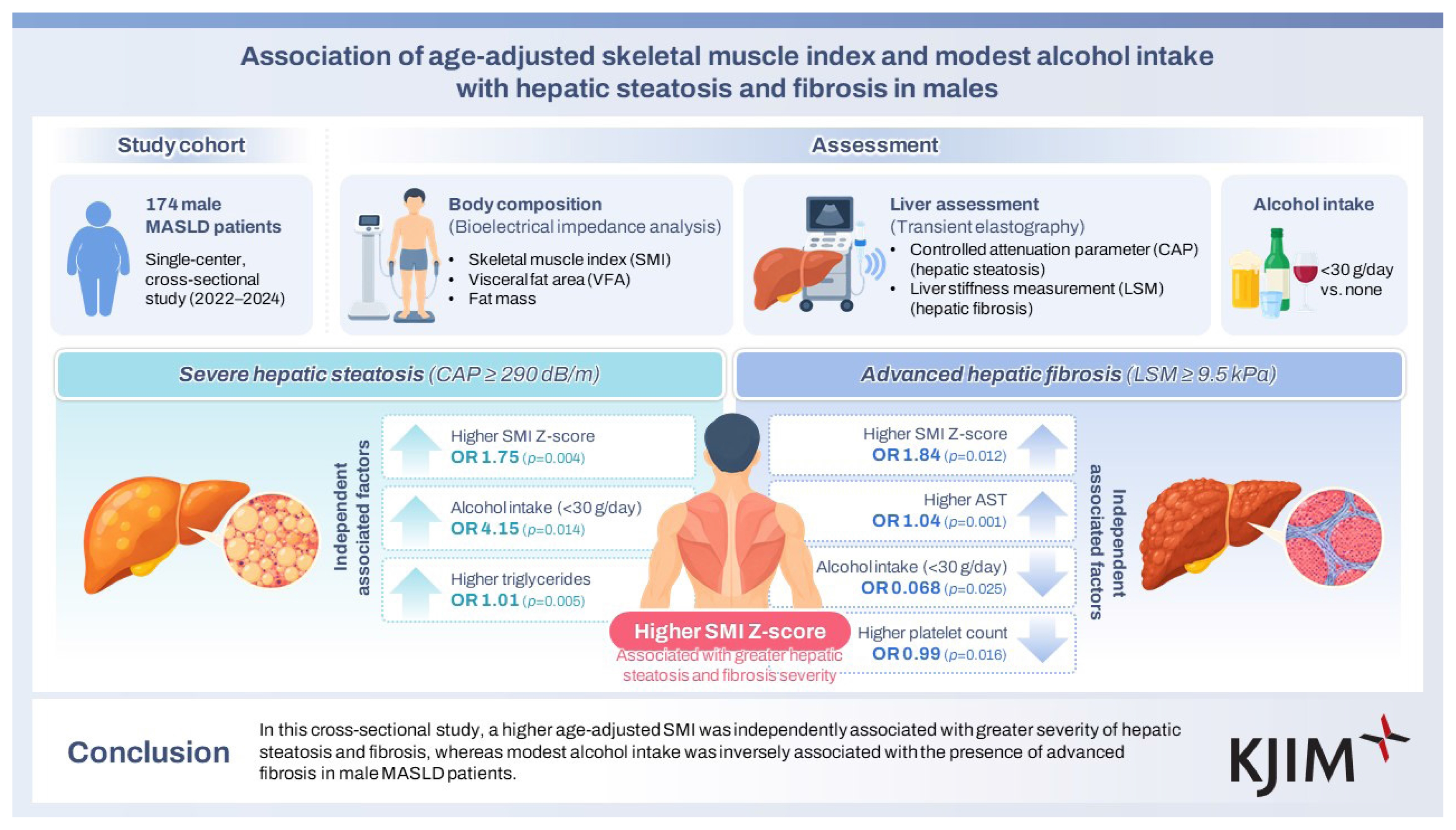
Metabolic dysfunction-associated steatotic liver disease (MASLD) is a growing global health concern linked to obesity, insulin resistance, and metabolic syndrome. Although body composition and alcohol intake influence disease progression, their specific associations with hepatic steatosis and fibrosis remain unclear.
Methods
Males (n = 174) with MASLD were evaluated. Hepatic steatosis and fibrosis were assessed using the controlled attenuation parameter (CAP) and liver stiffness measurement using transient elastography. Skeletal muscle index (SMI) and visceral fat area (VFA) were determined using bioelectrical impedance analysis. The participants were stratified according to alcohol consumption (< 30 g/d vs. none). Multivariate logistic regression was used to identify the factors independently associated with severe steatosis and advanced fibrosis.
Results
Severe steatosis was associated with higher body mass index, VFA, fat mass, and SMI. Factors independently associated with severe steatosis were modest alcohol intake, higher SMI Z-scores, and elevated triglyceride levels. In non-drinkers, a higher SMI Z-score remained significant. Advanced fibrosis was associated with a higher SMI Z-score and plasma aspartate aminotransferase levels, whereas moderate alcohol consumption and higher platelet count were inversely associated. The SMI Z-score positively correlated with both CAP and fibrosis severity.
Conclusions
A higher age-adjusted SMI was independently associated with a greater severity of hepatic steatosis and fibrosis, whereas modest alcohol intake was inversely associated with the presence of advanced fibrosis in male patients with MASLD.
INTRODUCTION
Metabolic dysfunction-associated steatotic liver disease (MASLD) has emerged as the most prevalent chronic liver disease worldwide, paralleling global obesity and metabolic syndrome epidemics [1,2]. Characterized by excessive hepatic fat accumulation, MASLD encompasses a broad histopathological spectrum, ranging from simple steatosis to steatohepatitis, advanced fibrosis, and cirrhosis. Given its strong association with cardiometabolic risk factors (CMRFs) such as obesity, insulin resistance, dyslipidemia, and hypertension, MASLD has become a critical public health concern [3].
Although the role of traditional metabolic risk factors in the development and progression of MASLD is well-established, the contribution of body composition, particularly skeletal muscle mass (SMM) and visceral adiposity, has gained increasing attention [4–6]. Recent evidence suggests that sarcopenia and alterations in muscle quality such as myosteatosis may be associated with the severity of hepatic steatosis and fibrosis [7]. However, these findings remain inconclusive, and the complex interplay between skeletal muscle, adipose tissue, and liver pathology warrants further investigation.
The impact of alcohol consumption on MASLD progression remains a subject of ongoing debate. Although MASLD is characterized by minimal or no alcohol intake, emerging data suggest that light to moderate alcohol consumption may exert differential effects on hepatic steatosis and fibrosis [8,9]. Clarifying these relationships is essential to refine risk stratification and management strategies in this population.
This study aimed to investigate the clinical, anthropometric, and biochemical determinants of hepatic steatosis and advanced fibrosis in individuals with MASLD, with a particular focus on the roles of SMM, visceral fat area (VFA), and light alcohol consumption. By integrating data from bioelectrical impedance analysis (BIA), liver elastography, and laboratory assessments, this study aimed to elucidate the key factors in males with MASLD associated with disease severity and explore the differential impact of light alcohol intake on hepatic fat accumulation and fibrosis progression.
METHODS
Study population
This single-center, cross-sectional study included 259 male patients who underwent diagnostic evaluation at the Department of Gastroenterology between January 1, 2022, and December 31, 2024. All participants showed evidence of hepatic steatosis on imaging studies. The exclusion criteria were as follows: (a) age under 20 years; (b) absence of elastography or BIA data; (c) positive serum hepatitis B surface antigen or hepatitis C virus antibody; (d) history or evidence of other chronic liver diseases, such as autoimmune hepatitis and primary biliary cholangitis; (e) excessive alcohol consumption (defined as > 30 g/d for males and > 20 g/d for females); and (f) current or prior use of steatogenic medications known to induce hepatic steatosis (e.g., corticosteroids, amiodarone, methotrexate, tamoxifen). MASLD was diagnosed based on the presence of hepatic steatosis and at least one of the following CMRFs: (a) overweight or central obesity, defined as a body mass index (BMI) ≥ 25 kg/m2 or waist circumference > 94 cm for males and > 80 cm for females; (b) hyperglycemia, defined as fasting plasma glucose ≥ 5.6 mmol/L (100 mg/dL), glycated hemoglobin (HbA1c) ≥ 5.7% (39 mmol/mol), or a prior diagnosis of diabetes or use of antidiabetic medications; (c) elevated blood pressure, defined as systolic/diastolic blood pressure ≥ 130/85 mm Hg or use of antihypertensive medications; (d) hypertriglyceridemia, defined as plasma triglycerides ≥ 1.70 mmol/L (150 mg/dL) or use of lipid-lowering therapy; and (e) low high-density lipoprotein cholesterol (HDL-C), defined as ≤ 1.0 mmol/L (40 mg/dL) in males and ≤ 1.3 mmol/L (50 mg/dL) in females, or use of lipid-lowering therapy. After applying the inclusion and exclusion criteria, 174 male patients diagnosed with MASLD were included in the final analysis.
Data collection
Clinical and laboratory data were collected at the time of initial evaluation. The demographic data included age, height, and body weight. Data regarding alcohol intake and medication history were collected from patients’ self-reports during standardized clinical assessments. Blood samples were collected after overnight fasting and analyzed for complete blood count (white blood cell count and platelet count), liver enzymes (aspartate aminotransferase [AST], alanine aminotransferase [ALT], gamma-glutamyl transferase), lipid profile (total cholesterol, triglycerides, HDL-C, and low-density lipoprotein cholesterol), and fasting glucose levels. The fibrosis-4 index was used to assess liver fibrosis. The number of CMRFs present in each patient was determined based on predefined criteria (as described in the MASLD diagnostic criteria), and patients were grouped accordingly. This study was approved by the Institutional Review Board (IRB) of Hallym University Dongtan Sacred Heart Hospital (IRB no. HDT 2025-04-002) and was conducted in accordance with the ethical principles of the Declaration of Helsinki. Given the retrospective design of the study and use of anonymized clinical data, the requirement for written informed consent was waived by the IRB.
Measurement and definition of body composition analysis
Body composition was assessed using a multi-frequency bioelectrical impedance analyzer (InBody 970; BioSpace Inc., West Des Moines, IA, USA) following an overnight fast of at least 8 h. The device uses hand-to-foot BIA, which sends varying frequencies of alternating currents through the body. BIA was performed, including measurements of percentage body fat, VFA, fat mass, muscle mass, skeletal muscle index (SMI), and derived VFA T/Z and SMI T/Z scores. In addition, the skeletal muscle-to-visceral fat ratio and SMM-to-body weight ratio were calculated. The SMI and VFA were standardized using both T- and Z-scores to evaluate each participant’s relative position compared with the reference populations. The T-score represents the deviation from the mean value in healthy young adults, whereas the Z-score reflects the deviation from age- and sex-matched population norms. These standardized scores were derived based on the reference values proposed by the Asian Working Group for Sarcopenia: 2019 consensus update on sarcopenia diagnosis and treatment [10].
Measurement and definition of severity of hepatic steatosis and fibrosis
Liver stiffness and steatosis severity were assessed using the FibroScan® 502 Touch model (Echosens, Paris, France). The device was equipped with both M and XL probes and included an integrated automatic probe selection algorithm, which recommended an appropriate probe based on the real-time assessment of the skin-to-liver capsule distance, thereby enhancing the accuracy and reliability of the measurements [11]. Certified nurses who were blinded to the patients’ clinical data performed all examinations. Patients were instructed to fast for at least 3 hours prior to the procedure. During the examination, patients were placed in a supine position with their right arm fully abducted to allow optimal access to the right hepatic lobe. Transient elastography was performed in the right intercostal space. Measurements were considered valid when at least 10 successful acquisitions were obtained, with a success rate of ≥ 60% and an interquartile range (IQR) to median liver stiffness measurement (LSM) ratio (IQR/M) ≤ 0.30. Examinations that did not meet these criteria were excluded from the analysis [12].
Severe hepatic steatosis was defined as a controlled attenuation parameter (CAP) value ≥ 290 dB/m, corresponding to histological steatosis involving ≥ 34% of hepatocytes (≥ S2), based on previously validated thresholds [13]. Significant fibrosis (F2) was defined as a LSM > 7.5 kPa, whereas advanced fibrosis (F3) was defined as LSM > 9.5 kPa. According to a meta-analysis of 33 studies, the cutoff values for advanced fibrosis ranged from 8.0 to 9.9 kPa, with an area under the receiver operating characteristic curve of 0.87 [12,13].
Statistical analysis
Continuous variables are expressed as mean ± standard deviation for normally distributed data or median with IQR for skewed data. The Shapiro–Wilk test was used to evaluate the normality of continuous variables. Categorical variables are summarized as frequencies and percentages. Between-group comparisons for continuous variables were conducted using Student’s t-test or the Mann–Whitney U-test, as appropriate. Chi-square test or Fisher’s exact test was used for categorical variables. Univariate logistic regression analysis was performed to identify the potential factors associated with severe hepatic steatosis and advanced hepatic fibrosis. Variables with a p value < 0.1 in the univariate analysis or those with established clinical relevance (such as platelet count for advanced fibrosis) were entered into multivariate logistic regression models to determine independent associated factors. Odds ratios (ORs) and 95% confidence intervals (CIs) were calculated. The relationship between the SMI Z-score and hepatic steatosis, as quantified by CAP, was assessed using Pearson’s correlation analysis. A linear regression model was fitted and visualized using a scatter plot. To explore the influence of low alcohol consumption (< 30 g/d) on hepatic steatosis and fibrosis, the CAP and LSM values were compared using boxplots stratified by alcohol intake status. All reported p values were two-sided, and statistical significance was defined as p < 0.05. All statistical analyses were performed using SPSS Statistics version 26.0 (IBM Corp., Armonk, NY, USA).
RESULTS
Baseline characteristics
Tables 1 and 2 present the baseline characteristics of 174 male participants diagnosed with MASLD. The mean age was 40.0 ± 10.4 years, and the median BMI was 28.8 kg/m2 (IQR 25.5–32.6). The most frequent number of coexisting CMRFs was four, observed in 59 participants (33.9%). Alcohol consumption of < 30 g/d was reported by 55 individuals (31.6%).

Baseline characteristics of demographics and clinical data

Baseline characteristics of body composition and laboratory test
The mean CAP score, reflecting hepatic steatosis, was 325.2 ± 37.3 dB/m, while the median LSM was 6.0 kPa (IQR 4.6–7.9). Based on a CAP threshold of 290 dB/m, the participants were categorized into the severe hepatic steatosis group and mild-to-moderate steatosis group. Compared to the mild-to-moderate group, individuals in the severe group demonstrated significantly higher body weight, BMI and waist circumference. A significantly greater proportion of participants in the severe group consumed < 30 g/d of alcohol (50 individuals, 35.7%, p = 0.018). The prevalence of significant fibrosis (LSM ≥ 7.5 kPa) was significantly higher in the severe group (46 patients, 32.9%; p = 0.015).
BIA revealed that the severe group had significantly greater VFA (116.8 cm2, IQR 88.9–155.6, p = 0.008) and fat mass (27.5 kg, IQR 21.6–34.0, p = 0.002). Muscle mass (34.6 kg, IQR 31.7–37.7, p = 0.001) and SMI (8.59 ± 0.76 kg/m2, p = 0.001) were also higher in the severe group. In the severe group, laboratory analyses revealed significantly elevated serum levels of AST (median 38.0 IU/L, IQR 28.0–52.0, p = 0.026) and ALT (median 69.0 IU/L, IQR 34.3–97.8; p = 0.011) compared to the non-severe group. In the lipid profile, triglyceride levels were significantly higher (167 mg/dL, IQR 128–227, p = 0.001), while HDL cholesterol levels were significantly lower (43.0 mg/dL, IQR 37.0–50.0, p = 0.033) in the severe group compared to the mild to moderate group.
Determinants and correlates of hepatic steatosis severity
Factors associated with hepatic steatosis severity among the study participants were analyzed (Table 3). Univariate regression analysis identified several variables that were significantly associated with hepatic steatosis severity, including BMI, alcohol intake, SMI Z-score, VFA Z-score, triglyceride level, and HDL cholesterol.

Factors associated with the severity of hepatic steatosis in male MASLD
Multivariate regression analysis incorporating these variables showed that alcohol intake (OR 4.15, 95% CI 1.33–12.95, p = 0.014), higher SMI Z-score (OR 1.75, 95% CI 1.23–2.70, p = 0.004), and elevated triglyceride levels (OR 1.01, 95% CI 1.00–1.02, p = 0.005) were independently associated with increased odds of severe hepatic steatosis. Additionally, when comparing CAP values stratified by alcohol intake status, the group with modest alcohol consumption (< 30 g/d) exhibited significantly higher CAP values compared to the non-drinking group (p = 0.018) (Fig. 3A). The p value of the univariate analysis of the interaction between the SMI Z-score and either alcohol intake or BMI with respect to the severity of hepatic steatosis was statistically significant; however, no statistical significance was observed in the multivariate analysis (p = 0.775 and p = 0.783, respectively). A subgroup analysis of participants without alcohol consumption demonstrated that a higher SMI Z-score was significantly associated with an increased likelihood of severe hepatic steatosis (OR 1.73, p = 0.008) along with elevated triglyceride levels (Supplementary Table 1).
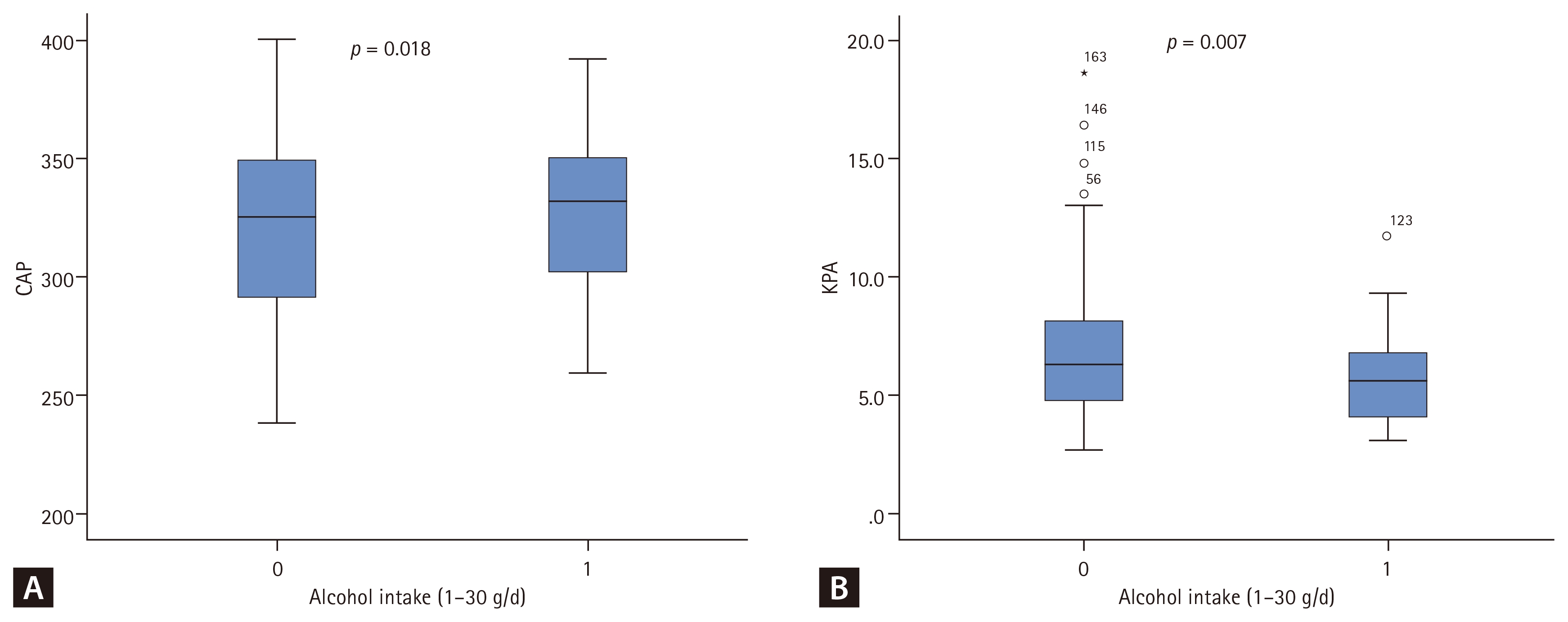
The comparison of alcohol consumption and (A) hepatic steatosis, (B) hepatic fibrosis in males with MASLD. MASLD, metabolic dysfunction-associated steatotic liver disease; CAP, controlled attenuation parameter.
A statistically significant positive correlation was observed between the SMI Z-score and CAP value (r = 0.328, p < 0.001), suggesting that a greater SMI was modestly associated with increased hepatic fat content (Fig. 1A). Among participants with severe steatosis, subgroup analysis stratified by BMI categories also revealed a positive correlation between BMI and SMI Z-score, with higher BMI associated with higher SMI Z-score values (p < 0.001) (Fig. 1B). Furthermore, when stratified by fibrosis stage, the positive correlation between the SMI Z-score and CAP remained robust and statistically significant in the mild-to-moderate fibrosis group (F0–F2 [< 9.5 kPa]; r = 0.350, p < 0.001). However, this correlation was attenuated and was not statistically significant in the advanced fibrosis group (F3, F4 [≥ 9.5 kPa]; r = 0.112, p = 0.670), likely because of the limited sample size of this subgroup (Supplementary Fig. 1).
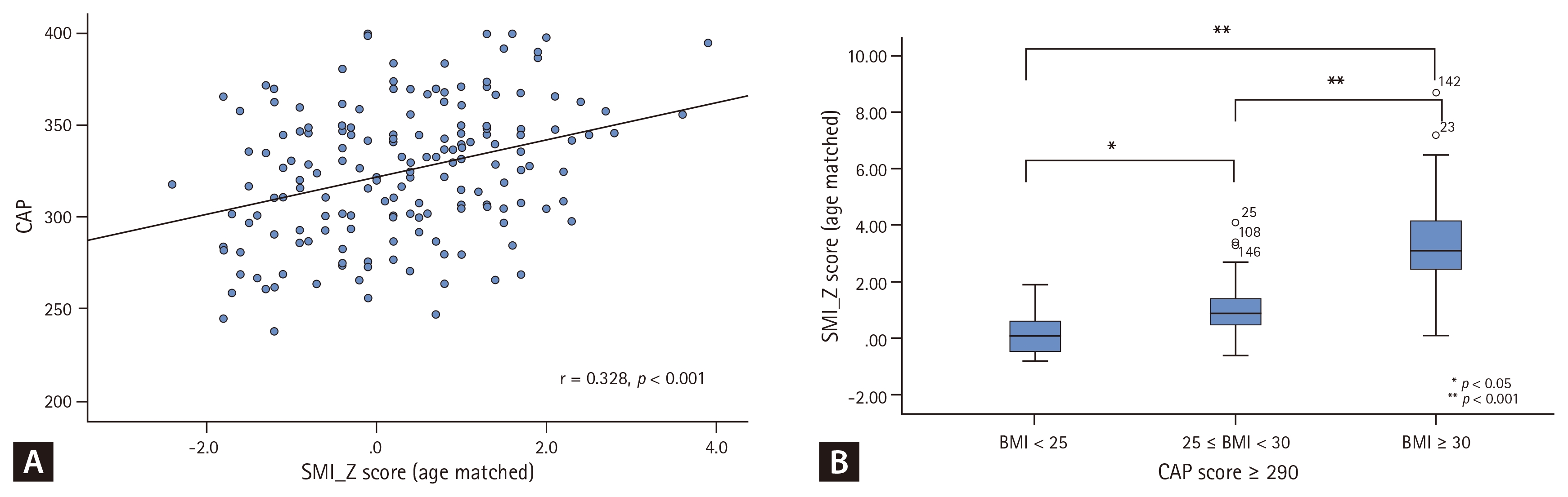
The correlation between SMI-Z score and hepatic steatosis in (A) all males with MASLD and (B) the severe hepatic steatosis group of males with MASLD. SMI, skeletal muscle index; MASLD, metabolic dysfunction-associated steatotic liver disease; CAP, controlled attenuation parameter; BMI, body mass index.
Determinants and correlates of advanced hepatic fibrosis
Factors associated with advanced hepatic fibrosis were analyzed in the study participants (Table 4). In univariate regression analysis, alcohol intake, SMI Z-score, and AST level were identified as significant predictors of advanced hepatic fibrosis.

Factors associated with the severity of hepatic fibrosis in male MASLD (F3, F4)
Multivariate regression analysis was subsequently performed, incorporating the significant factors from the univariate analysis along with platelet count, a well-established marker of hepatic fibrosis. In this model, elevated SMI Z-score (OR 1.84, 95% CI 1.14–2.97, p = 0.012) and increased AST levels (OR 1.04, 95% CI 1.02–1.05, p < 0.001) were independently associated with a higher likelihood of advanced hepatic fibrosis. In contrast, modest alcohol intake (OR 0.068, 95% CI 0.01–0.71, p = 0.025) and higher platelet count (OR 0.99, 95% CI 0.97–1.00, p = 0.016) were associated with a significantly decreased likelihood of advanced hepatic fibrosis. Consistent with these findings, the comparison of LSM stratified by alcohol consumption demonstrated that the modest alcohol intake group (< 30 g/d) had significantly lower stiffness values compared to the non-drinking group (p = 0.007) (Fig. 3B). The p value of the univariate analysis of the interaction between the SMI Z-score and BMI with respect to the severity of hepatic fibrosis was statistically significant; however, in the multivariate analysis, no statistical significance was observed between the SMI Z-score and either alcohol intake or BMI (p = 0.548 and p = 0.164, respectively). Additionally, a significant positive correlation was observed between the SMI Z-score and hepatic fibrosis severity (p = 0.001), further supporting the association between the SMI Z-score and liver fibrosis progression (Fig. 2).
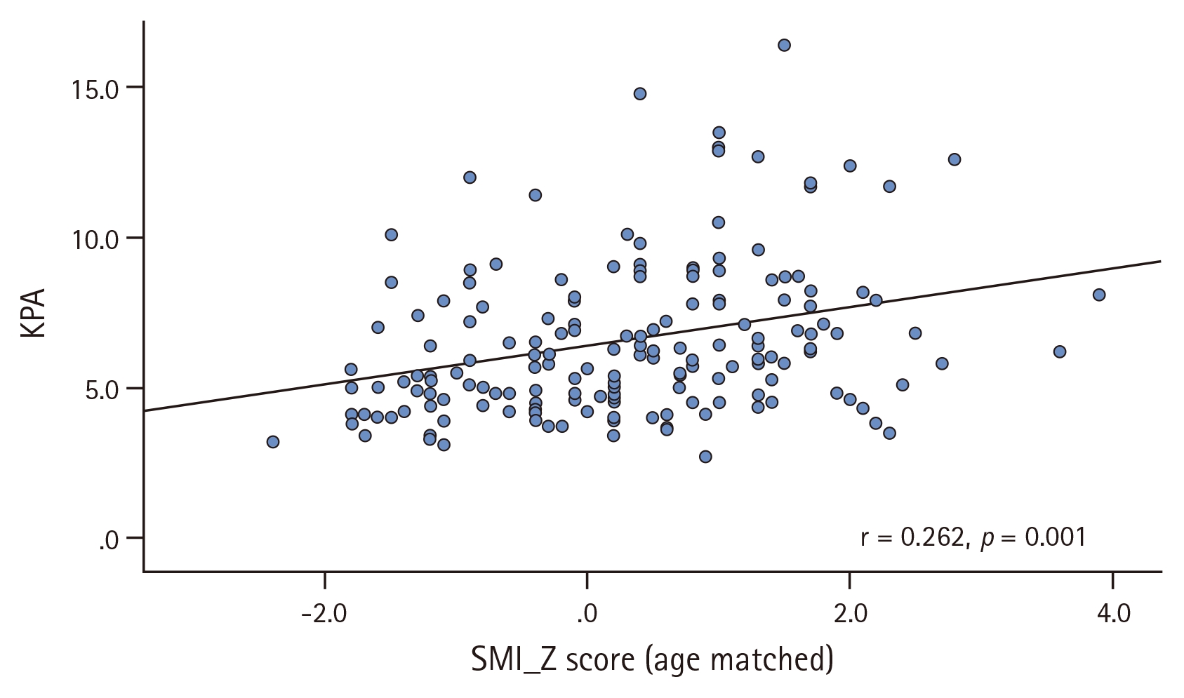
The correlation between SMI-Z score and hepatic fibrosis in all males with MASLD. SMI, skeletal muscle index; MASLD, metabolic dysfunction-associated steatotic liver disease; KPA,
DISCUSSION
This retrospective cross-sectional study aimed to analyze factors associated with the severity of hepatic steatosis and fibrosis in male patients with MASLD. In particular, we evaluated the correlation between body composition indices and clinical data. A key finding was that patients with severe hepatic steatosis exhibited higher values not only for obesity-related markers (weight, BMI, and waist circumference), VFA, and fat mass, but also for SMM and SMI than those in the mild-to-moderate group. These findings are consistent with the established characteristics of MASLD, where obesity-related indices and metabolic dysregulation, reflected by elevated AST and ALT levels, hypertriglyceridemia, and low HDL cholesterol levels, are commonly associated with disease progression. Through multivariate regression analysis, the SMI Z-score was identified as an independent factor associated with an increased severity of hepatic steatosis and a heightened risk of advanced hepatic fibrosis.
Conversely, the VFA Z-score was not an independent factor associated with the severity of steatosis or fibrosis. While the VFA is widely recognized as a factor closely associated with hepatic steatosis and fibrosis [14,15], the independent association between the VFA Z-score and severe hepatic steatosis was attenuated after multivariate adjustment in our study. This is likely due to multicollinearity between BMI and serum triglyceride levels, which are closely linked to visceral adiposity [16,17]. Additionally, because our cohort was predominantly obese with a high baseline median VFA (112.9 cm2), subtle variations in VFA may have lacked the capacity to differentiate steatosis severity. Rather, the robust independent association between the SMI Z-score and hepatic steatosis suggests that relative SMM and its qualitative changes are more critical and sensitive factors associated with MASLD severity than fat volume alone.
Of particular interest was the unexpected finding of increased muscle mass and higher SMI in the group with severe hepatic steatosis. Notably, the SMI Z-score, which accounts for age-related variations in muscle mass, was significantly associated with the severity of both steatosis and fibrosis. This observation contrasts with those of previous studies, including those involving biopsy-proven fatty liver disease and national cohort studies of non-alcoholic fatty liver disease, which reported that low SMI or sarcopenia is associated with an increased severity of steatosis and fibrosis [5,18,19]. However, these studies largely included female-dominant populations (approximately 53–55%) and participants with relatively low BMI (25.0 to 26.7 kg/m2). In contrast, our study focused exclusively on male patients with a higher average BMI of 28.8 kg/m2, thereby capturing a distinct clinical phenotype characterized by obesity and metabolic dysfunction. These results challenge the conventional emphasis solely on muscle loss and suggest that body composition patterns, such as the coexistence of visceral fat accumulation and preserved or even increased muscle mass, may influence disease progression differently in this specific population.
A plausible explanation for this paradoxical finding is the complex effects of obesity on muscle physiology. Previous research suggests that obese individuals may have greater absolute muscle mass than their normal-weight counterparts, likely due to chronic mechanical overload on anti-gravity muscles (e.g., the quadriceps and calves) [20,21]. However, adaptive hypertrophy may be accompanied by impaired muscle quality, contributing to reduced functional capacity and increased insulin resistance. In our study, the SMI Z-score increased in tandem with BMI in patients with severe steatosis (Fig. 1B). Therefore, it is plausible that the results reflect a transient increase in muscle mass characteristics in obese patients.
An important methodological consideration in our study was the use of BIA to estimate SMM. Although BIA is a highly practical and widely validated tool, it estimates fat-free mass indirectly based on total body water, unlike computed tomography or dual-energy X-ray absorptiometry (DXA), which directly visualizes tissue density and cross-sectional area [22,23]. While the SMI Z-scores were derived based on the Asian Working Group for Sarcopenia 2019 consensus, which incorporates both DXA and BIA criteria, it is acknowledged that BIA estimates can be influenced by fluid distribution, particularly in obese individuals. However, to mitigate this potential bias, a high-performance multi-frequency BIA device (InBody 970) was utilized, capable of distinguishing extracellular water, which has been shown to correlate strongly with DXA-measured appendicular skeletal mass. In cohorts characterized by obesity and metabolic dysfunction, such as patients with severe MASLD, altered hydration status, including subclinical fluid retention and an increased ratio of extracellular water, can frequently occur. Because BIA does not directly differentiate between functional contractile muscle fibers and non-contractile elements, such as intramuscular fat or edema, excess fluid or fatty infiltration might inadvertently contribute to a higher estimation of lean mass. As BIA measures muscle quantity rather than quality, the direct linear relationship between absolute muscle mass and liver steatosis may be inherently attenuated, which explains the modest correlation observed in our study (r = 0.328).
Taken together, although BIA has limitations in assessing muscle quality, the findings align with the concept that increased muscle mass accompanied by poor muscle quality (e.g., intramuscular fat) may negatively affect MASLD outcomes. This highlights the need for a more personalized approach for the management of MASLD, particularly in obese men with disproportionate visceral fat and varying muscle composition profiles. Supporting this notion, previous studies have emphasized the role of muscle quality, rather than quantity, in disease progression [7], demonstrating that myosteatosis assessed using computed tomography, rather than sarcopenia, is independently associated with non-alcoholic steatohepatitis and fibrosis progression [24].
To determine whether the effect of the SMI Z-score on disease progression was modified by lifestyle or baseline adiposity, interaction analyses using multivariate logistic regression models was performed. No significant interactions were observed between the SMI Z-score and alcohol intake or BMI with respect to the severity of hepatic steatosis (p = 0.775 and p = 0.783, respectively) or advanced hepatic fibrosis (p = 0.548 and p = 0.164, respectively). These findings indicate that the independent association between a higher SMI Z-score and worse MASLD outcomes is a consistent phenomenon in our cohort, rather than an artifact driven disproportionately by specific subgroups of alcohol consumption or degrees of obesity.
The effect of low alcohol consumption (< 30 g/d) on MASLD remains controversial. A positive association with steatosis severity is an intuitive finding because even small amounts of sustained alcohol intake can induce hepatic fat accumulation. However, the lack of a positive correlation with fibrosis severity differs from previous studies. This finding may be attributable to the predominantly low volume of self-reported alcohol intake (< 30 g/d) in our cohort, as well as the relatively small proportion of participants who reported consuming alcohol (n = 55, 31.6%), which inherently limited the statistical power of this analysis. Although some studies have suggested potential anti-inflammatory effects of low-dose alcohols, this remains speculative [25]. Considering the known hepatotoxic potential of alcohol, caution is warranted in the management of MASLD, even at low doses.
In summary, this study provides novel insights into the association between body composition, particularly increased SMI Z-scores, and severity of hepatic steatosis and fibrosis in male patients with MASLD. These findings diverge from those of previous research emphasizing sarcopenia and suggest that, in obese men, increased muscle mass may not confer metabolic protection, particularly when muscle quality is compromised. Our results underscore the importance of individualized clinical management based on body composition and lifestyle factors, including diet and alcohol intake.
This study had several limitations. These limitations include its single-center design, restriction to a male-only cohort, lack of direct assessment of muscle quality, reliance on self-reported alcohol consumption, and the cross-sectional nature of the analysis, which limits the ability to establish causality. Furthermore, the participants’ specific dietary habits or total caloric intake could not be evaluated, which are significant confounding factors for MASLD severity, owing to the lack of structured dietary assessments such as food frequency questionnaires. Another limitation was the relatively small number of patients with advanced fibrosis. While a higher SMI Z-score was independently associated with advanced fibrosis in the total study population, this association did not reach statistical significance in the multivariate analysis of the non-drinking subgroup. This discrepancy is likely attributable to the reduced statistical power in the subgroup analysis; when this already small subset of patients with advanced fibrosis was further stratified by alcohol consumption, the sample size was insufficient to detect a significant independent association or to determine if the impact of the SMI Z-score varies at different disease stages. Nevertheless, the borderline significance suggests a potential trend consistent with that of the total population. Further large-scale prospective studies are required to confirm whether the impact of age-adjusted muscle mass on liver fibrosis remains robust, particularly in non-drinking male patients with MASLD. Future prospective studies involving more diverse populations and incorporating imaging-based evaluations of muscle quality are warranted to confirm and explore these findings. Despite these limitations, this study had several strengths. First, it provides focused insight into a specific and underrepresented subgroup of obese male patients with MASLD, who have often been under-characterized in previous research. Second, the use of Z-scores for body composition parameters allowed age-adjusted interpretation of muscle mass, enhancing the clinical relevance of the findings. Finally, the integration of BIA-derived metrics with clinical and biochemical markers offers a practical and scalable approach for assessing metabolic risks in real-world settings.
Conclusion
This study demonstrated a significant association between increased SMI Z-scores and the severity of hepatic steatosis and fibrosis in male patients with MASLD. These findings highlight the importance of considering both muscle quantity and quality when evaluating MASLD-associated profiles and underscore the need for a personalized, body composition-informed approach to clinical management. Further prospective multicenter studies incorporating direct measures of muscle quality and diverse patient populations are warranted to validate and expand on these observations.
KEY MESSAGE
1. Higher age-adjusted skeletal muscle mass was independently associated with more severe hepatic steatosis and advanced fibrosis in male patients with MASLD.
2. Modest alcohol consumption was associated with increased hepatic steatosis but lower odds of advanced fibrosis, without suggesting a protective effect against liver disease progression.
3. These findings highlight the importance of assessing both muscle quantity and quality for a more comprehensive evaluation of MASLD severity.
Notes
CRedit authorship contributions
Janghan Jung: investigation, data curation, formal analysis, writing - original draft; Jin Lee: supervision; Dong Hee Koh: supervision; Jung Hee Kim: conceptualization, methodology, investigation, data curation, formal analysis, validation, writing - original draft, writing - review & editing
Conflicts of interest
The authors disclose no conflicts.
Funding
None