Pneumocystis jirovecii pneumonia in non-human immunodeficiency virus patients
Article information

Abstract
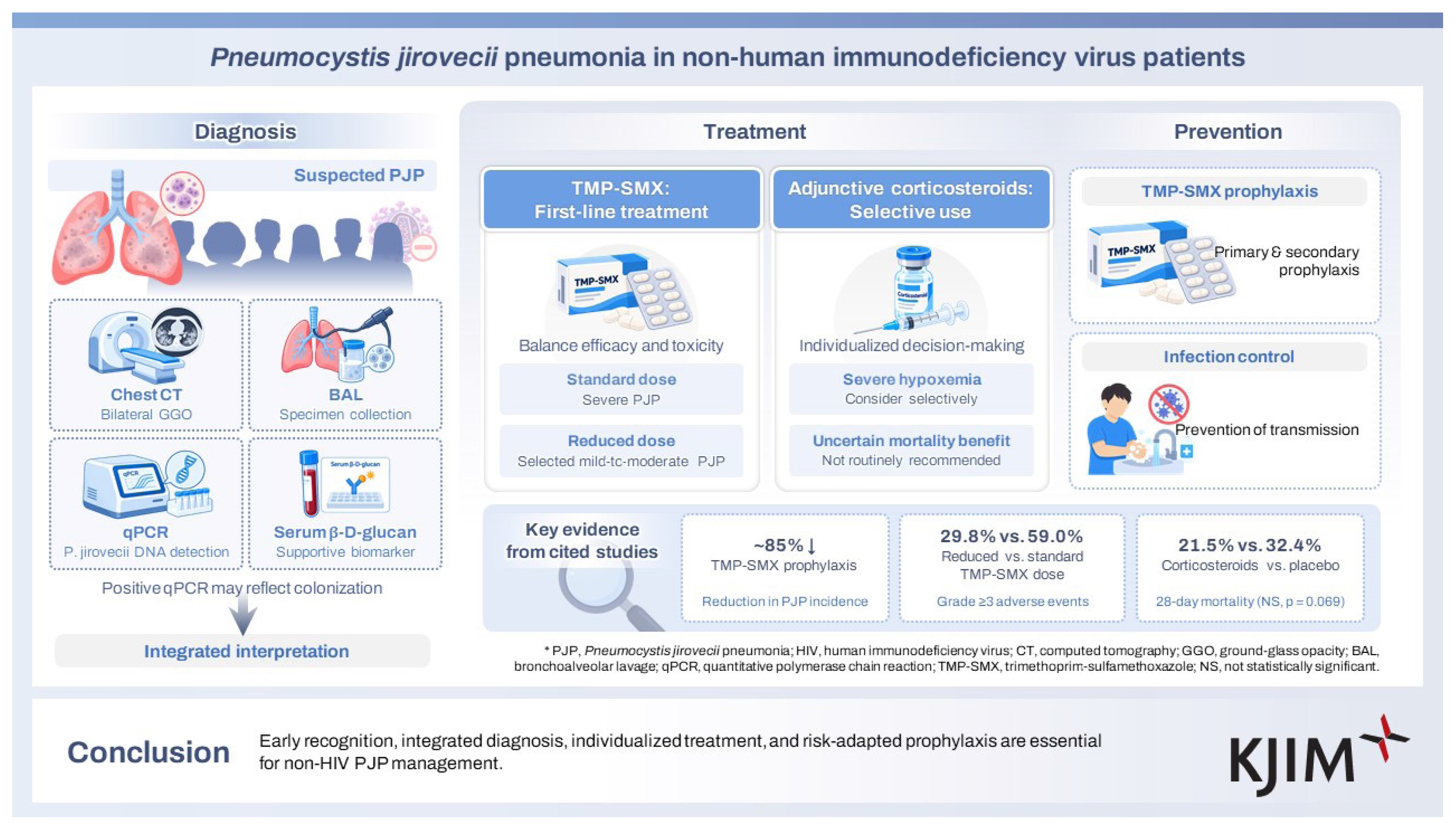
Pneumocystis jirovecii pneumonia (PJP) is an important opportunistic fungal infection in immunocompromised hosts. Although its incidence has declined markedly in people with human immunodeficiency virus (HIV) following the widespread use of antiretroviral therapy and prophylaxis, PJP is increasingly encountered in HIV-negative patients receiving immunosuppressive or targeted immunomodulatory therapies. In this population, PJP often follows a more fulminant course, with rapid progression to severe hypoxemia and acute respiratory failure. Diagnosis remains challenging because clinical manifestations are nonspecific and pulmonary organism burden is often lower than in HIV-associated disease. Accordingly, diagnosis requires integration of host risk factors, radiologic findings, microbiologic testing, and adjunctive biomarkers. Chest computed tomography commonly shows bilateral ground-glass opacities, while bronchoalveolar lavage with microscopy and, increasingly, quantitative PCR remains central to microbiologic confirmation. Serum β-D-glucan may provide useful supportive information but should be interpreted as an adjunct rather than a stand-alone test. Trimethoprim-sulfamethoxazole (TMP-SMX) remains the first-line treatment for PJP, but the optimal dose in HIV-negative patients remains uncertain. Emerging evidence suggests that reduced-dose TMP-SMX may preserve efficacy while improving tolerability in selected patients, whereas standard-dose therapy remains the reference regimen in severe disease. The role of adjunctive corticosteroids in HIV-negative PJP also remains unsettled, supporting a selective rather than routine approach. Prevention remains essential, and current evidence supports risk-adapted primary prophylaxis and individualized secondary prophylaxis for as long as clinically meaningful immunosuppression persists, complemented by infection-control measures in units caring for highly immunocompromised patients.
INTRODUCTION
Pneumocystis jirovecii pneumonia (PJP) remains a serious opportunistic fungal infection in immunocompromised individuals [1]. Although historically associated with acquired immunodeficiency syndrome (AIDS), the incidence of PJP in patients with human immunodeficiency virus (HIV) infection has declined substantially with the widespread adoption of antiretroviral therapy and prophylactic strategies [2]. By contrast, PJP is now encountered more frequently in HIV-negative patients, particularly in those with hematologic malignancies, solid tumors, solid organ transplantation (SOT) or hematopoietic cell transplantation (HCT), autoimmune or inflammatory disorders, and exposure to immunosuppressive or targeted immunomodulatory agents [3,4]. Compared with HIV-associated PJP, disease in HIV-negative patients usually follows a more fulminant course, with rapid progression to severe hypoxemia and acute respiratory failure, while lower organism burden may reduce the sensitivity of conventional diagnostic methods [5,6]. These features make timely diagnosis and management more difficult. Furthermore, many current treatment recommendations are still extrapolated largely from studies of HIV-associated PJP, despite important differences in pathophysiology and clinical course between HIV-positive and HIV-negative populations [7]. In particular, uncertainty persists regarding the optimal dose of trimethoprim-sulfamethoxazole (TMP-SMX) and the role of adjunctive corticosteroids in HIV-negative patients [1,8,9]. In this review, we summarize the evolving epidemiology, risk factors, diagnostic advances, treatment strategies, and prophylactic approaches for PJP in HIV-negative immunocompromised hosts, with particular emphasis on reduced-dose TMP-SMX and adjunctive corticosteroid therapy.
EPIDEMIOLOGY
Although Pneumocystis was first identified in the early 20th century, its clinical importance in humans was not clearly recognized until the mid-20th century, when clusters of disease were described in central and eastern Europe among premature and malnourished infants as plasma cell interstitial pneumonitis [1,10]. As clinical recognition broadened, PJP came to be understood as an opportunistic infection associated with impaired cellular immunity, including hematologic malignancy, transplantation, congenital immunodeficiency, cancer therapy, and corticosteroid exposure [1,10].
The next major epidemiologic transition occurred with the emergence of AIDS. In 1981, clusters of PJP in previously healthy young men were among the first sentinel events that defined the early AIDS epidemic [11]. Before the widespread use of effective antiretroviral therapy and prophylaxis, PJP developed in roughly 70–80% of patients with advanced HIV infection [12,13]. This burden subsequently declined sharply with modern HIV care, as illustrated by the EuroSIDA cohort, in which PJP incidence fell from 4.9 cases per 100 person-years before March 1995 to 0.3 cases per 100 person-years after March 1998 [2]. A similar decline was observed in the HIV Outpatient Study, where the incidence of first-episode PJP decreased to 3.9 cases per 1,000 person-years during 2003–2007 [2].
In parallel, the epidemiologic burden of PJP has progressively shifted toward HIV-negative immunocompromised hosts. Recent reviews consistently identify hematologic malignancies, solid tumors, SOT or HCT, autoimmune and inflammatory diseases, chronic lung disease, and exposure to corticosteroids, biologic agents, rituximab, and other targeted immunomodulatory therapies as the principal contemporary risk settings [3,7]. Population-based studies support a true increase in non-HIV PJP over the past two decades [14,15]. In England, laboratory-confirmed PJP increased by a mean of 7% per year between 2000 and 2010, largely driven by transplant recipients and patients with hematologic malignancies [14]. In Germany, the overall PJP incidence rose from 2.3 to 2.6 cases per 100,000 population between 2014 and 2019, while the annual number of PJP-associated deaths increased from 516 to 615 over the same period [15]. In that nationwide German analysis of 12,455 PJP cases, 10,331 cases (82.9%) occurred in HIV-negative patients, whereas annual HIV-associated cases decreased from 346 to 331 and annual non-HIV cases increased from 1,511 to 1,841 [15]. Notably, the increase was especially pronounced in patients with chronic lung disease, solid malignancies, and rheumatic disease, whereas case numbers declined in groups with more established prophylactic frameworks, including patients with HIV, hematologic malignancies, and transplantation [15].
The growing relative contribution of HIV-negative patients among PJP cases should be interpreted as the consequence of multiple, concurrent epidemiologic changes rather than a single explanatory mechanism. The burden of HIV-associated PJP has declined substantially with earlier HIV diagnosis, widespread access to effective antiretroviral therapy, and well-established preventive strategies for patients with advanced HIV-related immunosuppression [2,12,13]. In parallel, the population of HIV-negative patients at risk has expanded owing to aging, malignancy-directed therapy, transplantation, autoimmune and inflammatory diseases, chronic lung disease, and increasing exposure to corticosteroids, biologic agents, rituximab, and other targeted immunomodulatory therapies [3,4,7,14,15]. Cohort data further illustrate the heterogeneous clinical spectrum of contemporary non-HIV PJP. In the classic Mayo Clinic series of 116 non-AIDS patients, the major associated conditions were hematologic malignancy in 35 patients (30.2%), organ transplantation in 29 (25.0%), inflammatory disorders in 26 (22.4%), and solid tumors in 15 (12.9%), and 105 patients (90.5%) had received systemic corticosteroids within 1 month before diagnosis [16]. In a multicenter Quebec cohort of 129 non-HIV PJP cases, more than two-thirds of patients had either hematologic disease or autoimmune or inflammatory disease, 108 (83.7%) had received corticosteroids, 92 (71.3%) had received immunosuppressive agents, and only six patients (4.7%) had received prophylaxis [17]. Importantly, 63 of those 129 patients (48.8%) would not have met contemporary guideline-based or usual clinical criteria for prophylaxis, underscoring that current risk extends beyond traditionally recognized high-risk groups [17]. Thus, the contemporary epidemiologic shift toward non-HIV PJP likely reflects the combined effects of declining HIV-associated PJP, expansion of non-HIV immunocompromised populations, broader exposure to immunomodulatory therapies, and improved clinical recognition and diagnostic testing.
Korean nationwide data also support this epidemiologic transition. In an analysis using Health Insurance Review and Assessment Service and Korea Disease Control and Prevention Agency data from 2010 to 2021, annual non-HIV PJP cases increased from 60 in 2010 to 551 in 2021, and, using national population estimates as the denominator, the approximate crude annual incidence increased from 1.2 to 10.6 cases per 1,000,000 population during this period [18]. Distinct regional clustering was identified, with Seoul representing the primary cluster (relative risk [RR] 4.59), which was attributed to centralized tertiary referral centers, and Jeju-do representing the second cluster (RR 1.59), possibly favored by higher ambient temperature and humidity [18]. A 29.02% spatiotemporal increase was also observed across southern regions, predominantly among people older than 60 years, highlighting the growing clinical impact of immunosenescence [18].
Importantly, non-HIV PJP is not only increasingly frequent but also associated with worse outcomes than HIV-associated PJP. In a French single-center study of 604 PJP cases, including 143 HIV-positive and 461 HIV-negative patients, the ratio of non-HIV to HIV cases increased from 1.7 to 5.6 over the study period, and 14-day mortality was 20.6% in HIV-negative patients compared with 1.4% in patients with HIV [19]. In another study of 133 non-HIV patients with PJP, the majority had hematologic malignancies (80.5%), and the 30-day and 1-year mortality rates were 24.1% and 56.4%, respectively [20]. A recent Korean single-center study of 219 patients with PJP also reported lower 28-day survival in non-HIV PJP than in HIV-associated PJP (68.6% vs. 88.6%, p = 0.011) [21]. Contemporary studies and recent trial background data indicate that hospital mortality in HIV-negative PJP commonly remains in the range of 30–50% [5,22]. However, this poorer prognosis should not be interpreted solely as evidence of intrinsic pathogenetic differences between HIV-associated and non-HIV PJP. Mortality in HIV-negative patients also reflects host-related factors, including the underlying disease, age, comorbidities, severity of immunosuppression, concomitant organ dysfunction, coinfections, and delayed diagnosis [23–25]. Thus, the adverse outcomes observed in non-HIV PJP likely reflect a combination of rapid clinical deterioration, diagnostic difficulty related to lower organism burden, and competing mortality risks imposed by the host’s underlying condition and treatment context [19,23–25].
DIAGNOSIS
The diagnosis of PJP in HIV-negative immunocompromised patients requires integration of host risk factors, clinical presentation, radiologic findings, and microbiologic evidence, because no single clinical or imaging feature is sufficiently specific to establish the diagnosis in isolation [1,26,27]. In HIV-negative patients, PJP typically follows a more acute course than HIV-associated disease and often presents with fever, nonproductive cough, progressive dyspnea, and hypoxemia developing over days to 1–2 weeks [1,6,28]. Accordingly, PJP should be strongly suspected in patients receiving corticosteroids, transplantation-related immunosuppression, chemotherapy, rituximab, or other potent immunomodulatory therapies who present with otherwise unexplained diffuse pneumonia and impaired gas exchange [6,29].
Chest computed tomography (CT) is more sensitive than chest radiography and is particularly useful when the chest radiograph is normal or nondiagnostic despite high clinical suspicion [6]. Bilateral ground-glass opacities remain the dominant CT pattern, although consolidation (13.1–77.5%), septal thickening (75%), reticulation (21.5–45%), cysts (0.7–20%), nodules (10–16.1%), and pneumothorax (2.5%) may also be observed [20,30]. However, since imaging findings alone are not specific, careful evaluation for alternative or concomitant diagnoses is necessary if localized consolidation, pleural effusion, nodules, or marked asymmetry are observed [26].
Definitive diagnosis relies on demonstration of P. jirovecii in respiratory specimens or lung tissue [27]. Acceptable specimens include bronchoalveolar lavage (BAL) fluid, induced or spontaneous sputum, and transbronchial or open lung biopsy material [31]. Among these, BAL remains the most reliable diagnostic specimen in HIV-negative patients because organism burden is often lower than in HIV-associated disease; BAL also allows evaluation for competing or coexisting pulmonary infections [26]. Since the highest concentration of P. jirovecii is typically found within the alveoli, BAL fluid is superior to sputum samples; consequently, a negative PCR result from BAL fluid provides high diagnostic confidence for excluding the infection [32]. Induced sputum is less invasive and may be useful when bronchoscopy is not immediately feasible, but positive results should still be interpreted in clinical context and negative results do not reliably exclude disease in HIV-negative patients [33,34]. In the American Society of Transplantation (AST) guidance, the diagnostic yield of induced sputum was reported to be only 30–55%, compared with 80–95% for BAL, and a negative smear from a single respiratory specimen was considered insufficient to exclude PJP [6]. Specifically, HIV-negative patients exhibit a significantly lower diagnostic yield from the initial induced sputum test compared with HIV-positive patients; therefore, if PJP is clinically suspected, repeated testing should be considered [35].
When bronchoscopy is not feasible, upper respiratory tract specimens may retain considerable diagnostic value. A meta-analysis demonstrated that nasopharyngeal (NP) aspirate PCR has high diagnostic accuracy for PJP, with a pooled sensitivity of 89% and a specificity of 98%; notably, high sensitivity was maintained even among non-HIV patients (88%) [34]. In the same analysis, PCR substantially outperformed conventional microscopy-based methods, whose sensitivity (immunofluorescence approximately 71–74%; cytological staining approximately 50–58%) is considerably lower, underscoring the value of NP PCR as an initial, minimally invasive screening test when invasive sampling is not feasible [34].
In a paired-specimen analysis, the positivity rate of NP aspirate PCR was comparable to that of NP swabs, although BAL remained the most sensitive specimen (BAL 29.1% vs. NP 21.7%, p < 0.01) [36]. Notably, NP specimens showed an excellent negative percent agreement (100%) but a more limited positive percent agreement (72.2%) relative to bronchoscopic specimens [36]. These findings suggest that while a positive NP PCR can rapidly confirm the diagnosis, a negative result does not exclude PJP and should prompt lower respiratory tract sampling. Specimen selection and interpretation should therefore also take age into account. In children, from whom lower respiratory tract specimens are particularly difficult to obtain, NP aspirate PCR yielded detection rates comparable to those of lower respiratory tract specimens and was markedly superior to immunofluorescence assays, supporting its role for rapid and timely diagnosis in this population [37].
Microscopy remains clinically important because direct visualization of the organism provides strong microbiologic support for active disease, but its sensitivity is limited in HIV-negative patients, in whom fungal burden is often lower than in HIV-associated PJP [26,31]. Immunofluorescence-based methods are regarded as the most sensitive microscopic techniques and offer better yield than conventional stains, particularly in specimens with a low organism burden [38,39]. Nevertheless, a negative microscopy result in HIV-negative immunocompromised patients does not rule out PJP, which has accelerated the adoption of molecular testing in routine clinical practice [40].
PCR has become central to PJP diagnosis because it is substantially more sensitive than microscopy, particularly in HIV-negative patients [26]. Quantitative PCR (qPCR) on BAL fluid has shown excellent diagnostic performance across both HIV and non-HIV populations. A 2024 meta-analysis reported a pooled sensitivity of 98.7% and specificity of 89.3%, findings that were consistent with an earlier BAL-focused meta-analysis reporting a sensitivity of 98.3% and specificity of 91.0% [41,42]. However, its major limitation is that PCR may detect colonization rather than true pneumonia, especially when interpreted alone [41]. Consistent with this limitation, Robert-Gangneux and colleagues found in a 4-year prospective study that although BAL qPCR was more sensitive than microscopy, no single threshold could reliably distinguish infection from colonization in all immunocompromised patients [40]. PCR results should therefore be interpreted together with the clinical context, including symptoms, oxygenation, chest CT findings, and evaluation for alternative pathogens [27].
Contemporary practice increasingly uses two qPCR thresholds—with an intermediate “gray zone” between them—to separate colonization from active disease, although the reported cutoffs are assay-specific and not interchangeable (e.g., 120 and 1,900 trophic form equivalents/ mL [43]; 1,480 and 9,655 copies/μL in a non-HIV cohort [32]) [43–45]. A burden above the upper cutoff in a compatible clinical context supports active PJP and warrants prompt therapy, whereas a value below the lower cutoff favors colonization and may justify observation when suspicion is low; gray-zone values should be interpreted with adjunctive biomarkers, and—since HIV-negative patients carry a lower burden than those with HIV—a low result should not be used to withhold therapy when clinical suspicion is high. BAL metagenomic next-generation sequencing is also increasingly reported in non-HIV PJP and, despite cost and standardization concerns, is likely to become an important diagnostic adjunct [46].
Serum β-D-glucan (BDG) is best regarded as an adjunctive rather than a stand-alone diagnostic test for PJP [47,48]. In a systematic review and meta-analysis, serum BDG showed a pooled sensitivity of 91% and specificity of 79%, although sensitivity was lower in patients without HIV than in those with HIV (86% vs. 94%) [47]. In a 2025 systematic review and meta-analysis focused on the Fungitell assay, the conventional cutoff of 80 pg/mL yielded a pooled sensitivity of 83.5% and specificity of 75.5%, whereas a higher threshold of 400 pg/mL increased specificity to 93.6% at the expense of sensitivity [48]. These findings suggest that BDG may be most useful as a rule-out test in patients with low pretest probability, rather than as a definitive discriminator of active PJP on its own [49]. Because BDG is not specific for Pneumocystis, results should always be interpreted together with the clinical, radiologic, and microbiologic context [50]. This is particularly important in HIV-negative patients, in whom recent studies have shown that a negative BDG result alone is insufficient to exclude PJP, especially in patients with hematologic malignancy or other immunosuppression [51,52].
Overall, current evidence supports a practical diagnostic strategy in which PJP is suspected on the basis of host risk factors, acute respiratory symptoms, hypoxemia, and compatible CT findings, followed by prompt microbiologic testing using lower respiratory tract specimens whenever feasible [27]. In stable patients, induced sputum with immunofluorescence or PCR may serve as an initial test, but BAL is generally preferred in HIV-negative patients because of its greater diagnostic yield and its ability to identify coinfections [26]. Molecular assays have become central to diagnosis, but positive PCR results should not be interpreted in isolation; rather, they should be integrated with the clinical syndrome, chest CT pattern, and adjunctive biomarkers such as serum BDG to distinguish active pneumonia from colonization as accurately as possible [53].
Based on these considerations, we propose a risk-based and clinically integrated diagnostic algorithm for HIV-negative immunocompromised patients with suspected PJP (Fig. 1), adapted from the Korean expert consensus algorithm and contemporary diagnostic literature [26,27,29]. The diagnostic approach begins with identification of relevant host risk factors and compatible respiratory manifestations, followed by chest CT to determine radiologic compatibility. In at-risk patients, bilateral ground-glass opacities should prompt microbiologic testing, whereas atypical or incompatible CT findings should lead clinicians to evaluate alternative or concomitant pulmonary processes [29,30]. When PJP remains suspected, bronchoscopy with BAL should be prioritized when feasible because organism burden is often low in HIV-negative patients and BAL provides the highest diagnostic confidence for microscopy, immunofluorescence, PCR-based testing, and coinfection evaluation [26,27,41,42]. If bronchoscopy is not feasible, non-BAL specimens such as induced sputum, expectorated sputum, oral wash, or NP specimens may be used for PCR according to local availability, but negative results should be interpreted cautiously in high-risk patients [29,33–35]. Serum BDG should be used as an adjunctive biomarker rather than as a stand-alone diagnostic test, because positive results are not specific for PJP and negative results may not fully exclude PJP in selected HIV-negative immunocompromised patients [47–52]. The key interpretive step is to distinguish active PJP from colonization: microscopy or immunofluorescence positivity from BAL or lung tissue provides the strongest microbiologic support for proven PJP, whereas PCR positivity, particularly from non-BAL or low-burden specimens, should be interpreted according to host risk, clinical severity, CT pattern, degree of hypoxemia, quantitative organism burden when available, BDG results, and competing diagnoses [27,32,41]. Active PJP is favored by a high-risk host, compatible CT findings, clinically significant hypoxemia, positive BDG, high organism burden, and absence of a more plausible alternative diagnosis, whereas colonization is more likely when symptoms are mild or absent, CT findings are atypical, BDG is negative, organism burden is low, or another diagnosis better explains the pulmonary findings [27,29,32,47]. In patients with relevant host risk factors, compatible CT findings, and respiratory compromise, empiric anti-Pneumocystis therapy should not be delayed while confirmatory microbiologic results are pending [7,26].
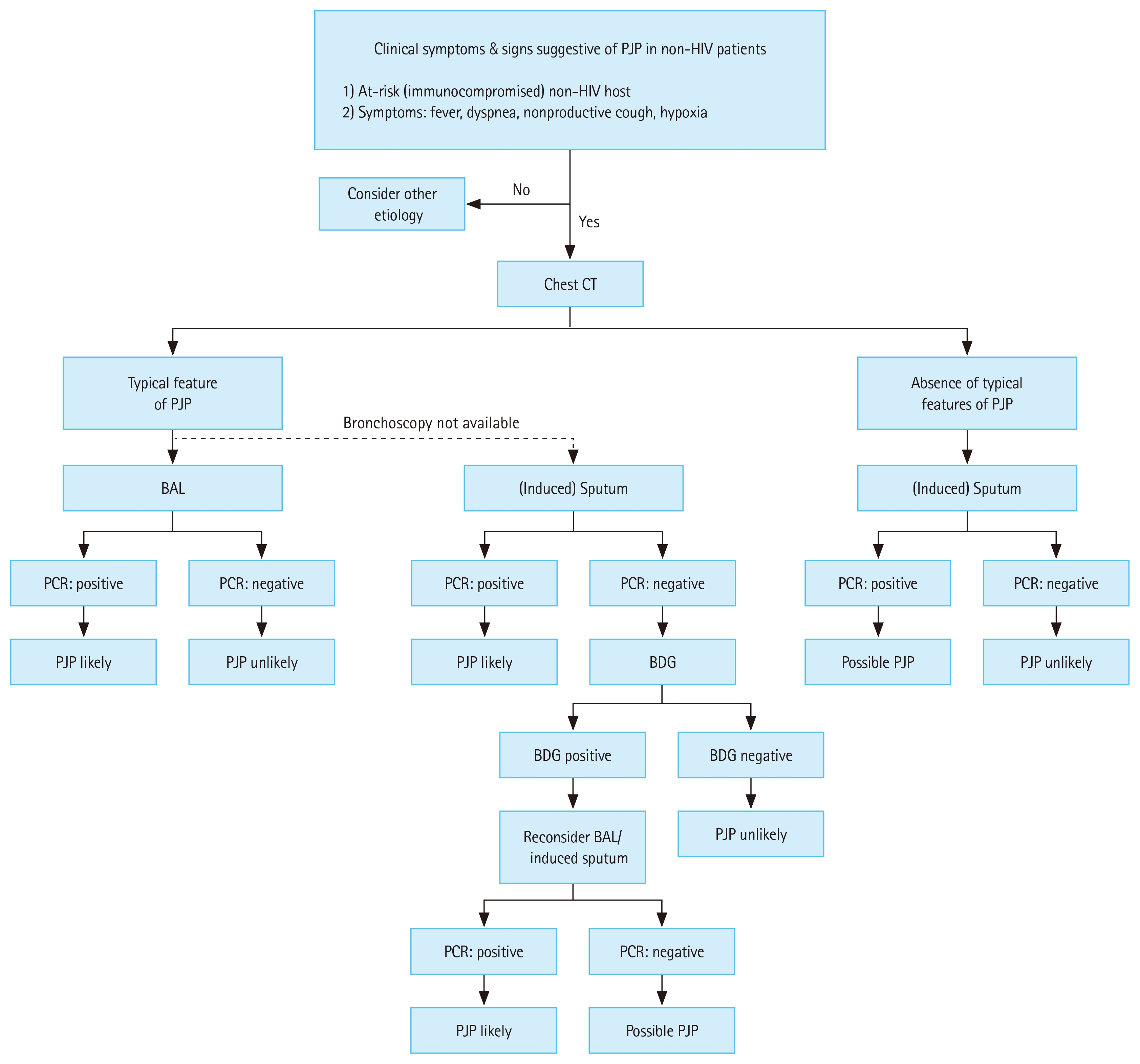
Proposed risk-based and clinically integrated diagnostic algorithm for Pneumocystis jirovecii pneumonia (PJP) in HIV-negative immunocompromised patients, adapted from the Korean expert consensus algorithm and contemporary diagnostic literature [29]. HIV, human immunodeficiency virus; BAL, bronchoalveolar lavage; BDG, (1→;3)-β-D-glucan; CT, computed tomography; PCR, polymerase chain reaction.
TREATMENT
Prompt initiation of treatment is a central determinant of outcome in PJP, particularly in HIV-negative immunocompromised patients, in whom delayed treatment has been associated with higher rates of respiratory deterioration, mechanical ventilation, and death [23,24]. Treatment should therefore be started as soon as PJP is clinically suspected in an at-risk patient with compatible symptoms and radiologic findings, and it should not be deferred while awaiting diagnostic confirmation [7]. Severity assessment should not rely solely on oxygenation indices, because conventional community-acquired pneumonia scores may underestimate disease severity in non-HIV PJP; rather, oxygenation should be interpreted together with respiratory rate, age, comorbidities, and evidence of extrapulmonary organ dysfunction [7,54].
TMP-SMX remains the preferred first-line treatment for PJP. Current guideline-based dosing is TMP 15–20 mg/kg/ day plus SMX 75–100 mg/kg/day for 14–21 days, and a total duration of 21 days is typically used in HIV-negative patients [1,55]. In patients with mild disease and preserved enteral absorption, oral therapy may be used from the outset, whereas moderate-to-severe disease should generally be treated intravenously, with step-down to oral therapy after clear clinical improvement [1,55]. Issues related to reduced-dose TMP-SMX strategies are discussed separately below.
Alternative regimens are primarily reserved for patients with contraindications, major intolerance, or failure of TMP-SMX [1]. Acceptable alternative treatment options include intravenous pentamidine (4 mg/kg/day), primaquine (30 mg base/day) plus clindamycin (600 mg three times daily), and atovaquone (750 mg twice daily with food) [56–58]. Among these, primaquine plus clindamycin is particularly important because the European Conference on Infections in Leukemia (ECIL-6) identifies it as the preferred alternative in patients with documented TMP-SMX intolerance and the preferred second-line or salvage regimen when a change in therapy is required [7]. Before primaquine is prescribed, glucose-6-phosphate dehydrogenase (G6PD) deficiency should be excluded because of the risk of hemolysis [1]. Atovaquone may be considered in mild-to-moderate disease, but the absence of an intravenous formulation limits its role in severe PJP [1]. Although TMP-SMX combined with caspofungin has been reported in limited case series, the supporting evidence remains weak [53]. Because echinocandins inhibit β-1,3-D-glucan synthesis and therefore mainly target the cystic rather than the trophic form of Pneumocystis, echinocandin monotherapy should not be used, as adequate anti-PJP efficacy has not been demonstrated [59,60]. Treatment regimens, alternative options, and adjunctive corticosteroids are summarized in Table 1.

Treatment of Pneumocystis jirovecii pneumonia in HIV-negative immunocompromised patients
Response assessment should be guided primarily by the clinical course rather than by early time points alone [7,61]. Early worsening can occur despite appropriate therapy, particularly in patients with severe inflammatory lung injury; therefore, formal reassessment is generally recommended after approximately 1 week of full-dose therapy [7,62]. Persistent or progressive respiratory dysfunction should prompt repeat chest CT and bronchoscopy with BAL to evaluate for coinfection or an alternative diagnosis [61]. Persistently positive P. jirovecii PCR in BAL fluid should not be interpreted as proof of treatment failure, because organism detection may persist for days to weeks despite effective therapy [63]. However, a recent study suggests that P. jirovecii PCR-negative conversion predicts the prognosis of HIV-negative patients with PJP and acute respiratory failure; follow-up PJP PCR monitoring may therefore be carefully considered in specific clinical contexts to assist in prognostic assessment [64]. Likewise, serial serum BDG measurement is not recommended for routine response assessment because available data are inconsistent [25,65].
Drug-related toxicity is a major practical issue during PJP treatment and must be actively monitored throughout therapy. TMP-SMX is associated with rash, fever, nephrotoxicity, electrolyte disturbances, bone marrow suppression, and hepatotoxicity; these adverse effects appear to be at least partly dose dependent [8,66]. Clindamycin-primaquine may cause nausea, vomiting, neutropenia, Clostridioides difficile–associated diarrhea, hemolysis in patients with G6PD deficiency, and bone marrow suppression [6]. Pentamidine is associated with nephrotoxicity, electrolyte abnormalities, dysglycemia including insulin-dependent diabetes mellitus, pancreatitis, and QT prolongation [1]. Because many patients are simultaneously receiving chemotherapy, corticosteroids, calcineurin inhibitors, or other immunosuppressive agents, drug–drug interactions and cumulative toxic effects must be monitored closely throughout treatment [7]. Particular caution is warranted with concomitant medications common in this population: calcineurin inhibitors (tacrolimus, cyclosporine) amplify nephrotoxicity and hyperkalemia—especially in transplant recipients and when combined with renin–angiotensin system blockers—while methotrexate myelotoxicity and warfarin anticoagulation are also potentiated, warranting early international normalized ratio monitoring (data for direct oral anticoagulants are limited) [67]. TMP also inhibits tubular creatinine secretion and may raise serum creatinine without a true fall in glomerular filtration rate, so pseudo-nephrotoxicity should not be mistaken for genuine renal injury in SOT recipients receiving calcineurin inhibitors [67]. Treatment-dose TMP-SMX should be reduced by 50% at a creatinine clearance of 15–30 mL/min and generally avoided below 15 mL/min [1].
In severe PJP, timely intensive care unit (ICU) monitoring and early recognition of impending respiratory failure are critical [19]. Noninvasive ventilation has not shown clear superiority over oxygen therapy alone in patients admitted to the ICU with hypoxemic acute respiratory failure [68,69]. Accordingly, if noninvasive ventilation is attempted, patients should be monitored closely for ongoing respiratory deterioration, and prompt transition to invasive mechanical ventilation should be considered when failure becomes likely. The question of adjunctive corticosteroids in HIV-negative PJP remains controversial and is addressed separately below.
Reduced-dose TMP-SMX
TMP-SMX remains the first-line therapy for PJP, and current guidelines still recommend TMP 15–20 mg/kg/day plus SMX 75–100 mg/kg/day as the standard regimen [1,7]. However, the evidence for this conventional dose is limited, particularly in HIV-negative patients, and concern has increased regarding dose-dependent toxicities such as nephrotoxicity, electrolyte disturbances, hepatotoxicity, rash, and bone marrow suppression [8,66,70,71]. These concerns have led to growing interest in reduced-dose TMP-SMX strategies that aim to preserve clinical efficacy while improving tolerability.
A systematic review and meta-analysis by Butler-Laporte et al. [8] suggested that reduced-dose TMP-SMX may provide similar efficacy with fewer severe adverse events. In that analysis, reduced-dose regimens, defined as TMP 15 mg/kg/day or less, were not associated with a statistically significant increase in mortality compared with standard-dose therapy, while grade 3 or higher adverse events were reduced by 18% [8]. However, the included studies were heterogeneous with respect to study design, underlying host population, severity of illness, timing of dose reduction, and the definition of reduced-dose therapy [8]. In particular, reduced-dose regimens varied substantially, with TMP doses ranging from approximately 7.5 to 15 mg/kg/day across studies, limiting the ability to identify a single optimal reduced-dose regimen [8,70,71].
Subsequent observational studies have provided more specific clinical data supporting this dose-sparing approach while also helping to define its appropriate scope [70,71]. In a multicenter Swedish study of 113 patients with hematologic malignancies, reduced-dose TMP-SMX (TMP 7.5–15 mg/kg/day) was not associated with day-8 clinical failure or 30-day mortality compared with standard-dose therapy [70]. Notably, among patients with mild-to-moderate pneumonia, defined by a baseline PaO2/FiO2 > 200 mm Hg, all 44 patients treated with the reduced-dose regimen were alive at day 30 [70]. In a multicenter Japanese cohort of patients with non-HIV PJP, low-dose TMP-SMX, defined as TMP less than 12.5 mg/kg/day, was associated with similar 30-day and 180-day mortality after adjustment for baseline differences, whereas grade 3 or higher adverse events were significantly less frequent with low-dose therapy than with conventional dosing (29.8% vs. 59.0%), particularly nausea and hyponatremia [71]. These findings suggest that reduced-dose TMP-SMX may be clinically useful in selected HIV-negative patients, especially those with mild-to-moderate disease or substantial concern for treatment-limiting toxicity [8,70,71].
Nevertheless, reduced-dose TMP-SMX should not be generalized to all HIV-negative immunocompromised patients. Data remain limited for several clinically important high-risk groups, including SOT recipients, patients who have undergone chimeric antigen receptor (CAR) T-cell therapy, and patients with profound lymphopenia or severe cellular immune dysfunction [6,8,70,72,73]. Interpretation is also complicated by the absence of a standardized definition of severe PJP across studies, with variable use of PaO2/FiO2 ratio, alveolar–arterial oxygen gradient, oxygen requirement, mechanical ventilation, or composite clinical criteria [7,8,70,71]. Thus, reduced-dose TMP-SMX may be considered in carefully selected HIV-negative patients with mild-to-moderate disease, advanced age, renal dysfunction, or a high risk of treatment-limiting toxicity [8,70,71]. By contrast, standard-dose TMP-SMX should remain the reference regimen for severe PJP in accordance with current guideline recommendations, including the 2016 ECIL guidance, until prospective studies and future guideline updates better define the patient populations, severity thresholds, and dosing strategies in which reduced-dose therapy can be used safely and effectively [7].
Adjunctive corticosteroids in HIV-negative patients
The role of adjunctive corticosteroids in HIV-negative PJP remains considerably more uncertain than in HIV-associated disease. Earlier guidance recommended that glucocorticoid use in non-HIV PJP be decided on a case-by-case basis rather than routinely, reflecting the absence of randomized trials and conflicting findings across several retrospective studies [7].
Observational evidence regarding adjunctive corticosteroids in HIV-negative PJP remains inconsistent, largely because available studies differ in patient populations, disease severity, corticosteroid indication, timing, dose, and adjustment for treatment-selection bias. An earlier systematic review by Injean et al. found no clear association between adjunctive corticosteroids and survival in non-HIV PJP, emphasizing the small sample sizes, heterogeneity in steroid definitions, and high risk of treatment-selection bias across available studies [74]. A subsequent meta-analysis by Ding et al. [9], which included 16 retrospective studies and 2,518 patients, reported that adjunctive corticosteroids were associated with increased mortality in the overall non-HIV PJP population (odds ratio [OR] 1.37, 95% confidence interval [CI] 1.07–1.75). However, subgroup analysis suggested a possible severity-dependent effect, with lower mortality in hypoxemic patients (OR 0.69, 95% CI 0.47–1.01) and significantly lower mortality in those with respiratory failure (OR 0.63, 95% CI 0.41–0.95). Individual retrospective cohort studies have similarly failed to demonstrate a consistent overall benefit. In a cohort of 323 hospitalized adults without HIV, Wieruszewski et al. [75] found that early corticosteroid administration within 48 hours was not associated with better respiratory outcomes (OR 0.76, 95% CI 0.24–2.28), lower mortality, shorter hospital stay, reduced ICU admission, or reduced need for mechanical ventilation. Similarly, Assal et al. [20] reported that early adjunctive corticosteroid therapy in non-HIV cancer patients was not associated with improvement in 30-day mortality (hazard ratio [HR] 1.45, 95% CI 0.7–3.04), 1-year mortality (HR 1.25, 95% CI 0.75–2.09), respiratory deterioration, intubation, or coinfections after propensity weighting [20]. In contrast, Lemiale et al. [22] observed that high-dose steroids (≥ 1 mg/kg/day prednisone equivalent) were independently associated with increased ICU mortality in HIV-negative patients with severe PJP. In summary, these data do not support routine adjunctive corticosteroid therapy for all HIV-negative patients with PJP, but they leave open the possibility of selective use in patients with marked hypoxemia or respiratory failure, while underscoring the need to avoid indiscriminate or high-dose corticosteroid exposure.
The most important recent evidence comes from the Effect of Steroids During Pneumocystis Infection Among Non-HIV Immunocompromised Patients (PIC) trial, the first large randomized, double-blind, placebo-controlled trial of adjunctive corticosteroids (intravenous methylprednisolone 30 mg twice daily on days 1–5, 30 mg once daily on days 6–10, then 20 mg once daily until day 21) in HIV-negative immunocompromised patients with P. jirovecii pneumonia and acute hypoxemic respiratory failure [5]. The trial did not meet its primary endpoint: 28-day mortality was not significantly reduced (21.5% vs. 32.4%; p = 0.069). Among the prespecified secondary outcomes, 90-day mortality was lower with corticosteroids (28.0% vs. 43.2%; p = 0.022), and in the subgroup not intubated at randomization there was less progression to invasive mechanical ventilation (10.1% vs. 26.1%; p = 0.020), without a significant increase in secondary infections or insulin requirement; randomization was stratified by oxygen needs (< 6 L/min vs. ≥ 6 L/min), among other factors. Because the primary endpoint was not met, these secondary findings are hypothesis-generating and should be interpreted with caution. Nevertheless, they are shifting the discussion away from blanket avoidance of corticosteroids in HIV-negative PJP with severe hypoxemia toward a more selective approach.
Overall, the current evidence does not support routine adjunctive corticosteroid therapy for all HIV-negative patients with PJP. A more defensible approach is to reserve corticosteroids for carefully selected patients with severe hypoxemic respiratory failure, while avoiding indiscriminate use in mild disease and remaining cautious with very high-dose regimens.
Clinical significance and management of pathogen codetection
With the widespread adoption of multiplex molecular assays and next-generation sequencing, concomitant pathogens—such as cytomegalovirus (CMV), herpes simplex virus (HSV), and various bacteria—are increasingly detected in HIV-negative patients with PJP. The literature indicates that pulmonary CMV codetection occurs in 20–65% of non-HIV PJP cases, and its presence is frequently associated with a more fulminant clinical course, a higher incidence of acute respiratory distress syndrome, and significantly increased short-term mortality [76,77].
However, whether the isolated viruses represent active pathogenic coinfection requiring targeted antiviral therapy or merely incidental benign shedding driven by profound iatrogenic immunosuppression remains a subject of intense debate. Retrospective cohorts have yielded conflicting results; some suggest that adjunctive ganciclovir therapy may be warranted in critically ill patients with high pulmonary CMV viral loads [78], whereas others caution against routine antiviral initiation because of the risk of cumulative toxicities such as myelosuppression and nephrotoxicity [76].
In the authors’ opinion, a risk-stratified, quantitative approach is warranted. Routine antiviral treatment for all bystander viral codetections should be avoided. Instead, preemptive therapy (e.g., ganciclovir) should be restricted to patients exhibiting severe respiratory failure (PaO2/FiO2 ratio < 200 mm Hg) coupled with high-load pulmonary replication (≥ 3–4 log10 copies/mL in BAL fluid) or documented systemic CMV antigenemia [76,79]. For HSV, given that its detection in respiratory specimens predominantly represents proximal airway reactivation rather than true parenchymal pneumonitis, concomitant acyclovir therapy should not be routinely administered unless visible mucosal ulceration or histopathological evidence of tissue invasion is present.
PREVENTION
Primary prophylaxis
Primary prophylaxis is intended to prevent a first episode of PJP in patients whose underlying disease or immunosuppressive treatment confers a clinically meaningful risk of infection [4]. TMP-SMX remains the agent of choice for primary prophylaxis in non-HIV populations because it has the strongest evidence base and the greatest preventive efficacy [4,66]. In the Cochrane review of non-HIV immunocompromised patients, TMP-SMX prophylaxis reduced documented PJP by 85% and lowered PJP-related mortality, with a pooled RR of 0.17 for PJP-related death [66].
In contemporary practice, primary prophylaxis is recommended for several clearly defined non-HIV populations, including patients with acute lymphoblastic leukemia receiving antileukemic therapy, recipients of allogeneic HCT, and SOT recipients [4,6,80]. It is also recommended for selected patients receiving alemtuzumab, purine analogs, phosphatidylinositol 3-kinase inhibitors, temozolomide with concurrent radiotherapy, autologous HCT for hematologic malignancy, or CAR T-cell therapy [4,6,73]. PJP prophylaxis is also recommended for patients with glioma receiving concurrent temozolomide and radiotherapy, particularly during the concomitant chemoradiotherapy phase and until recovery from treatment-related lymphopenia or other clinically relevant immunosuppression [73,80]. For patients receiving prolonged high-dose corticosteroids, commonly defined as a prednisone-equivalent dose of ≥ 20 mg/day for at least 1 month, PJP prophylaxis is generally considered when additional risk factors for immunodeficiency are present [73,81].
In transplant recipients, PJP prophylaxis should be regarded as standard care [6]. AST guidance recommends routine prophylaxis for all SOT recipients for at least 6–12 months after transplantation, with lifelong prophylaxis often favored for lung and small bowel transplant recipients and for patients with prior PJP [6,82]. Risk is greatest in the first 6 months after transplantation but can recur later during periods of intensified immunosuppression, particularly after graft rejection, CMV infection, higher-dose corticosteroid exposure, or prolonged neutropenia [6,82,83]. In kidney transplant recipients who developed PJP after completing a standard 6-month prophylactic course, rejection and CMV infection remained independent risk factors, suggesting that an additional 6–9 months of prophylaxis may be warranted after these events [84–86].
Among patients with rheumatic diseases, the available evidence supports a risk-adapted rather than universal approach [81,87,88]. In patients receiving prolonged high-dose glucocorticoids, TMP-SMX significantly reduced 1-year PJP incidence and PJP-related mortality, with adjusted HRs of 0.07 and 0.08, respectively, and the number needed to treat was lower than the number needed to harm for a serious adverse reaction [88]. In contrast, in patients receiving prolonged medium-dose glucocorticoids, overall PJP incidence was low, and the potential benefit of prophylaxis appeared to be concentrated in those with additional risk factors such as baseline lymphopenia or concomitant steroid-pulse therapy [87].
Rituximab-treated patients represent another heterogeneous group in whom primary prophylaxis should be individualized. In a cohort of 3,524 rituximab-treated patients across hematologic, rheumatic, and transplant-related indications, TMP-SMX prophylaxis significantly reduced 1-year PJP incidence (HR 0.20, 95% CI 0.10–0.42), and only one infection occurred during active TMP-SMX exposure in the time-varying analysis [89]. However, among patients with rheumatic diseases receiving rituximab, the risk-benefit balance was driven largely by concomitant high-dose glucocorticoid use, with a PJP incidence of 7.93 (95% CI 2.91–17.25) per 100 person-years in this subgroup, compared with 0.40 (95% CI 0.01–2.25) in the subgroup without high-dose glucocorticoid use [90]. These findings argue against universal prophylaxis for every rituximab-treated patient and instead support prophylaxis particularly when rituximab is combined with high-dose glucocorticoids or other major risk modifiers.
TMP-SMX remains the preferred agent for PJP prophylaxis [66]. Commonly used adult regimens include one single-strength (SS; TMP 80 mg/SMX 400 mg) tablet daily or one double-strength (DS; TMP 160 mg/SMX 800 mg) tablet three times weekly, with dose adjustment according to renal function; some guidelines and clinical settings also use one DS tablet daily [91]. Available comparative data suggest that daily and intermittent TMP-SMX schedules provide broadly similar preventive efficacy, allowing regimen selection to be individualized according to tolerability, adherence, and clinical context [66,92]. If TMP-SMX cannot be used, commonly used alternatives include atovaquone, dapsone, and aerosolized pentamidine, although these agents are generally regarded as second-line options because TMP-SMX has the strongest efficacy data [93,94]. G6PD deficiency should be excluded before dapsone is prescribed [95].
The duration of primary prophylaxis should generally parallel the duration of clinically relevant immunosuppression rather than follow a single universal stopping rule. Thus, patients with acute lymphoblastic leukemia continue prophylaxis through maintenance therapy, and allogeneic HCT recipients continue prophylaxis for at least 6 months after transplant or longer if immunosuppression persists [73,80]. SOT recipients usually receive prophylaxis for 6–12 months, with reinitiation during periods of intensified immunosuppression such as graft rejection, CMV infection, or prolonged neutropenia [6]. The duration after agents such as rituximab, temozolomide, or idelalisib should be individualized according to the expected duration of immune dysfunction [4]. Prophylactic indications, preferred agents, and duration recommendations for primary and secondary prophylaxis are outlined in Table 2.

Primary and secondary prophylaxis of Pneumocystis jirovecii pneumonia in HIV-negative immunocompromised patients
Secondary prophylaxis
Secondary prophylaxis refers to PJP prophylaxis administered after successful treatment of a prior episode to prevent recurrence in patients who remain immunocompromised [4]. In non-HIV immunocompromised patients, secondary prophylaxis is generally recommended after completion of treatment when clinically meaningful immunosuppression persists, but the optimal duration and criteria for discontinuation have not been firmly established [7,84]. Unlike HIV-associated PJP, in which discontinuation can be guided by validated immune reconstitution criteria such as sustained CD4 recovery and virologic suppression, no universally accepted immune marker has been validated to guide discontinuation of secondary prophylaxis in non-HIV patients [28]. Indeed, CD4-positive T-cell count alone appears insufficient for guiding prophylaxis decisions in autoimmune and inflammatory diseases, because PJP may occur despite CD4 counts above conventional HIV-based thresholds [96].
Available recurrence data in non-HIV populations remain sparse. In a retrospective cohort of SOT recipients who recovered from PJP, no recurrence was observed among 41 patients during follow-up, although only 22 patients received secondary prophylaxis [97]. This finding suggests that recurrence may be uncommon in some settings, but it should not be generalized to all non-HIV immunocompromised populations, because underlying diseases, intensity of immunosuppression, and prophylaxis practices differ substantially across clinical settings. Persistent severe lymphopenia may support continued or reinstated prophylaxis in selected populations such as SOT recipients, as a low absolute lymphocyte count, especially ≤ 500 cells/mm3, has been associated with late-onset PJP after transplantation [98].
Special caution is required for patients treated with rituximab, as immune dysfunction may persist after treatment; cases of PJP associated with rituximab exposure have generally occurred in the context of concomitant cytotoxic chemotherapy or administration of significant doses of corticosteroids [89,90,99]. B-cell depletion may persist for 6–12 months after treatment discontinuation, and in rituximab-treated cohorts many breakthrough PJP cases occurred after prophylaxis had been stopped, with longer prophylactic courses associated with lower subsequent PJP incidence [89,90]. In rituximab-exposed patients who have recovered from PJP, prolonged secondary prophylaxis may therefore be reasonable until immune reconstitution is clinically assured [99]. Discontinuation may be considered after an adequate interval from the last rituximab dose, preferably with documented peripheral B-cell reconstitution, usually assessed by CD19-positive B-cell recovery when available, and after resolution or substantial reduction of concomitant high-risk immunosuppression such as high-dose corticosteroids, cytotoxic chemotherapy, maintenance rituximab, or persistent severe lymphopenia [89,90,99,100]. However, no validated B-cell or lymphocyte threshold has been established for discontinuing secondary prophylaxis in this setting.
Current expert guidance therefore favors continuing secondary prophylaxis for as long as clinically meaningful immunosuppression persists, especially in patients with hematologic malignancy, allogeneic HCT, SOT with recent rejection treatment, prolonged corticosteroid exposure, persistent lymphopenia, or ongoing immunomodulatory therapy [7,101]. TMP-SMX remains the preferred agent for secondary prophylaxis, using the same prophylactic regimens commonly used for primary prevention [7]. If TMP-SMX cannot be used, alternative agents such as atovaquone, dapsone, or aerosolized pentamidine may be considered, although these agents are generally regarded as second-line options [7].
Transmission and infection control
Although PJP has traditionally been conceptualized as reactivation of latent infection in an immunocompromised host, accumulating epidemiologic and molecular evidence indicates that de novo acquisition and healthcare-associated transmission may also occur [102]. Clusters and outbreaks have been described in transplant units, hematology wards, and other settings caring for highly immunocompromised patients, and molecular typing studies have supported possible person-to-person transmission in several investigations [102–104]. Consistent with these observations, a Korean single-center study at a 2,700-bed tertiary hospital classified 16 of 95 analyzable PJP episodes (16.8%) as hospital-onset disease, with a median interval of 38 days from admission to diagnosis [105]. In that study, molecular analysis of mitochondrial large subunit rRNA and dihydropteroate synthase genes, combined with hospitalization history, identified four possible cases of nosocomial transmission [105]. Together, these epidemiologic and molecular observations support the clinical relevance of healthcare-associated acquisition. Detection of P. jirovecii DNA in exhaled breath or face mask samples from HIV-uninfected patients further provides a plausible biological mechanism for respiratory shedding and transmission, although the infectious dose, duration of contagiousness, and optimal isolation strategy remain uncertain [106].
Therefore, infection-control measures should be considered as a complementary component of PJP prevention, particularly in units caring for highly immunocompromised patients. Practical measures may include early recognition of suspected cases, avoidance of room sharing between patients with suspected or confirmed PJP and highly immunocompromised patients, application of institutional respiratory precautions during evaluation or outbreak investigations, reassessment of prophylaxis indications in exposed high-risk patients, and epidemiologic investigation with molecular typing when clusters occur [102–104,106,107]. However, current evidence remains insufficient to define a universal isolation policy for all patients with PJP; therefore, infection-control strategies should be adapted to the local patient population, outbreak context, and institutional policy [107].
CONCLUSION
PJP remains a severe opportunistic infection in HIV-negative immunocompromised patients, in whom disease is often more acute, more difficult to diagnose, and associated with worse short-term outcomes than in HIV-associated disease. As the population exposed to corticosteroids, biologic agents, rituximab, and other targeted therapies continues to grow, improving recognition of non-HIV PJP has become increasingly important. Current evidence supports an integrated approach based on early clinical suspicion, multimodal diagnosis, TMP-SMX-based therapy, selective consideration of reduced-dose treatment and adjunctive corticosteroids, and risk-adapted prophylaxis. In practical terms, PJP management begins with prompt empiric TMP-SMX-based therapy, is refined by severity-based decisions regarding dose intensity and adjunctive corticosteroids, extends into individualized primary and secondary prophylaxis for as long as clinically meaningful immunosuppression persists, and is complemented by infection-control measures in settings caring for highly immunocompromised patients. Further studies focused specifically on HIV-negative patients are needed to refine diagnostic strategies, optimize treatment intensity, and better define prophylactic indications and duration.
Notes
CRedit authorship contributions
Jinnam Kim: resources, investigation, writing - original draft, funding acquisition; Choseok Yoon: writing - review & editing, visualization; Bongyoung Kim: writing - review & editing, supervision; Se Yoon Park: conceptualization, resources, investigation, writing - review & editing, supervision
Conflicts of interest
The authors disclose no conflicts.
Funding
Jinnam Kim was supported by the research fund of Hanyang University (HY-202500000001284). The funders had no role in the study design, data collection, data analysis, data interpretation, manuscript preparation, or the decision to submit.