Diagnosis and treatment of extrapulmonary tuberculosis in HIV-infected patients: a persistent challenge
Article information

Abstract
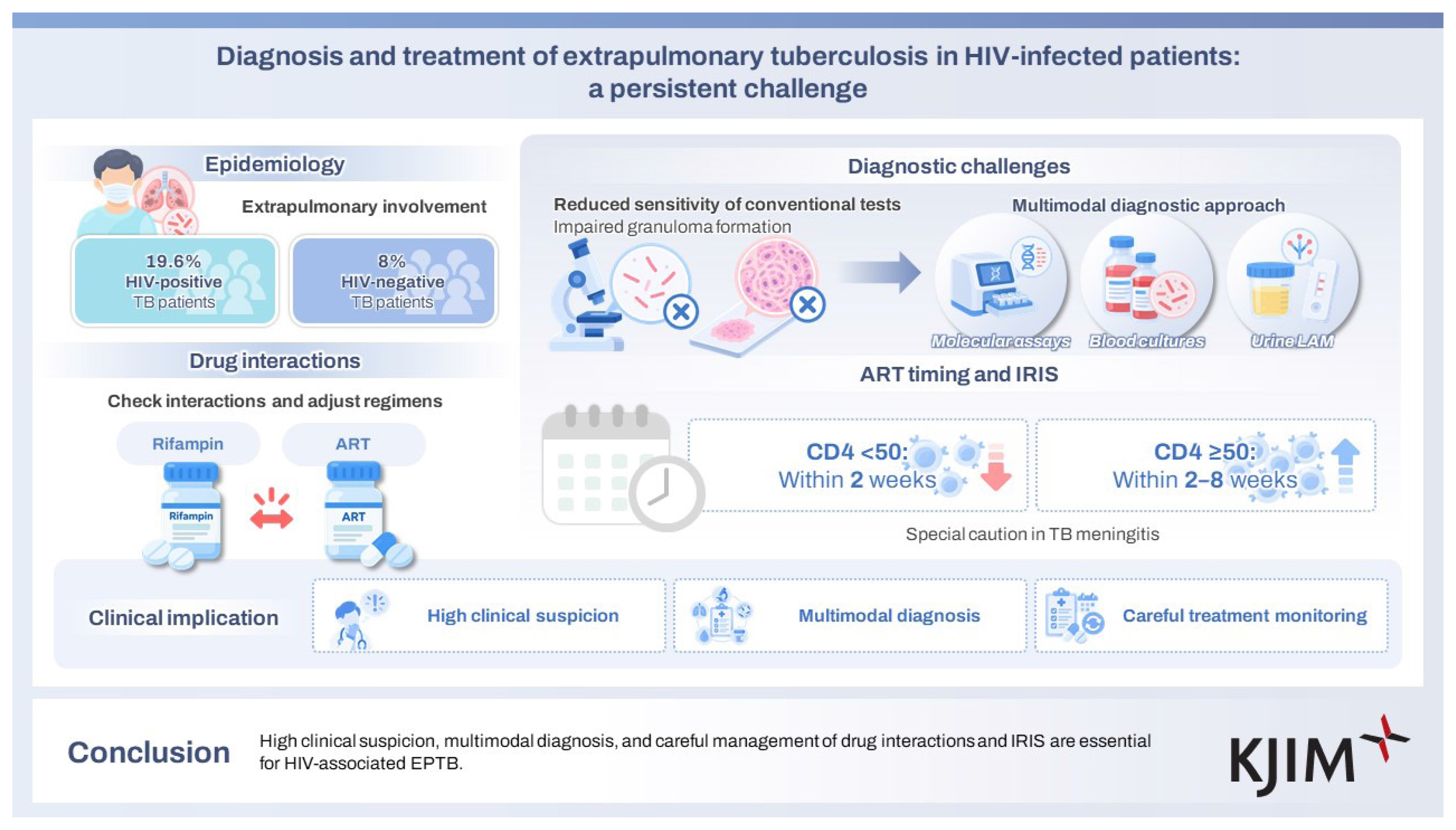
This review summarizes the current knowledge regarding the epidemiology, diagnostic challenges, and therapeutic considerations of extrapulmonary tuberculosis (EPTB) in HIV-infected patients. Individuals with advanced HIV infection are particularly vulnerable to EPTB and disseminated disease; extrapulmonary involvement is observed more frequently than in non-HIV-infected populations. Diagnostic evaluation is complicated by the reduced sensitivity of smear microscopy and histopathology due to impaired granuloma formation. Therefore, molecular assays, blood cultures, and urine lipoarabinomannan testing may serve as useful adjuncts. Therapeutic management requires careful attention to drug–drug interactions between rifamycins and antiretroviral agents, as well as the risk of immune reconstitution inflammatory syndrome and timing of initiating antiretroviral therapy. Although many uncertainties remain, maintaining a high index of suspicion and adopting a multifaceted diagnostic and therapeutic approach may improve outcomes in patients with HIV-associated EPTB.
INTRODUCTION
Extrapulmonary tuberculosis (EPTB) is an opportunistic infection that occurs in individuals with AIDS. This review provides practicing clinicians with a comprehensive understanding of EPTB in the context of HIV infection. Specifically, it addresses the epidemiological characteristics, diagnostic strategies, fundamental principles of treatment, and the critical considerations required during therapeutic management. By synthesizing the current evidence and clinical experience, this study aimed to enhance the capacity of healthcare professionals to recognize, diagnose, and effectively manage EPTB in HIV-infected patients.
EPIDEMIOLOGY
According to the Joint United Nations Programme on HIV/ AIDS (UNAIDS), the global incidence of new HIV infections has declined since approximately 1996 [1]. In contrast, data from the Korea Disease Control and Prevention Agency (KDCA) indicate that a reduction in newly confirmed HIV cases in Korea has only been observed since 2020 [2]. However, it is essential to note that the HIV/AIDS statistics reported by the KDCA are based on the number of confirmed cases rather than the total number of infections; therefore, these statistics require cautious interpretation [3–6].
Globally, in 2024, approximately 5.8% of incident tuberculosis cases occurred in people living with HIV [7]. Moreover, tuberculosis is the most common AIDS-defining opportunistic infection worldwide, with approximately 30% of cases presenting with EPTB [8,9]. Patients with advanced immunosuppression frequently develop EPTB, including disseminated tuberculosis, as their CD4 cell counts decline [10–14]. Regarding the most common sites of EPTB, lymphatic involvement is common in both HIV-infected and HIV-uninfected individuals. However, disseminated disease and central nervous system or meningeal involvement occur relatively more frequently in HIV-infected patients, whereas pleural involvement is proportionally more common in patients without HIV infection [10,15,16].
In Korea, tuberculosis accounts for 10–25% of all opportunistic infections, and together with pneumocystis pneumonia and candidiasis, it represents one of the most frequent opportunistic diseases among individuals with HIV [17–20]. According to a study by Lee et al. [21], 19.0% of HIV-infected patients with tuberculosis in Korea had extrapulmonary involvement, a proportion notably higher than the 8.0% observed in non-HIV-infected individuals. However, the study by Lee et al. was relatively small-scale and was conducted among patients enrolled between 2001 and 2005, which limits its applicability in capturing more recent trends. According to nationwide data released by the KDCA, approximately 22% of tuberculosis cases identified between 2020 and 2024 were extrapulmonary [22]. However, this study did not provide separate statistics regarding the HIV infection status, thereby limiting its ability to assess the overall burden of HIV-associated EPTB. Given that the proportion of EPTB is higher among HIV-infected patients than among HIV-uninfected individuals [10,12–14], HIV-associated EPTB can be regarded as a clinical scenario commonly encountered in practice. It should also be noted that pulmonary tuberculosis and EPTB frequently occur simultaneously in HIV-infected patients [23–25].
Nevertheless, with the widespread implementation of antiretroviral therapy (ART), the incidence of opportunistic infections, including tuberculosis, has markedly declined compared to that in previous decades [8,9,26,27].
Opportunistic infections, including tuberculosis, are also associated with failure of ART and therefore warrant careful attention [28,29].
DIAGNOSIS
The diagnostic principles of EPTB in HIV-infected individuals are largely similar to those in non-HIV-infected populations. However, several important points must be considered.
First, the sensitivity of smear microscopy can be reduced in HIV-infected patients (Table 1). Even in non-HIV-infected individuals, EPTB often exhibits a paucibacillary nature; this inherently lowers the sensitivity of smear testing [30]. Additionally, the HIV-associated impairment of cell-mediated immunity leads to the formation of incomplete granulomas. As a result, localized bacillary containment within specific lesions may be limited, particularly in advanced immunosuppression, whereas the risk of systemic dissemination of Mycobacterium tuberculosis is increased. Importantly, immunosuppression may lead to an overall increase in the bacillary burden in HIV-infected patients. However, due to impaired granuloma formation associated with immune deficiency, the bacillary burden within specific lesions may paradoxically be lower. In pulmonary tuberculosis, HIV-infected patients frequently demonstrate a lower bacillary burden than their non-HIV-infected counterparts [31]. Collectively, these observations underscore the need for clinicians to recognize that the sensitivity of smear microscopy in diagnosing EPTB may be further diminished in patients with HIV. Diagnostic strategies should therefore be designed to address these limitations. When acid-fast bacilli are observed on smears, the possibility of a nontuberculous mycobacterial infection should also be considered.

Advantages and disadvantages of diagnostic tests for EPTB
Second, the sensitivity of histopathological diagnosis may be reduced. As noted earlier, in patients with HIV, decreased CD4 T-cell counts and immune dysregulation impair the formation and maintenance of tuberculous granulomas. Consequently, histological specimens often show incomplete or atypical patterns instead of well-structured granulomas. Indeed, lower CD4 counts have been associated with diminished structural integrity of granulomas [32,33]. Therefore, clinicians should be mindful of these limitations when attempting histopathological diagnoses in patients with advanced HIV infection. The absence of the classic granulomatous architecture or caseous necrosis should not be considered sufficient evidence to exclude the possibility of tuberculosis.
Third, molecular diagnostic techniques such as polymerase chain reaction (PCR), Xpert® MTB/RIF (Cepheid, Sunnyvale, CA, USA), Xpert® MTB/RIF Ultra (Cepheid, Sunnyvale, CA, USA), and loop-mediated isothermal amplification should be actively considered. These diagnostic assays can be particularly valuable for assessing specimens such as lymph node aspirates, cerebrospinal fluid (CSF), pleural fluid, ascitic fluid, and tissue biopsies, where the sensitivity of conventional AFB smears is relatively low. However, it is important to note that these tests also have limitations in sensitivity. For example, the sensitivity of Xpert MTB/RIF, which is widely used in Korea, has been reported to be approximately 90% in lymph node aspirates, 70% in CSF, 50% in pleural fluid, and 60% in ascitic fluid. Similarly, PCR assays demonstrate variable sensitivity depending on the specimen type: approximately 40% in lymph node aspirates, 50–100% in CSF, 80–95% in pleural fluid, and 35–75% in ascitic fluid [34–40].
Fourth, drug susceptibility testing should be performed. Although current evidence remains inconclusive, HIV infection may increase the risk of drug-resistant tuberculosis [41,42], and among patients with advanced HIV infection, the likelihood of multidrug-resistant tuberculosis is particularly elevated [43]. Therefore, when managing tuberculosis in HIV-infected individuals, clinicians should proactively employ molecular-based drug susceptibility assays such as Xpert® MTB/RIF (Cepheid, Sunnyvale, CA, USA), Xpert® MTB/RIF Ultra (Cepheid, Sunnyvale, CA, USA), BD MAX™ MDR-TB (Becton, Dickinson and Company, Franklin Lakes, NJ, USA), and Anyplex™ II (Seegene Inc., Seoul, Republic of Korea), in addition to conventional culture-based susceptibility testing.
Fifth, blood cultures can be particularly useful in patients with advanced immunosuppression (e.g., CD4 < 100 cells/ μL) and suspected disseminated diseases. Due to HIV-associated immunosuppression, the likelihood of isolating M. tuberculosis from the blood of HIV-infected patients with tuberculosis is relatively high. In some cases, the organism may be identified exclusively in blood cultures while remaining undetected in specimens from other sites [44,45]. Moreover, repeated blood cultures have been reported to increase diagnostic yield [46].
Recently, a method was developed to detect the lipoarabinomannan (LAM) antigen in M. tuberculosis urine for the rapid diagnosis of tuberculosis. Although urinary LAM assay demonstrates high specificity, its sensitivity remains limited [47,48]. However, in patients with advanced HIV infection (CD4 ≤ 100 cells/mm3), the sensitivity of the assay is relatively higher, particularly for next-generation tests, allowing it to serve as a useful adjunct for rapid diagnosis [49,50]. Nevertheless, although some data suggest potential diagnostic utility for HIV-associated EPTB, direct evidence supporting this application remains limited.
As an adjunct to the diagnosis of EPTB, measurement of adenosine deaminase (ADA) levels in the CSF, pleural fluid, ascitic fluid, synovial fluid, or pericardial fluid may provide supportive information [51–56]. However, ADA levels can also be elevated in certain rheumatological diseases, malignancies such as lymphoma, and some cases of empyema. Moreover, ADA elevation may be less pronounced in HIV-infected patients; therefore, clinicians should interpret these results with caution [57–59].
Although the interferon gamma release assay (IGRA) test may serve as an adjunctive tool in the evaluation of EPTB, its limited sensitivity and specificity, particularly in immunocompromised patients, precludes its use as a standalone diagnostic test. Thus, IGRA results must always be interpreted in conjunction with clinical findings [60–62].
TREATMENT
The principles and treatment regimens for EPTB in HIV-infected patients are generally similar to those for HIV-negative individuals and HIV-positive patients with pulmonary tuberculosis. In patients with HIV-TB co-infection, two critical issues require special attention: (1) potential drug–drug interactions (DDIs) between antiretroviral agents and anti- tuberculosis medications, and (2) complications related to immune reconstitution inflammatory syndrome (IRIS).
DDIs are particularly problematic with rifampin. Although rifampin is an essential component of tuberculosis treatment, it is a potent inducer of several drug-metabolizing enzymes, including CYP3A4, CYP2C9, CYP2C19, and UGT. Additionally, rifampin affects drug transporters such as organic anion transporting polypeptides and P-glycoprotein (P-gp). These properties confer high potential for clinically significant drug interactions.
Rifampin does not exhibit clinically significant interactions with nucleoside reverse transcriptase inhibitors (NRTIs). However, rifampin induces P-gp, which reduces the efficacy of tenofovir alafenamide (TAF). However, co-administration of TAF with rifampin has been shown to maintain adequate intracellular concentrations of the active metabolite, tenofovir diphosphate [63]. Therefore, although TAF may be used concomitantly with rifampin when clinically necessary, careful monitoring is required. For similar reasons, TAF may also be administered to patients receiving rifabutin when clinically necessary, albeit with close monitoring [64].
Other agents, including bictegravir, cobicistat-boosted elvitegravir, cabotegravir (both oral and injectable formulations), rilpivirine (both oral and injectable formulations), doravirine, etravirine, protease inhibitors, and lenacapavir, should not be coadministered with rifampin. When rifampin is used, dose adjustments are required; dolutegravir should be increased to 50 mg twice daily and raltegravir to 800 mg twice daily (Table 2) [65,66].

Information on DDIs involving antiretroviral agents continues to evolve as new agents are introduced and additional data become available. For the most up-to-date guidance, clinicians can refer to the “HIV Drug Interactions” website maintained by the University of Liverpool, as well as the corresponding smartphone application (HIV iChart) [67].
In patients not receiving ART at the time of tuberculosis diagnosis, initiating ART concurrently with anti-tuberculosis therapy or within the first few weeks of treatment for tuberculosis increases the risk of IRIS [68]. Conversely, deferring ART until tuberculosis therapy is completed is not recommended as this approach has been associated with increased mortality [69].
Patients with EPTB or disseminated tuberculosis are known to be at relatively high risk of developing paradoxical tuberculosis-associated IRIS or paradoxical reactions following the initiation of ART [70–72]. This phenomenon is presumed to result from the combination of a high mycobacterial antigen burden before treatment and the widespread distribution of lesions, which together trigger excessive inflammatory responses during immune recovery. Lymph node tuberculosis may present with worsening lymphadenopathy, abscess formation, or fistula development, whereas central nervous system tuberculosis can lead to increased intracranial pressure, tuberculoma enlargement, and neurological deterioration, necessitating special caution.
When the peripheral blood CD4 T-cell count is < 50 cells/ mm3, initiation of ART within 2 weeks after starting tuberculosis treatment is recommended. For patients with CD4 T-cell counts ≥ 50 cells/mm3, definitive data regarding the optimal timing of ART initiation are lacking. Nevertheless, given the overall benefit of early ART initiation in individuals with HIV infection, current guidelines recommend starting ART within 2–8 weeks of commencing anti-tuberculosis therapy in such patients [65].
Tuberculous meningitis is a condition in which IRIS poses a serious concern and requires careful management. Initiating ART concurrently with anti-tuberculosis therapy in patients with tuberculous meningitis has been associated with increased mortality [73]. Therefore, to mitigate the risk of life-threatening IRIS, ART should be initiated only after clinical improvement of meningitis has been achieved with anti-tuberculosis therapy, and after at least 2 weeks of anti-tuberculosis treatment [65]. If profound immunosuppression necessitates earlier initiation of ART, close monitoring is essential [74].
IRIS typically develops within the first 3 months of ART initiation. The risk is particularly high in patients with low CD4 T-cell counts, a high mycobacterial burden, or when ART is started shortly after the initiation of anti-tuberculosis therapy [70,71,75,76]. Mild IRIS is often managed symptomatically with nonsteroidal anti-inflammatory drugs and careful observation. In contrast, severe IRIS may require treatment with corticosteroids (such as prednisolone, 1 mg/ kg daily) with gradual tapering as clinical improvement occurs [72,77].
In patients with advanced HIV infection, opportunistic infections may present with absent or mild symptoms, owing to profound immunosuppression. Following the initiation of ART, restoration of immune function can lead to the emergence of clinical manifestations of previously unrecognized opportunistic infections, a phenomenon referred to as unmasking IRIS. Therefore, when new signs suggestive of infection appear within the first few months of ART, the possibility of unmasking IRIS should be considered.
Another important consideration is the possibility of drug-resistant tuberculosis. IRIS is characterized by clinical deterioration despite the administration of appropriate therapy; however, a lack of improvement may also occur when first-line anti-tuberculosis agents are ineffective owing to drug resistance. In practice, distinguishing between these two scenarios is challenging. Therefore, in HIV-infected patients with tuberculosis co-infection, drug susceptibility testing using both molecular and conventional methods should be performed proactively.
CONCLUSIONS
EPTB is a major complication in HIV-infected patients. In this population, establishing a diagnosis of EPTB can be challenging because the histopathological findings or smear results for M. tuberculosis may be inconclusive. Therefore, clinicians should maintain a high index of suspicion for EPTB, even in the absence of definitive diagnostic findings. When managing these patients, careful attention must be directed toward potential DDIs, the risk of IRIS, and the possibility of drug-resistant tuberculosis.
Notes
CRedit authorship contributions
Jihwan Bang: conceptualization, writing - review & editing
Conflicts of interest
The author discloses no conflicts.
Funding
None