Gastroesophageal reflux disease as a comorbidity in chronic lung diseases: mechanism, clinical impacts and therapeutic uncertainty
Article information

Abstract
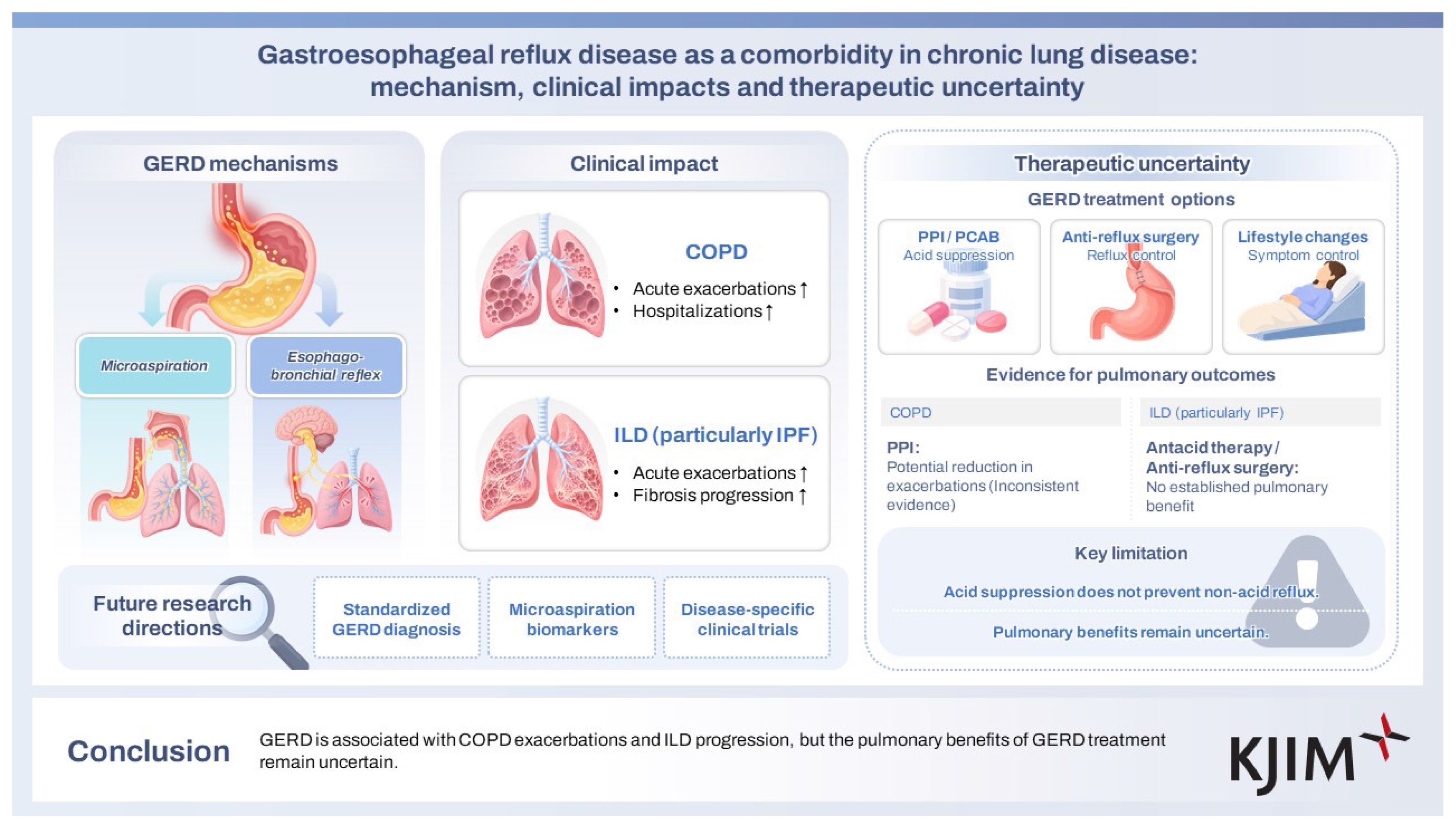
Chronic lung diseases, particularly chronic obstructive pulmonary disease and interstitial lung disease, are major contributors to global morbidity and mortality. Gastroesophageal reflux disease (GERD) is highly prevalent in these populations and has been implicated in exacerbations and disease progression. This review examined recent guideline statements for these diseases, as well as observational, physiological, and interventional studies evaluating the association between GERD and chronic lung diseases. The prevalence of GERD in chronic lung diseases has been reported to be as high as 87%. Plausibly, GERD contributes to respiratory morbidity through microaspiration and neurogenic mechanisms. Although associations with acute exacerbations and disease progression are consistent, high-quality evidence demonstrating that GERD treatment modifies long-term pulmonary outcomes remains limited. Future research priorities include standardized diagnosis and classification of GERD phenotypes and their impact on lung disease, validated noninvasive biomarkers of microaspiration, and rigorously designed trials targeting reflux mechanisms with disease-specific endpoints.
INTRODUCTION
Chronic lung diseases, particularly chronic obstructive pulmonary disease (COPD) and interstitial lung disease (ILD), are major contributors to global morbidity and mortality. COPD is characterized by persistent airflow limitation [1,2], whereas ILD or interstitial pneumonia encompasses a group of disorders that cause inflammation or fibrosis of the lungs [3,4]. Both diseases significantly impair lung function [5,6], reduce quality of life [7,8], and are associated with acute exacerbations, placing a heavy burden on healthcare systems.
Gastroesophageal reflux disease (GERD) is a common comorbidity in patients with chronic lung diseases. GERD results from the retrograde flow of stomach acid into the esophagus, causing symptoms such as heartburn and regurgitation [9]. GERD has been observed to negatively impact the disease course in patients with COPD and ILD. Microaspiration of gastric contents into the airways is believed to link GERD with respiratory pathophysiology.
The relationship between chronic lung diseases and GERD is multifaceted, with shared mechanisms such as inflammation [10], microaspiration [11], and neural reflexes [12] contributing to exacerbations of both COPD and ILD. GERD has been reported to increase the frequency of acute exacerbations in patients with COPD [13,14]. In ILD, GERD has been associated with progression of pulmonary fibrosis, particularly in idiopathic pulmonary fibrosis (IPF) [11,15].
Although the prevalence of GERD in patients with chronic lung diseases is high, the clinical benefit of acid suppression therapy on respiratory outcomes remains controversial, representing a significant unmet clinical need. In this review, we focus on the relationship between GERD, COPD, and ILD. We summarize the current evidence regarding epidemiology, pathophysiological mechanisms, clinical consequences, and therapeutic implications, and we highlight the major limitations of existing studies and priorities for future research.
COPD
Definition, types & causes
COPD is characterized by persistent respiratory symptoms and airflow limitation resulting from airway and/or alveolar abnormalities [2]. Smoking, biomass exposure, and occupational exposure to dust or smoke are associated with increased COPD risk [16]. Genetic susceptibility, impaired lung development, and environmental exposures across the life course also contribute to COPD development [18–20].
Clinical features
The typical respiratory symptoms of COPD are caused by persistent airflow limitation and pathological changes. The most common symptoms include dyspnea, chronic cough, sputum production, and exercise intolerance [1]. In advanced disease, COPD causes systemic manifestations such as weight loss, skeletal muscle dysfunction, and cardiovascular comorbidities [20]. Patients with emphysema may display hyperinflation, decreased breath sounds, and prolonged expiration on physical examination, whereas those with chronic bronchitis may display coarse crackles, wheezing, or rhonchi. Symptoms are often slowly progressive [1]. Patients with COPD may present with acute exacerbations, which are characterized by acute worsening of respiratory symptoms requiring additional therapy [1,21]. Frequent acute exacerbations are associated with accelerated lung function decline [22] and higher mortality [23].
Diagnosis
The diagnosis of COPD depends on a combination of history, risk factor exposure, and objective demonstration of airflow limitation on spirometry [24]. A diagnosis should be considered in individuals with chronic respiratory symptoms and a history of exposure to risk factors [1,25]. On spirometry, a post-bronchodilator forced expiratory volume in 1 second/forced vital capacity (FVC) < 0.7 confirms airflow obstruction consistent with COPD [1,19,20].
Treatment
The treatment of COPD comprises pharmacologic and non-pharmacologic therapies. Long-acting bronchodilators are the cornerstone of COPD management [1]. Both long-acting muscarinic antagonists and long-acting β2-agonists are recommended for patients with COPD who have significant respiratory symptoms or a history of acute exacerbations [1,2], often in combination [2]. In patients who experience frequent or severe exacerbations, inhaled corticosteroids can be used to reduce exacerbation risk when blood eosinophil counts are increased [1,2,26]. Other pharmacologic options include phosphodiesterase-4 inhibitors [27], macrolides [28], and mucolytics [29]. The most important non-pharmacologic intervention is smoking cessation [30]. Pulmonary rehabilitation improves dyspnea, exercise tolerance, fatigue, and overall health status [31,32]. It is recommended for all symptomatic patients across disease severities [1]. Selected patients with severe, heterogeneous emphysema and hyperinflation may benefit from lung volume reduction by surgery [33] or endobronchial valve placement [34,35].
ILD
Definition & classification
ILD indicates a broad and heterogeneous group of disorders that affect the lung interstitium, alveolar structures, and small airways, leading to a spectrum of inflammatory and fibrotic changes [36]. A recently updated statement from the European Respiratory Society and American Thoracic Society defines the term “interstitial pneumonia” as an umbrella term encompassing approximately 200 rare diseases involving these compartments [4]. The statement acknowledges that the term “ILD” can also be used to refer to these disorders. The updated classification system stratifies diseases primarily according to major histologic and radiologic patterns, such as usual interstitial pneumonia, nonspecific interstitial pneumonia (NSIP), and the newly recognized bronchiolocentric interstitial pneumonia (BIP). In addition, the term desquamative interstitial pneumonia has been replaced by alveolar macrophage pneumonia, and acute interstitial pneumonia has been replaced by idiopathic diffuse alveolar damage [4]. Another crucial classification is based on fibrotic and non-fibrotic presentations. These two forms differ in their prognostic and therapeutic implications [37].
Etiology and risk factors
Historically, ILDs have been categorized as idiopathic or non-idiopathic [38]. Current understanding of ILD etiology considers it a continuum involving genetic susceptibility, environmental exposures, immune-mediated processes, and age-related biological pathways [39]. A major category of ILD etiology involves connective tissue diseases, including rheumatoid arthritis, systemic sclerosis, myositis, and Sjögren’s disease [40–42]. Hypersensitivity pneumonitis (HP) is another major etiologic category caused by inhalational exposure to organic antigens such as molds, birds, or agricultural particulates [43–45]. The newly categorized BIP pattern on high-resolution computed tomography (HRCT) suggests high confidence for HP [4]. Drug-related ILD is associated with various medications, and the list is growing with the increasing use of novel drugs [46]. Environmental and occupational exposures, such as asbestos, silica, metal dusts, and agricultural agents, are also critical etiologic contributors to secondary ILD [23]. Infections and conditions associated with immune deficiency are also recognized causes of ILD, with lymphoid interstitial pneumonia representing a characteristic manifestation [36].
Clinical features
The clinical presentation of ILD is heterogeneous and influenced by the underlying pattern of injury, rate of progression, and associated etiologies [36]. The most common symptoms include progressive exertional dyspnea and persistent cough, which is typically nonproductive [4]. Symptoms often evolve insidiously over months to years, particularly in fibrosing ILDs [47,48]. Non-fibrotic inflammatory patterns, including organizing pneumonia (OP) or non-fibrotic BIP, may manifest with more acute or subacute respiratory and constitutional symptoms, such as fever, fatigue, and malaise [44].
Diagnosis
HRCT remains central to ILD evaluation and frequently provides pattern-level diagnostic information. The final diagnosis requires an integrated approach combining clinical history, exposure assessment, serologic evaluation, imaging, and multidisciplinary discussion [49]. A detailed exposure history is crucial for identifying HP and occupational ILD. Serologic testing is essential for evaluating CTD-ILD. Antinuclear antibodies, rheumatoid factor, anti-cyclic citrul-linated peptide antibodies, and myositis-specific antibodies are key elements [4]. When a confident diagnosis cannot be established through clinical, radiological, and serologic evaluation, lung biopsy may be required [49].
Treatment
Management of ILD is guided by the etiology, underlying disease pattern, and presence of progressive fibrosis [37]. In fibrotic ILDs with progression of fibrosis, antifibrotics play an important role. In IPF, antifibrotic therapy with nintedanib [50] or pirfenidone [51] represents the standard of care. In patients with non-IPF fibrosing ILD who exhibit progression of fibrosis, nintedanib has been shown to significantly slow the rate of FVC decline, supporting its use in progressive pulmonary fibrosis (PPF) [52]. In exposure-related and secondary forms of ILD, identification and elimination of the causative exposure remain critical. Avoidance of occupational and environmental antigens is essential in conditions such as HP and pneumoconiosis and may stabilize or improve disease when implemented early [43–45]. Immunosuppression is central to the management of inflammatory and immune-mediated ILDs, including CTD-ILDs, cryptogenic OP, and cellular NSIP. Regardless of the type of ILD, supportive and adjunctive care should also be optimized [42,53,54].
GASTROESOPHAGEAL REFLUX
Definition & pathophysiology
GERD is a chronic condition characterized by the reflux of gastric contents into the esophagus, resulting in symptoms, mucosal injury, or complications [12,55]. It is now understood as a clinical and pathophysiological spectrum rather than a single disease entity [12]. Recent definitions emphasize that GERD is a disorder involving esophagogastric junction (EGJ) barrier dysfunction, reflux burden, and visceral sensitivity [56], with significant heterogeneity across patients [57].
The anti-reflux barrier comprises the lower esophageal sphincter (LES), crural diaphragm, intra-abdominal segment of the esophagus, and geometric configuration of the EGJ [58]. Transient LES relaxations are the most common mechanism in healthy individuals; however, increased frequency or altered triggering of these events can result in excessive reflux episodes [59]. Hiatal hernia structurally weakens the EGJ barrier by altering the pressure gradient between the abdomen and thorax, thereby enabling retrograde flow of gastric contents into the esophagus [60]. Recent findings highlight impaired EGJ contractile integral and delayed EGJ relaxation as key physiological correlates of abnormal reflux [61].
Another aspect of reflux events is esophageal clearance mechanisms, including primary and secondary peristalsis [58]. Ineffective esophageal motility prolongs mucosal exposure to acidic or non-acidic gastric contents. Delayed gastric emptying, increased intra-abdominal pressure, obesity, and dietary triggers further contribute to the pathophysiology [12]. Both acidic and non-acidic reflux and mixed reflux can cause symptoms in patients, including persistent cough [62], impaired mucosal integrity [63], and altered sensory processing [12].
Clinical features
The clinical presentation of GERD is broad and variable, reflecting the heterogeneous causative mechanisms of reflux and esophageal sensitivity [12]. Symptoms have traditionally been divided into typical esophageal symptoms and extra-esophageal manifestations [64]. However, recent phenotype-based frameworks emphasize that symptom profiles do not reliably predict the severity of underlying reflux physiology, thereby reinforcing the need for individualized assessment [12].
Patients with GERD may present with respiratory, laryngeal, or other symptoms even in the absence of typical symptoms [12]. The most common extra-esophageal symptoms include chronic cough, laryngitis, hoarseness, throat clearing, globus sensation, asthma-like symptoms, wheezing, episodic dyspnea, and dental erosions or halitosis [64]. In many patients, extra-esophageal symptoms arise from non-acid reflux, aerosolized refluxate, or esophagobronchial reflex pathways rather than classic acid-mediated mucosal injury [65]. This explains the persistence of symptoms even with high-dose proton pump inhibitor (PPI) therapy and why physiological testing is required to establish a causal relationship [12,66].
Contemporary GERD phenotyping recognizes that patients with similar symptoms may have different physiological abnormalities [16]. Patients with non-erosive reflux disease (NERD) have normal endoscopic findings but demonstrate abnormal esophageal acid exposure associated with symptoms [12,67]. In contrast, patients with reflux hypersensitivity experience typical symptoms triggered by physiological reflux events despite normal acid exposure [12,68]. Patients with functional heartburn have symptoms unrelated to reflux events and typically lack a therapeutic response to acid suppression [12,69]. These distinctions are clinically relevant because treatment strategies vary substantially between phenotypes [12].
Diagnosis
The diagnosis of GERD involves clinical assessment, endoscopic evaluation, and physiological reflux monitoring [66]. Current diagnostic frameworks emphasize that no single test is definitive for all patients; rather, the clinical context and symptom phenotype determine the appropriate approach [12]. Patients with extra-esophageal or respiratory manifestations often require objective testing because symptoms alone are insufficient to establish GERD as an etiology [12].
In patients with typical symptoms of heartburn and regurgitation, in the absence of alarm features, an empirical trial of a PPI is a reasonable initial diagnostic and therapeutic approach [12,55,66]. A symptomatic response suggests a diagnosis of GERD, although placebo effects and overlap with functional disorders limit the diagnostic precision of empirical therapy [70].
The role of upper gastrointestinal endoscopy in the diagnosis of GERD is to identify reflux-related mucosal injury and associated complications [12]. However, most patients with extra-esophageal manifestations have NERD; thus, a normal endoscopy does not exclude pathological reflux [67].
Ambulatory reflux monitoring is crucial for patients with ambiguous, refractory, or extra-esophageal symptoms. The Lyon consensus established thresholds for pathological acid exposure and introduced impedance-based metrics that quantify both acid and non-acid reflux [12]. Conventional pH monitoring is indicated in patients with suspected GERD but no objective evidence on endoscopy, particularly when symptoms are atypical [12]. Impedance-pH monitoring is preferred for evaluating extra-esophageal symptoms such as chronic cough, laryngeal symptoms, asthma, or persistent symptoms despite PPI therapy [71]. This is because it detects weakly acidic and non-acid reflux [12,55]. These features may be relevant in chronic respiratory diseases, where non-acid reflux may play a pathogenic role even in the absence of erosive esophagitis.
Treatment
Treatment strategies encompass lifestyle measures, pharmacologic acid suppression, and endoscopic or surgical interventions [55,66]. Increasingly, therapy is individualized based on the symptom profile, presence of erosive or extra-esophageal manifestations, and objective evidence of pathological reflux [66,71].
Lifestyle modification remains one of the foundations of GERD management [72,73]. First, weight reduction in overweight or obese patients can substantially reduce esophageal acid exposure and improve symptoms [55,66]. Other recommended interventions include avoiding meals within 2–3 hours of bedtime, limiting patient-identified triggers such as alcohol, chocolate, caffeine, carbonated drinks, spicy foods, and high-fat foods, elevating the head of the bed for nocturnal symptoms, and minimizing activities or conditions that increase intra-abdominal pressure [73,74].
PPIs remain the most effective pharmacologic agents for healing erosive esophagitis and controlling typical reflux symptoms [66,75]. They are commonly used as first-line therapy in patients with classic symptoms or endoscopically confirmed GERD [66,72]. However, a substantial proportion of symptomatic patients do not respond completely to PPIs, particularly those with non-acid reflux, reflux hypersensitivity, or functional heartburn [12,57]. H2-receptor antagonists (H2RAs) are less effective than PPIs [76] but may be used in patients with milder symptoms [66] or as adjunctive therapy for nocturnal acid breakthrough [77]. Alginate-based formulations, which form a non-systemic floating gel barrier, can reduce postprandial reflux and regurgitation [78,79] and may be particularly helpful in patients with troublesome regurgitation or during pregnancy [80,81].
Emerging pharmacologic therapies increasingly aim to overcome the limitations of PPIs, particularly their slow onset of action, which requires approximately three days to reach steady state, incomplete acid suppression, and inter-individual variability in efficacy [82]. Potassium-competitive acid blockers (PCABs), including vonoprazan, tegoprazan, and the more recently introduced fexuprazan, are new additions to the pharmacologic management of GERD [83]. PCABs directly and reversibly inhibit the gastric H+/K+-ATPase, producing rapid, potent, and sustained acid suppression independent of parietal cell activation [82].
Vonoprazan is the most extensively evaluated PCAB, with multiple randomized trials demonstrating superior or non-inferior healing of erosive esophagitis, especially in patients with severe disease [73,84]. Early real-world and clinical data suggest potential benefits for patients with PPI-refractory GERD or those with rapid CYP2C19 metabolism, as PCABs provide more predictable pharmacologic effects [55].
Anti-reflux surgery is an important therapeutic option in patients with documented GERD who have severe esophagitis, large hiatal hernias, and/or persistent symptoms [66]. Laparoscopic Nissen fundoplication remains the gold standard surgical treatment [17,75], providing durable symptom relief when appropriate candidates are selected [75].
Magnetic sphincter augmentation has emerged as an effective alternative that reinforces the LES while preserving the physiology of the reflux barrier and improving quality of life [85,86]. Endoscopic interventions such as transoral incisionless fundoplication [74,87] and radiofrequency energy delivery [88] may benefit select patients [72,75].
RELATIONSHIP BETWEEN GERD & CHRONIC LUNG DISEASES
Epidemiology of GERD in chronic lung diseases
Epidemiologic studies demonstrate a higher prevalence of GERD in patients with chronic lung diseases compared with the general population [13,89], although the prevalence varies widely depending on the diagnostic criteria used [12]. In patients with COPD, the prevalence of GERD is estimated to range between 17% and 78% [14]. The ECLIPSE study, a large international observational cohort study of more than 2,164 patients with COPD, identified GERD as one of the most common comorbidities, present in 29% of patients [90]. A nationwide study in Korea using the Health Insurance Review and Assessment database found that the prevalence of GERD was significantly higher in the COPD group than in age- and sex-matched controls, with an odds ratio of approximately 1.4 [89].
GERD is prevalent in ILD, particularly IPF, in which GERD is one of the most frequently identified comorbidities [91,92]. In a prospective study including 65 patients with IPF, 87% demonstrated abnormal esophageal acid reflux on 24-hour pH monitoring [92]. More recently, a population-based case-control study found that patients with IPF had 1.78 times higher odds of GERD compared with matched controls [93].
Association with disease development: causation vs. correlation
Epidemiologic studies consistently demonstrate a high prevalence of GERD in patients with chronic lung diseases. However, the directionality and causality of this association remain inconclusive [94,95]. Physiological changes due to chronic lung diseases may themselves promote reflux. In COPD, hyperinflation of the lungs leads to flattening of the diaphragm, which compromises the sphincter-like function of the crural diaphragm, predisposing patients to reflux events [96]. In fibrotic lung diseases, reduced lung compliance causes stronger negative intrathoracic pressure during inspiration, which facilitates the reflux process [97].
Conversely, a potential role for GERD in the development of chronic lung diseases has been suggested by various studies. Population-based cohort analyses have shown that individuals with GERD have a higher subsequent risk of developing COPD compared with matched controls without GERD [13]. Similarly, reflux symptoms often precede the diagnosis of IPF, suggesting a temporal relationship [97]. A recent large-scale bidirectional Mendelian randomization study demonstrated that genetic susceptibility to GERD was causally associated with an increased risk of IPF after adjustment for confounders [95]. Despite these findings, residual confounding factors, surveillance bias, and shared risk factors, such as smoking, obesity, and medication use, continue to limit definitive conclusions regarding the causal relationship between GERD and chronic lung diseases [94,95,98].
The bidirectional relationship between GERD and chronic pulmonary diseases is summarized in Fig. 1.
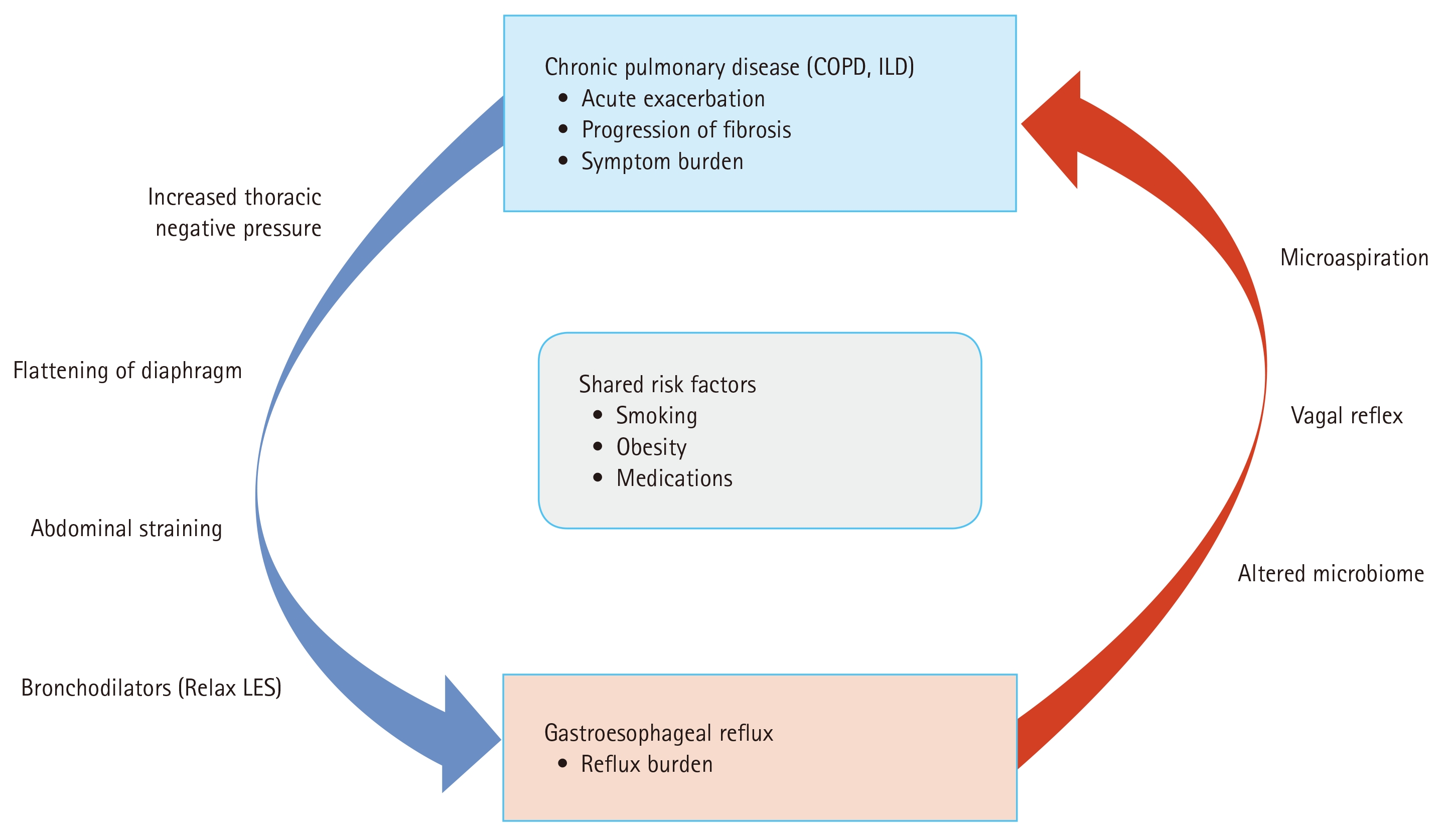
Bidirectional relationship between GERD and chronic pulmonary diseases. GERD, gastroesophageal reflux disease; COPD, chronic obstructive pulmonary disease; ILD, interstitial lung disease; LES, lower esophageal sphincter.
Mechanisms linking GERD to lung injury
The most direct and extensively studied mechanism by which GERD contributes to lung injury is microaspiration of gastric contents [92,99]. Microaspiration refers to the repeated, often subclinical entry of small volumes of gastric or esophageal contents into the lower respiratory tract, in contrast to overt aspiration [100]. The aspirated material includes acid, pepsin, and bile acids [101]. These components cause direct chemical injury to the lung parenchyma, disrupting tight junctions, impairing mucociliary clearance, and activating immune responses [102]. Pepsin, produced exclusively in the stomach, can be detected in bronchoalveolar lavage fluid and is considered a specific biomarker of gastric aspiration [103]. Pepsin exposure has been linked to alveolar epithelial injury, surfactant dysfunction, and profibrotic pathways, suggesting a potential role in the development or progression of pulmonary fibrosis [97,103].
Microaspiration may also alter the pulmonary microbiome, leading to dysbiosis [104]. The concept of the gut–lung axis has emerged, whereby reflux-related changes in pulmonary microbial ecology trigger chronic airway inflammation and accelerate fibrotic remodeling in susceptible individuals [105].
In addition to microaspiration, respiratory symptoms can be caused by vagally mediated esophagobronchial reflex pathways. Embryologically and anatomically, the esophagus and bronchial tree share common vagal innervation, providing a neuroanatomic basis for reflex-mediated respiratory effects of esophageal stimuli [106]. Acid exposure in the distal esophagus can activate vagal afferent nerves, triggering bronchoconstriction and cough via central vagal pathways without refluxate entering the airway [106,107]. Human physiological studies using intraesophageal acid perfusion have demonstrated measurable bronchoconstriction and increased airway responsiveness [108], supporting the existence of this reflex mechanism. Furthermore, activation of these neural pathways can lead to the release of neuropeptides such as tachykinins in the airways, resulting in neurogenic inflammation, mucus secretion, and heightened airway responsiveness [109]. Collectively, these observations suggest that vagal reflex mechanisms may contribute to the respiratory manifestations of GERD.
Impact of GERD on acute exacerbations and disease progression
In COPD, GERD has been reported as an independent predictor of frequent acute exacerbations. A prospective study demonstrated that patients with COPD and reflux symptoms were more likely to experience exacerbations (relative risk, 6.55; 95% confidence interval, 1.86–23.11; p < 0.001) compared with those without reflux symptoms [110]. Multiple observational studies and meta-analyses have also consistently reported associations between GERD and higher frequencies of acute exacerbations and hospitalization rates compared with those without GERD [13,14,111]. GERD has also been linked to worse health-related quality of life, with increased chronic cough and nocturnal respiratory symptoms in COPD [13,112,113].
Similarly, GERD has been associated with acute exacerbations of IPF. Acute exacerbation refers to an acute, clinically significant respiratory deterioration with new widespread alveolar abnormalities [114] that may also occur in non-IPF ILDs [115]. Microaspiration is hypothesized to be one of the triggering factors for acute exacerbations [15]. A study analyzing bronchoalveolar lavage fluid detected a significant elevation in pepsin levels in patients with acute exacerbations compared with those with stable disease [103]. GERD has also been associated with more rapid decline in FVC and shorter radiological fibrosis-free survival [103,116].
Effects of GERD treatment on lung diseases
Evidence regarding the effects of PPIs on COPD outcomes remains mixed. A single-blind randomized trial in older patients with COPD, which excluded patients with GERD, reported fewer exacerbations with lansoprazole than with usual care [117]. In the prospective PROMISE-COPD cohort, patients with GERD receiving PPIs had a higher risk of severe exacerbations than patients without GERD who were not receiving PPIs, although this observational comparison could not establish the effect of PPI treatment itself [118]. Conversely, a Taiwanese retrospective cohort found that higher-dose PPI exposure was associated with lower risks of exacerbation and mortality than lower-dose exposure among patients with COPD and symptomatic GERD [119]. An analysis of 104,439 patients from South Korean reimbursement data demonstrated a decreased risk of exacerbation with PPI treatment [120]. A meta-analysis of 15 randomized controlled trials reported that PPI therapy was associated with fewer acute exacerbations and lower mortality rates, although the trials included in the analysis were limited by small sample sizes and variable diagnostic criteria for GERD [121]. On the other hand, results regarding PPI therapy for COPD remain mixed. PPI treatment was associated with a reduction in moderate exacerbations, but the effects on severe exacerbations were less consistent [13]. Concerns regarding an increased pneumonia risk with PPIs also persist, as this issue has not been extensively studied in the COPD subgroup [122,123]. Under these circumstances, current guidelines do not recommend the routine use of PPIs to reduce exacerbation risk in COPD [1,20]. Key studies are summarized in Table 1.

GERD-directed treatment and acute exacerbations of COPD
In ILDs, particularly IPF, the role of antacid therapy has been controversial in the absence of large randomized controlled trials. A landmark study in 2011 analyzed a retrospective cohort of patients with IPF and comorbid GERD and reported that GERD medication use was associated with longer survival and less radiologic fibrosis [116]. A subsequent post-hoc analysis of three clinical trials evaluating antifibrotic treatment for IPF reported slower FVC decline and less frequent acute exacerbations with GERD treatment [15]. Based on these results, regular antacid therapy was conditionally recommended for the treatment of IPF in an international clinical practice guideline [124]. However, subsequent post-hoc analyses of similar randomized controlled trials of other antifibrotic agents failed to reproduce these findings and suggested an increased risk of infection [125]. An analysis of Australian IPF registry data also did not demonstrate benefits of antacid treatment [126]. The effect of surgical intervention for reflux has also been studied in patients with IPF. The WRAP-IPF study, a multicenter phase 2 study comparing laparoscopic anti-reflux surgery with standard care, demonstrated that surgery was safe and showed a trend toward fewer acute exacerbations and deaths but did not meet its primary endpoint of slowing FVC decline over 48 weeks [127]. These studies are summarized in Table 2. As a result, the 2022 update of the international guideline made conditional recommendations against antacid medication and anti-reflux surgery for IPF [42].

GERD-directed treatment and IPF outcomes
LIMITATIONS AND KNOWLEDGE GAP
Despite the epidemiologic link and biological mechanisms connecting GERD with chronic lung diseases, significant knowledge gaps remain. A major limitation across studies has been heterogeneity in the definition of GERD and diagnostic confirmation methods. Studies have not consistently confirmed the presence of GERD using objective reflux testing, particularly impedance-pH monitoring.
COPD and ILD are themselves heterogeneous disease groups, and the impact of GERD may differ according to disease phenotype, severity, exacerbation risk, and the presence of fibrosis. Pulmonary endpoints, such as definitions of acute exacerbations of chronic lung diseases, have not been standardized across studies, making direct comparisons difficult and increasing the risk of inconsistent interpretations. Moreover, definitions of acute exacerbations in COPD [1] and IPF [114] have been updated, and application of new criteria to older data may yield different results.
Another limitation is that previous studies evaluating GERD treatments often grouped H2RAs and PPIs together without addressing differences in their efficacy. Additionally, despite maximal acid suppression with PPIs, non-acid refluxates may still be present and cause persistent lung injury. This persistent mechanical reflux may partly explain the trend toward benefit observed with laparoscopic anti-reflux surgery in patients with IPF. Furthermore, PCABs have not been comprehensively evaluated for their effects on lung diseases. Comparative data between PPIs and PCABs in patients with chronic lung diseases, particularly regarding clinical outcomes and cost-effectiveness, are warranted.
New classes of medication, PCABs, have not been comprehensively evaluated for their effects on lung diseases. Comparative data between PPIs and PCABs in patients with chronic lung diseases, particularly in terms of clinical outcomes and cost-effectiveness, are warranted. Furthermore, despite maximal acid suppression, non-acid refluxates may still be present and cause persistent lung injury. This may partly explain the trend toward benefit observed with laparoscopic surgery in patients with IPF [49].
To address these unmet needs, future research on the development of noninvasive biomarkers of microaspiration would facilitate further investigation into the impact of GERD on the lungs. Validation of markers such as exhaled breath condensates is essential. In clinical trials, GERD should be diagnosed using mandatory 24-hour multichannel impedance-pH monitoring to accurately quantify both non-acid and weakly acidic reflux. More standardized definitions of disease-specific endpoints should be applied. In ILD, studies investigating the relationship between GERD and disease progression have been conducted mostly in patients with IPF. With the new categorization of PPF, which shows a disease course similar to that of IPF, further studies are warranted. Ideally, therapeutic measures that block reflux mechanisms themselves without complications need to be evaluated to determine whether GERD treatment can benefit patients with chronic respiratory diseases.
Notes
CRedit authorship contributions
Jin-Young Huh: methodology, investigation, writing - original draft, writing - review & editing, visualization; Jin Woo Song: conceptualization, methodology, validation, writing - review & editing, supervision, project administration
Conflicts of interest
The authors disclose no conflicts.
Funding
None