Epidemiology of interstitial lung diseases in Asia
Article information

Abstract
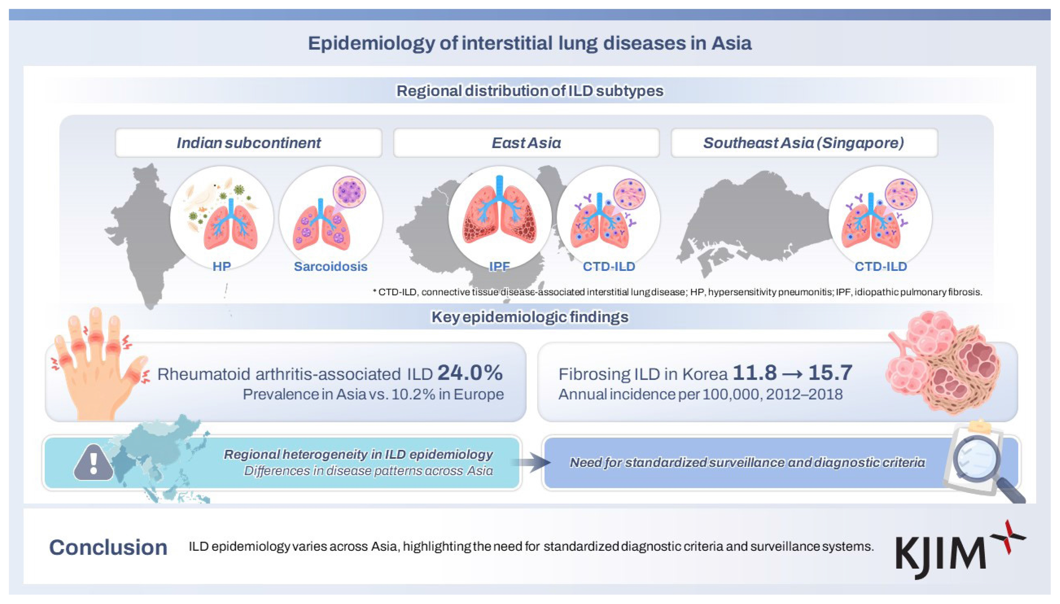
The epidemiology of interstitial lung diseases (ILDs) in Asia has gained increasing attention with the emergence of new population-based datasets and national registries. Current evidence demonstrates that the burden and subtype distribution of ILDs vary considerably across regions, reflecting differences in genetic susceptibility, environmental or occupational exposure, and healthcare infrastructure. IPF is the commonest subtype of ILD in Europe and North America. However, Asian epidemiologic data suggest a more heterogeneous distribution, including hypersensitivity pneumonitis and sarcoidosis across the Indian subcontinent, and connective tissue disease-associated ILD among East Asians, comprising substantial disease burden. Although these studies provide important insights, they also reveal substantial methodological heterogeneity, including variable diagnostic code-based disease ascertainment, inconsistent case definitions, and limited availability of longitudinal follow-up. Environmental risk factors unique to Asian settings remain poorly characterized, and molecular or genomic data are sparse despite the fact that regional differences in disease phenotype and severity are evident. This review synthesizes and critically appraises the available epidemiological evidence on major ILD subtypes across Asia, summarizes emerging trends, and highlights key sources of variability in reported incidence and prevalence.
INTRODUCTION
Interstitial lung diseases (ILDs) represent a heterogeneous group of diffuse parenchymal lung disorders characterized by varying degrees of inflammation and fibrosis [1]. Although their individual incidence is low, ILDs collectively impose a substantial disease burden, contributing to significant morbidity, mortality, and healthcare utilization worldwide [2]. Idiopathic pulmonary fibrosis (IPF), connective tissue disease-associated ILD (CTD-ILD), hypersensitivity pneumonitis (HP), and sarcoidosis are the most prevalent and clinically significant subtypes [3]. Over the past two decades, epidemiological studies provided insights into the incidence, prevalence, and natural history of ILDs, particularly in Western populations [4]. Analyses of large-scale population-based studies and national registries in North America and Europe have provided robust estimates and informed healthcare planning [5,6]. However, extrapolating these findings to Asian populations is limited by several factors. Asia is home to more than half of the global population, with genetic heterogeneity [7], environmental exposure to biomass fuel [8,9], avian antigens [10] and occupational dust [11], and heterogeneous healthcare systems and diagnostic practices [12,13]. These factors likely shape both the burden and subtype distribution of ILDs in ways that are distinct from those in Western cohorts.
Asian databases [13–15], hospital registries [16], and collaborative initiatives [12] have expanded population-level ILD data; however, non-uniform diagnostic practices and coding inconsistencies exist. This review synthesizes the current evidence on IPF, HP, CTD-ILD, and sarcoidosis in Asia, highlighting regional differences and knowledge gaps to guide future research.
ILD IN ASIA: HOW DO WE COMPARE WITH THE REST OF THE WORLD?
Previous data on the relative frequency of ILD subtypes suggest that IPF and sarcoidosis are the most common worldwide. IPF accounted for 11.5–38.6% and 22.5–51.9% of all ILD cases in Europe and North America, respectively, while sarcoidosis accounted for 14.9–42.6% of ILD cases in Europe [2]. In contrast, HP and sarcoidosis predominated in Asia, accounting for 47.3% and 42.2% of ILD cases, respectively, except in Pakistan and China, where IPF was most prevalent [2]. However, most North American and European data were analyzed between 1988–2013 [2], prior to the 2011 IPF guidelines, which refined the diagnostic criteria to emphasize the necessity of a multidisciplinary approach involving clinicians, radiologists, and pathologists [17]. In contrast, epidemiology of ILD in Asia evolved between 2007–2018, and it remained unclear whether the shift in emphasis from histopathological to radiological diagnosis affected the relative incidence of ILD subtypes. An important limitation of the Asian data between 2007–2018 was that most centers were of South Asian (Indian subcontinent) origin, namely India and Pakistan, with only two East Asian centers (both from China) [2]. Recent data on ILD epidemiology in Southeast Asia suggest that CTD-ILD is the most common subtype, accounting for up to 52% of total ILD cases (Fig. 1) [18]. In contrast, CTD-ILD accounted for 7.2–12.8% of total ILD cases in Europe and North America, except France (17.1%) and one Canadian center (33.1%) [2]. Similarly, one center in Australia reported that CTD-ILD accounted for 17.7% of total ILD cases. Asians represent 19.3% and 17.4% of Canadian [19] and Australian [20] populations, respectively. This suggests a genetic predisposition for the East Asian population to develop CTD-ILD.
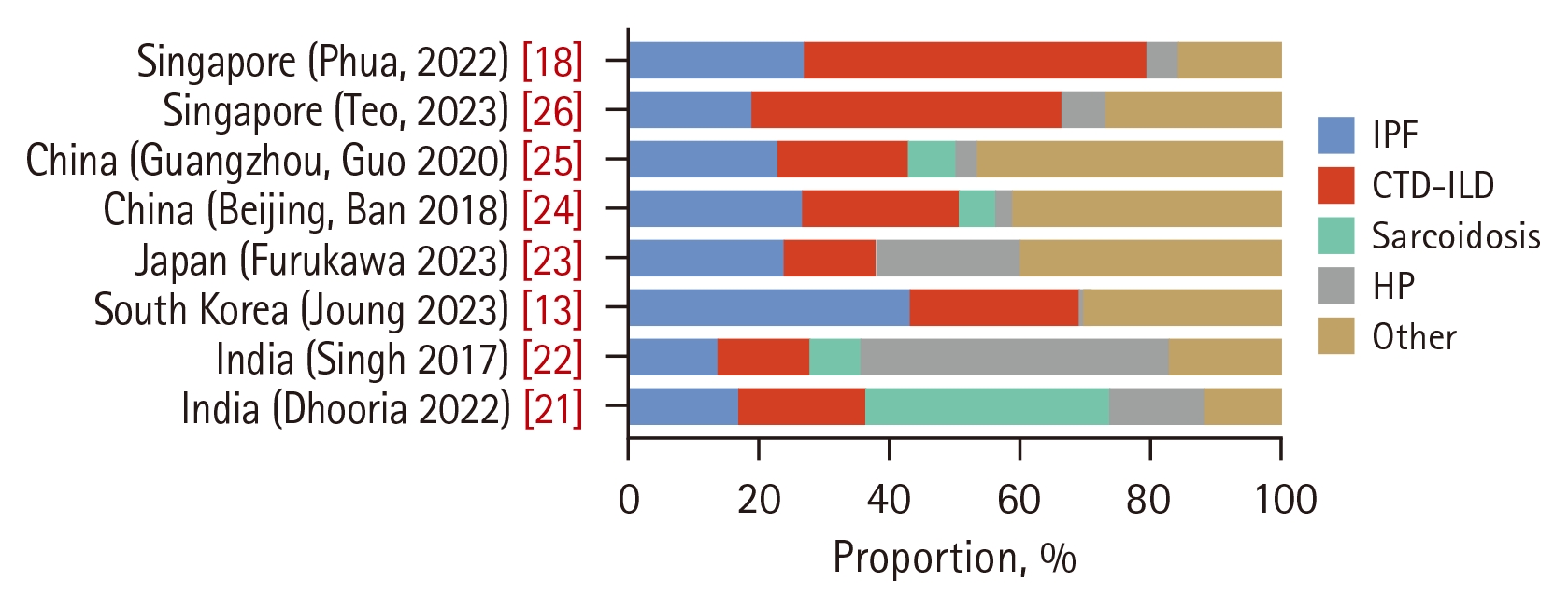
Distribution of ILD subtypes in Asian cohorts. Relative proportions of IPF, CTD-ILD, sarcoidosis, HP, and other ILDs across cohorts from India, South Korea, Japan, China (Beijing and Guangzhou), and Singapore. Values were normalized to 100% for each cohort. “Other” conditions include unclassifiable ILD, nonspecific interstitial pneumonia, cryptogenic organizing pneumonia, acute interstitial pneumonia, respiratory bronchiolitis/desquamative interstitial pneumonia, lymphoid interstitial pneumonia, pulmonary alveolar proteinosis, lymphangioleiomyomatosis, pulmonary Langerhans cell histiocytosis, and drug-induced ILD. For harmonized comparison across cohorts, interstitial pneumonia with autoimmune features and other infrequent or unclassified ILD entities were grouped into the “Other ILD” category. ILD, interstitial lung disease; IPF, idiopathic pulmonary fibrosis; CTDILD, connective tissue disease-associated ILD; HP, hypersensitivity pneumonitis.
DISTRIBUTION OF ILD SUBTYPES ACROSS ASIA
The distribution of ILD subtypes varied substantially across Asia (Fig. 1). In India, two recent cohorts showed a predominance of sarcoidosis and HP [21,22]. Sarcoidosis accounted for 37.3% and HP for 14.4% of total ILD cases in the Dhooria cohort (n = 2,005) [21], whereas the Singh registry (n = 1,084) reported an even higher proportion of HP (47.3%) [22]. The relative contributions of IPF (13.7–17.0%) and CTD-ILD (13.9–19.3%) were modest compared to exposure-related and granulomatous ILD, reflecting the influence of local environmental and occupational exposures.
In several East Asian cohorts, IPF, unclassifiable ILD, and CTD-ILD constituted the majority of ILD subtypes. A Korean nationwide cohort (n = 19,994) showed that IPF was the most common subtype (43.3%), followed by unclassifiable ILD (29.4%), CTD-ILD (25.8%), and ILD due to external factors (1.1%) [13]. Sarcoidosis and HP were reported to be 0.3% and 0.2%, respectively, in this cohort, which may partially reflect the study design and the case-ascertainment algorithm that primarily captured fibrotic ILD cases. The PROMISE study in Japan, a prospective national ILD registry with a centralized multidisciplinary diagnosis, also demonstrated an overall predominance of fibrotic-ILD cases (n = 2,741) [23]. Unclassifiable ILD was the most common subtype (24.9%), followed by IPF (23.7%), HP (22.1%), and CTD-ILD (13.8%) [23]. Distinctively, HP accounted for a considerably larger proportion of cases in this study than in the Korean nationwide cohort. Chinese cohorts displayed additional variability. In the cohort from Beijing (n = 2,615), IPF (26.5%) and CTD-ILD (24.1%) were the major subtypes, with smaller contributions from sarcoidosis (5.6%) and HP (2.4%) [24]. The Guangzhou cohort (n = 1,945) similarly demonstrated notable proportions of IPF (20.3%) and CTD-ILD (18.3%), along with a broader distribution of non-IPF idiopathic interstitial pneumonia, interstitial pneumonia with autoimmune features (IPAF), and rare ILDs. These entities were grouped as “other ILD” category, resulting in a relatively larger proportion (46.8%) compared with other cohorts [25].
Data from Singapore further illustrate how referral patterns may shape apparent subtype distributions. In a retrospective cohort of patients with ILD referred to a tertiary healthcare center at the National University Hospital, Singapore (2016–2022; n = 238), CTD-ILD accounted for 47% of the total cases, followed by IPF (19%), HP (7%), and IPAF (8%) [26]. These proportions reflect referral bias (personal communication with the author), with patients suspected of developing autoimmune or complex ILD phenotypes preferentially referred to specialized ILD services, while typical IPF cases may be managed by general respiratory physicians outside such clinics rather than representing population-level prevalence.
Taken together, the ILD subtype distribution in Asia shows clear regional patterns. India is characterized by a higher proportion of sarcoidosis and HP, whereas East Asian cohorts demonstrate substantial contributions from IPF and CTD-ILD. Interestingly, the Singapore cohort reported a predominance of Indian ethnicity in patients with fibrotic HP (70%), despite accounting for only a minority of the population [26,27]. This suggests a predisposition for Indians to develop HP beyond environmental factors.
POPULATION-BASED EPIDEMIOLOGY OF ILD IN ASIA
Population-based estimates from Asian countries showed considerable geographical variation in ILD burden (Table 1). Nationwide data from India (n = 2,005) suggested an overall ILD incidence of 10–20 per 100,000 person-years and a prevalence of 49–98 per 100,000 population during 2015–2020 [21], indicating a substantial ILD burden that may have been underestimated in earlier reports. In South Korea, nationwide data (n = 19,994) demonstrated a steady increase in fibrosing ILD, with incidence rising from 11.8 in 2012 to 15.7 in 2018 per 100,000 person-years and prevalence increasing from 18.8 in 2011 to 39.0 in 2018 per 100,000 population [13]. In Hong Kong, long-term nationwide data (n = 5,924) showed a declining standardized incidence (from 5.4 to 2.6 per 100,000 person-years), but a gradual increase in prevalence (from 23.1 to 32.6 per 100,000 population) between 2005 and 2020, which may be due to cumulative case accrual and longer disease duration [14].

Population-based epidemiologic studies of ILD in Asia
Model-based estimates from the Global Burden of Disease (GBD) project also mirror the geographic contrasts observed in real-world data [28,29]. In the GBD 2019 study, high-income Asia Pacific countries, including Japan, South Korea, and Singapore, showed high age-standardized ILD prevalence, with estimates exceeding 100 cases per 100,000 population (Japan, 162.6; South Korea, 102.2; Singapore, 109.1) [28]. China had a lower estimate (35.6 cases per 100,000 population), and several Southeast Asian countries, including Indonesia, Malaysia, the Philippines, Thailand, and Vietnam, reported even smaller burden, ranging from 10.5 to 23.9 per 100,000 population [28]. India occupied an intermediate position, with an estimated prevalence of 73.6 per 100,000 population [28]. Although these values rely on models rather than directly observed data, the overall pattern is consistent with population-based findings and underscores pronounced regional heterogeneity in the ILD burden across Asia.
IPF
IPF is diagnosed based on the guidelines of the American Thoracic Society (ATS), European Respiratory Society (ERS), Japanese Respiratory Society (JRS), and Latin American Thoracic Association (ALAT) [30,31]. Diagnosis requires the exclusion of known causes of ILD and the identification of usual interstitial pneumonia (UIP) using high-resolution computed tomography (HRCT) or histopathology. When a probable UIP pattern is present on HRCT, diagnosis is made through multidisciplinary discussion. Population-based studies from Japan, South Korea, and Taiwan have consistently reported an increasing incidence and prevalence of IPF over time, although the absolute estimates vary substantially (Table 2). This increase likely reflects improved recognition of IPF, the adoption of the 2011 and 2018 international guidelines [17,32], population ageing, and the introduction of reimbursement for antifibrotic therapy, which may have encouraged IPF-specific coding [33]. When compared with Western populations, the overall burden of IPF in Asia appears broadly comparable to that in Europe but lower than that in North America, with pooled incidence estimates of 4.4 per 100,000 person-years in Asia, 5.1 in Europe, and 9.0 in North America, and prevalence rates of 14.8, 14.6, and 27.2 per 100,000 population, respectively [34].

Population-based incidence and prevalence of IPF in Asia
The reported estimates depend largely on the case definition and coding system. Broad case definitions based on diagnostic codes alone capture fibrotic ILD with limited specificity for IPF, whereas narrow definitions require additional evidence from imaging, bronchoalveolar lavage, lung biopsy, or specialist assessment. In South Korea, Gjonbrataj et al. [35] reported an annual incidence of 48.5 per 100,000 population when all ILD codes (J84) were included, 16.2 when the analysis was restricted to ILD with fibrosis (J84.1), and 1.7 when the IPF-specific code (J84.1A) was applied. In Taiwan, the standardized incidence of IPF increased approximately sevenfold, from 1.66 to 11.35 per 100,000 population, and the standardized prevalence increased more than tenfold, from 1.98 to 27.25 per 100,000 population, between 2011 and 2019 [33]. This increase partly reflects changes in case definitions after the 2011 guidelines and the transition from ICD-9-CM to ICD-10-CM coding in 2016, which introduced an IPF-specific code (J84.112) [33]. In Japan, a claims-based analysis estimated the prevalence of IPF at 27 per 100,000 population [36]. In Hong Kong, the ICD-9-CM codes 515 and 516.3 had overlapping nomenclature, and the incidence of IPF could not be ascertained separately and was therefore estimated for IIP as a group [14]. Most nationwide estimates end around 2019, and data covering the period after the COVID-19 pandemic and the 2022 guideline update [30] remain limited. A recent Korean nationwide study extended the observation period to 2023 and reported that the crude incidence and prevalence of IPF among adults aged ≥ 50 years increased from 6.07 per 100,000 person-years in 2011 to 16.86 in 2023 and from 8.91 per 100,000 population in 2010 to 63.78 in 2023, respectively [37]. Because the denominator was restricted to adults aged ≥ 50 years, these estimates are not directly comparable with those based on the total population.
HP
HP is an exposure-related ILD diagnosed according to the established ATS and American College of Chest Physicians guidelines, which emphasize the identification of a causative agent together with compatible radiologic findings, supported by bronchoalveolar lavage or histopathology when necessary [38–40].
Studies from Asia highlight the diverse environmental exposures associated with HP across the region. In a prospective multicenter study of 380 patients with HP across 27 centers in India, identifiable exposures were documented in 78.9% of cases [41]. Common exposure included air coolers (48.1%), air conditioners (26.3%), birds (21.4%), molds (20.7%), and occupational exposures such as farming and cotton work (5.8%). A Japanese single-center study through a multidisciplinary discussion, identified 334 antigens in total (average 1.9 antigens per case), with avian-related antigens accounting for 47.3%, molds 34.4%, humidifier-related antigens 17.1%, and others 1.2% [42]. Similarly, a Korean single-center cohort of 101 patients with biopsy-proven fibrotic HP demonstrated that microbial exposure (including mold and humid hay) was the most common (49.5%), followed by chemical exposure (17.8%), animal proteins (7.9%), and plant proteins (6.9%), while 14.9% had no identifiable antigen [43]. Collectively, these studies highlight the diverse environmental exposures associated with HP in Asia, with microbial and avian antigens as the frequent identifiable exposures. This heterogeneity may partly explain the variability in reported epidemiological estimates across countries.
Population-based estimates of HP in Asia are limited, with only South Korea and Japan reporting nationwide data (Table 3). In South Korea, a claims-based analysis using the Health Insurance Review and Assessment Service (HIRA) database (n = 8,678) demonstrated a declining incidence from 2.16 per 100,000 person-years in 2012 to 1.14 in 2020, whereas prevalence was not reported [44]. In Japan, a nationwide stratified hospital survey (575 responding hospitals) estimated a cross-sectional incidence of 2.5 per 100,000 person-years for fibrotic, and 2.0 for non-fibrotic HP, with prevalence estimates of 6.3 and 3.6 per 100,000 population, respectively [45].

Population-based incidence and prevalence of HP in Asia
Estimates from Asia are broadly comparable with those reported in Western data, with the exception of India, where the estimated incidence and prevalence of HP were higher (1.4–2.9 per 100,000 person-years and 6.2–12.3 per 100,000 population, respectively) [21]. U.S. administrative claims-based analysis reported 1-year cumulative incidence rates of 1.28–1.94 per 100,000 person-years and prevalence up to 2.71 per 100,000 population [46], while a Danish nationwide registry study found an average incidence of 1.16 per 100,000 person-years [47]. Overall, these findings indicate that the epidemiologic burden of HP in Asia is similar in magnitude to that observed in Western populations, with differences reflecting variations in environmental exposures, diagnostic practices, and recognition of occupational risk. In addition, beyond global common exposures (e.g., birds and mold), certain region-specific exposure clusters have been described (e.g., summer-type HP reported in Japan), and variations in exposure ascertainment may contribute to differences in the reported phenotypes and rates [48].
CTD-ILD
CTD-ILD refers to diffuse parenchymal lung disease occurring in patients who fulfill the established classification criteria, with pulmonary involvement identified using HRCT and/or histopathology. Data from Asia across multiple autoimmune diseases consistently show a higher prevalence and more severe phenotype of CTD-ILD than those reported in the Western populations. Systemic sclerosis (SSc), rheumatoid arthritis (RA), idiopathic inflammatory myopathy (IIM), and Sjögren’s syndrome (SjS) demonstrate greater ILD penetrance in East Asia, accompanied by distinctive autoantibody patterns (e.g., higher frequencies of anti-MDA5 and anti-Scl-70 antibodies), higher rates of fibrotic or rapidly progressive (RP) disease, and substantial variation in diagnostic access across countries [49–52]. Although most estimates are derived from hospital-based cohorts, the above mentioned consistent findings highlight the urgent need for standardized ILD screening, harmonized diagnostic criteria, and robust population-based studies to more accurately characterize the CTD-ILD burden in Asia.
Population-based estimates of SSc-ILD in Asia are scarce, with Japan providing the only nationwide assessment to date (Table 4). A claims-based analysis integrating Japan Medical Data Center and Medical Data Vision database, reported an incidence of 1.9 per 100,000 person-years and a prevalence of 13.9 per 100,000 population for SSc-associated ILD, with markedly higher rates among women (25.1 [female] vs. 4.5 [male] per 100,000) and older adults (82.1 [≥ 65 years] vs. 12.4 [< 65 years] per 100,000) [15]. These data represent the first large-scale population-level quantification of CTD-ILD burden in the region.

Population-based incidence and prevalence of CTD-ILD in Asia
Global data on RA-associated ILD corroborates the high autoimmunity-related ILD burden in Asia. A recent systematic review of 33 studies (14,281 patients) reported that the pooled global prevalence of ILD among patients with RA is 21.4%, with higher estimates in South America (24.6%) and Asia (24.0%) than in Europe (10.2%) and North America (18.0%) [49]. Within Asia, the prevalence of ILD among patients with RA ranges 5–17% in Japan and Korea, and 30–40% or higher in several Chinese cohorts, demonstrating substantial regional heterogeneity. Although these studies are not population-based, they consistently show a predominance of fibrotic ILD patterns, particularly UIP (11.0%), compared with nonspecific interstitial pneumonia (6.7%) and organizing pneumonia (1.7%). These studies collectively underscore that RA-ILD, similar to SSc-ILD, represents a major autoimmune-related ILD burden across Asian populations [49].
IIM-ILD burden is disproportionately high in Asia. A global meta-analysis of 34 studies involving 10,130 patients with polymyositis or dermatomyositis reported an overall ILD prevalence of approximately 41%, with the highest pooled estimates in Asian cohorts (~50%), exceeding those typically observed in Europe or North America [50]. Across Asian cohorts, ILD prevalence in polymyositis/dermatomyositis is high (often ~40–70%), with anti-MDA5-associated phenotypes contributing to severe RP-ILD in East Asia [50,51]. A complementary systematic review of 6,058 cases of dermatomyositis/polymyositis estimated the global prevalence of RP-ILD at 8.9%, with multiple Asian cohorts reporting frequencies above the global average (7.3% [Asian] vs. 3.5% [non-Asian]) [51]. This pattern aligns with the regional enrichment of anti-MDA5 positive dermatomyositis in East Asia, a phenotype closely associated with fulminant ILD. Consistent with this biology, short-term mortality in RP-ILD associated with anti-MDA5 positive ILD frequently exceeded 40–60% in Japanese, Korean, and Chinese studies despite intensive immunosuppression [51].
Evidence from a large systematic review (23 studies, 6,157 patients) indicated that the pooled prevalence of ILD among patients with primary SjS was higher in Asia (20%; 95% CI, 12–33%) than in Europe (10%; 95% CI, 6–16%) [53]. Most Asian estimates come from China and Japan, where hospital-based cohorts commonly report ILD in 20–40% of patients, with several Chinese studies exceeding 50%. Japanese cohorts generally report a lower prevalence (~10–30%) than Chinese cohorts, but the rates remain consistently higher than those observed in the European population. Overall, these findings consistently demonstrate that ILD in SjS is more frequent in Asia than in the Western regions, aligning with the broader pattern of higher autoimmunity-related ILD burden across East Asia.
The relatively high ILD burden caused by SjS is noteworthy. It is well known that a significant proportion of patients have features of autoimmunity but do not fulfill the established criteria for CTD-ILD. They may often be classified as undifferentiated CTD-ILD or as a research tool with defined criteria in three domains: IPAF [54]. One criticism of the IPAF criteria is that the SjS syndrome is not included within the clinical domain of IPAF. This may have important implications in the Asian context, where the disease burden of SjS is relatively high.
SARCOIDOSIS
Sarcoidosis is diagnosed based on compatible clinical and radiologic findings with histologic confirmation of non-caseating granulomas after the exclusion of alternative causes, such as infection or malignancy, based on the international guidelines [55].
Recent global estimates suggest substantial geographic variation in the epidemiology of sarcoidosis, with high incidence rates of 10–20 per 100,000 person-years in Western and Northern European populations, exceeding 30–40 per 100,000 in African-American populations. Low incidence rates of sarcoidosis, usually below 2 per 100,000, have been reported across most of Asia [56], with the exception of a recent Japanese claims study [57]. However, considerable heterogeneity remained evident within Asia.
Population-based data remain limited; however, studies have demonstrated meaningful regional differences (Table 5). In Singapore, a single-center cohort extrapolated to the national population yielded an estimated incidence of 0.56 per 100,000 person-years, accompanied by striking ethnic gradients ranging from 0.23 in Chinese to 1.30 in Malays, and 4.57 in Indians [58]. Ethnicity-related differences were also observed in clinical profiles. Chinese patients were more often asymptomatic (57.9%) and had better preserved lung function, whereas Indian patients exhibited more symptomatic disease and accounted for most cases of radiologic progression. These findings suggest that ancestry influences both disease occurrence and phenotype, even within shared environments.

Population-based incidence and prevalence of sarcoidosis in Asia
Beyond the differences in incidence, Japanese cohorts also demonstrate distinct patterns of organ involvement compared to Western populations [59]. In particular, Japanese studies have reported a higher frequency of ocular involvement (50–54% vs. 4–25% in Western cohorts), neurosarcoidosis (6–7% vs. 2–4%), and cardiac sarcoidosis (20–23% vs. 2–5%) [57,59–62]. In contrast, cutaneous involvement also appears to be less frequent (11–35% vs. 34–70%) in Asian cohorts than in Western populations [59–61,63]. Taken together, these findings indicate that the clinical manifestation of sarcoidosis in Asia is quantitatively and qualitatively distinct, likely reflecting a combination of genetic susceptibility, immune response patterns, and environmental exposure.
Epidemiological estimates of the relative disease burden and attributable mortality of Asian sarcoidosis suggest mild disease, but are limited. Many studies lacked detailed information on disease subtype or stage and did not clearly distinguish between fibrotic and non-fibrotic pulmonary involvement [57,58,61,62,64–66]. In the Singapore series, 74.4% of pulmonary sarcoidosis cases were stage 0 or 1 [58]. In an Indian series by Madan et al. [67], most (47.7%) patients had stage 1 sarcoidosis. Limited evidence from a study involving 437 patients with pulmonary sarcoidosis from Finland and 457 from Japan followed up for 5 years, reported chest X-ray normalization within 1 year in 46% of patients from Japan compared to 16% of patients from Finland, suggesting that geographic differences may be involved in the prognosis of pulmonary sarcoidosis [68].
Taken together, the available data suggest that the burden of fibrotic sarcoidosis is relatively lower in Asian populations than in Western populations; however, this should be interpreted cautiously, as differences in clinical presentation and potential under-ascertainment may influence these estimates.
LIMITATIONS OF CURRENT EVIDENCE ON ILD EPIDEMIOLOGY
This review has some limitations that should be considered when interpreting the findings of previous epidemiological studies conducted in Asia. Diagnostic frameworks for major ILD subtypes have evolved over time and differences in how the updated criteria have been adopted across regions may have resulted in heterogeneous case ascertainment between studies and periods. Particularly, for IPF, successive revisions of diagnostic guidelines [17,32] and evolving interpretations of HRCT and pathological criteria likely influence case ascertainment over time. In support of this, a national survey from Singapore demonstrated substantial differences in diagnostic evaluation practices between ILD- and non-ILD trained physicians, including variability in the consistency of supportive testing, confidence in HRCT interpretation, and inter-rater agreement for radiological and clinical diagnoses [69]. This reflects how real-world diagnostic processes remain heterogeneous, even within a single healthcare system. In addition, much of the available epidemiological evidence in Asia is derived from administrative claims-based databases and medical benefit registries [13,33,35,70–76] in which disease ascertainment is influenced by national reimbursement policies, coding practices, and healthcare system structures. Consequently, the reported incidence and prevalence estimates may not represent fully accurate absolute numbers, but rather reflect patterns of diagnostic recognition within specific policy environments.
FUTURE DIRECTIONS AND RESEARCH GAPS
Despite ongoing epidemiological studies across Asia, substantial knowledge gaps remain, limiting comprehensive understanding and regional harmonization of the ILD burden. First, standardized diagnostic criteria and coding algorithms for ILD subtypes are urgently required to reduce heterogeneity across national claims databases and registries. This is particularly relevant for subtypes such as fibrotic HP and CTD-ILD, where diagnostic challenges overlap with other fibrotic ILDs, along with variability in case ascertainment and coding practices. This variability may contribute to differences in epidemiological estimates. Second, longitudinal population-based cohorts with systematic follow-up are rare in Asia, restricting their ability to evaluate the natural history, progression trajectories, and survival trends. Third, environmental and occupational exposures unique to Asia remain incompletely characterized. Integrating geospatial exposure metrics, occupational surveillance, and molecular allergen profiling (e.g., antigen-specific IgG panels and proteomic identification of environmental antigens) into ILD cohorts would strengthen the causal inference. Fourth, the genetic and immunological determinants of ILD subtypes appear to differ across Asian populations; however, large-scale genomic, epigenomic, and multi-omics studies remain sparse and largely limited to single countries. Finally, access to anti-fibrotic therapy, immunomodulatory agents, and multidisciplinary ILD care varies widely across Asia, highlighting the need for health-system level research to evaluate disparities in diagnosis, treatment, and outcomes.
CONCLUSION
Emerging epidemiological data from Asia reveal that the burden, subtype distribution, and clinical expression of ILDs across the region differ substantially from Western populations. While IPF and CTD-ILD predominate in East Asia, sarcoidosis and HP predominate in the Indian subcontinent. CTD-ILDs across diverse autoimmune diseases consistently demonstrate a higher prevalence and severe phenotype in East Asian cohorts. These regional differences reflect a complex interplay among genetic susceptibility, environmental exposure, healthcare access, and methodological variations in case ascertainment. Despite the notable progress enabled by national claims databases, hospital registries, and multinational collaborations, significant gaps persist, including inconsistent diagnostic coding, limited population-based data for several ILD subtypes, and limited longitudinal follow-up data capable of defining the natural history and outcomes. Strengthening harmonized surveillance systems, improving diagnostic standardization, and expanding the prospective multicenter research infrastructure will be essential to accurately characterize ILD epidemiology in Asia and inform tailored clinical and public health strategies for this increasingly important group of diseases.
Notes
CRedit authorship contributions
Hee-Young Yoon: conceptualization, methodology, resources, investigation, data curation, formal analysis, validation, software, writing - original draft, writing - review & editing, visualization; Felicia S. W. Teo: conceptualization, investigation, data curation, supervision, project administration; Ju Hyun Oh: conceptualization, methodology, writing - review & editing; Jin Woo Song: conceptualization, resources, writing - review & editing, visualization, supervision, project administration, funding acquisition
Conflicts of interest
The authors disclose no conflicts.
Funding
This study was supported by Bio & Medical Technology Development Program (NRF-2022M3A9E4082647) of the National Research Foundation of Korea (NRF), funded by the Ministry of Science and ICT, Republic of Korea; the National Institute of Health Research Project (2024ER090500); and the Korea Environment Industry & Technology Institute through the Core Technology Development Project for Environmental Diseases Prevention and Management Program, funded by the Korean Ministry of Environment (RS-2022-KE002197), Republic of Korea.