Concomitant cryoglobulinaemic vasculitis alongside ANCA-associated vasculitis: concurrence or coincidental individual occurrence
Article information

Abstract
Background/Aims
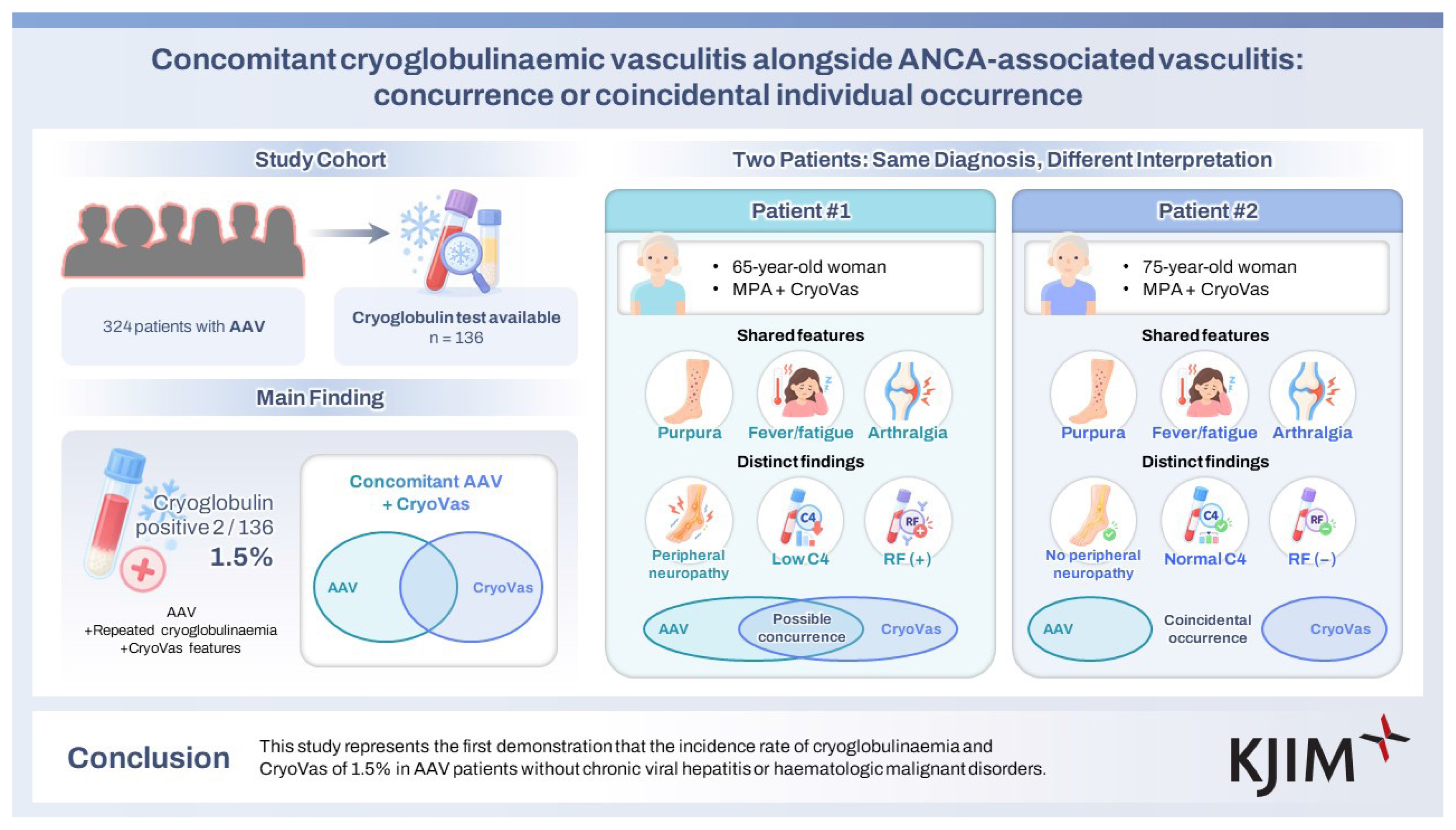
Cryoglobulinaemic vasculitis (CryoVas) and antineutrophil cytoplasmic antibody-associated vasculitis (AAV) share the common characteristic of being small vessel vasculitides but exhibit significant differences in pathologic findings. This study investigated the incidence rate of cryoglobulinaemia and the concurrence of AAV and CryoVas in AAV patients without chronic viral hepatitis or haematologic malignancies.
Methods
Among the 324 patients diagnosed with AAV, 136 with available cryoglobulin test results were included. Their clinical data were reviewed, and clinical and laboratory data, including AAV-specific indices at diagnosis, were recorded. Repeated cryoglobulinaemia was defined as cryoglobulin positivity in sera on at least two occasions separated by an interval of ≥ 12 weeks. The classification criteria for CryoVas were applied to all 136 patients with AAV.
Results
The median age of the patients was 61.0 years (50.0−69.0), and 41.2% were male patients. Of the 136 patients, 75, 31, and 30 were diagnosed with microscopic polyangiitis (MPA), granulomatosis with polyangiitis (GPA), and eosinophilic GPA, respectively. Cryoglobulin was detected in sera of only two patients, yielding a detection rate of 1.5%. Both patients exhibited repeated cryoglobulinaemia and presented with red skin spots, constitutional symptoms, articular involvement, and vascular involvement; enabling their classification as having CryoVas alongside MPA. Additionally, one patient displayed peripheral neuropathy, reduced serum C4 levels, and rheumatoid factor positivity.
Conclusions
This study represents the first demonstration that the incidence rates of cryoglobulinaemia and CryoVas are 1.5% in patients with AAV without chronic viral hepatitis or haematologic malignancies.
INTRODUCTION
Cryoglobulin refers to a group of circulating immunoglobulins (Igs) that precipitate in vitro at temperatures below 37°C and may redissolve at ≥ 37°C. From a laboratory perspective, this term does not denote solely isolated or specified immunoglobulins targeting a defined series of antigens [1]. Cryoglobulinaemia is defined as a medical condition characterised by the presence of cryoglobulin in sera and is classified into three types according to the characteristics of the antibodies: type I, comprising monoclonal IgM, or monoclonal IgG; type II, comprising monoclonal IgM, and polyclonal IgG; and type III, comprising polyclonal IgM + polyclonal IgG. Types II and III are categorised as mixed cryoglobulinaemia due to their composition of varying mono- or polyclonal IgM and polyclonal IgG [1,2]. In most cases of mixed cryoglobulinaemia, rheumatoid factor (RF) constitutes the IgM fraction exhibiting binding affinity for the Fc fragment of IgG [3]. Type I cryoglobulinaemia is frequently associated with haematological diseases, whereas, type II and III are commonly linked to infectious and inflammatory conditions [1,2].
Most patients with cryoglobulinaemia remain asymptomatic; however, a small proportion may infrequently present with diverse systemic symptoms, including cryoglobulinaemic vasculitis (CryoVas) [4]. CryoVas, a small vessel vasculitis, is characterised by vasculitis with cryoglobulin immune deposits in capillaries, venules, or arterioles, and associated with cryoglobulins in sera [5]. This condition predominantly affects three major organ systems: the skin, kidneys, and peripheral nervous system [6,7]. By contrast, antineutrophil cytoplasmic antibody (ANCA)-associated vasculitis (AAV) is also a small vessel vasculitis, distinguished by fibrinoids in predominantly small vessels and occasionally medium-sized arteries, and is associated with ANCAs in sera [5]. Although AAV and CryoVas share similarities in the size of affected vessels, their histological features differ markedly. Unlike CryoVas, AAV primarily exhibits few or no immune deposits in affected tissues, a key feature distinguishing it from Cryo-Vas [8,9]. Consequently, simultaneous occurrence of AAV and CryoVas has been considered unlikely.
Nevertheless, the potential concurrence of AAV and Cryo-Vas warrants reconsideration for two reasons: AAV is an uncommon cause of cryoglobulinaemia [2], and the systemic involvement and clinical manifestations of the two vasculitides are notably similar [6,7,10–14]. To date, few reports have documented ANCA-positive patients with cryoglobulinaemia alongside underlying conditions such as viral hepatitis, or conversely, cryoglobulin-positive patients with AAV [15,16]. However, the concurrence of AAV and CryoVas, or the differentiation of their clinical features, has not been investigated in a substantial cohort of patients with AAV. Therefore, this study examined the incidence of cryoglobulinaemia and concurrence of CryoVas in 324 patients with AAV.
METHODS
Patients
In this study, medical records of 324 patients with AAV enrolled in the Severance Hospital ANCA-associated VasculitidEs (SHAVE) cohort were retrospectively reviewed. Patients were included based on the following criteria: i) initial classification of AAV was established at this hospital by rheumatologists between November 2005 and June 2024; ii) fulfilment of the 1990 American College of Rheumatology (ACR) classification criteria for eosinophilic granulomatosis with polyangiitis (EGPA), the 2007 European Medicine Agency algorithm for AAV, and the 2012 revised international Chapel Hill Consensus Conference nomenclature of vasculitides at diagnosis [5,10]; iii) fulfilment of the 2022 ACR/European Alliance of Associations for Rheumatology (EULAR) classification criteria for AAV at diagnosis [11–14]; iv) availability of well-documented medical records sufficient to collect clinical data at diagnosis; v) availability of the results of ANCA tests performed within 4 weeks before or at diagnosis [17,18]; vi) availability of the results of cryoglobulin tests performed at diagnosis, with additional results from repeated tests approximately 12 weeks later in cases of cryoglobulin-positive patients [6]; vii) follow-up duration of 6 months or longer after diagnosis; viii) absence of concomitant serious infectious diseases or malignancies at diagnosis (including no evidence of hepatitis B or hepatitis C viral infection) [11–13]; and ix) no history of immunosuppressive drug use within 4 weeks before diagnosis.
This study was approved by the Institutional Review Board (IRB) of Severance Hospital (Seoul, Korea; IRB No. 4-2020-1071), and conducted in accordance with the Declaration of Helsinki. Due to the retrospective design of the study and use of anonymised patient data, the requirement for written informed consent was waived.
Clinical data at diagnosis
Demographic including age, sex, body mass index (BMI), and smoking history were collected. AAV-related variables, such as AAV subtype, ANCA type and positivity, AAV-specific indices, and systemic manifestations, were reviewed. Myeloperoxidase (MPO)-ANCA and proteinase 3 (PR3)-ANCA were measured using an immunoassay, whereas, perinuclear (P)-ANCA and cytoplasmic (C)-ANCA were detected using an indirect immunofluorescence assay with ethanol fixation. According to the 2022 ACR/EULAR classification criteria for AAV, both MPO-ANCA/PR3-ANCA and P-ANCA/ C-ANCA were recognised as ANCA results [11–13]. AAV-specific indices included the Birmingham vasculitis activity score (BVAS), and the Five-factor score (FFS) at diagnosis [19,20]. Systemic manifestations were investigated based on the systemic items of BVAS [19]. The results of laboratory tests performed at diagnosis including erythrocyte sedimentation rate (ESR) and C-reactive protein (CRP) levels were also documented.
Detection of cryoglobulin
Whole blood was collected in a plain tube (without an anticoagulant), pre-warmed to 37°C, and immediately centrifuged at 3,500 rpm for 15 minutes at 37°C to separate the serum. The serum was then aliquoted into two tubes: one tube was stored in a refrigerator at 4°C, and the other was incubated at 37°C for 3 days. After this period, the tube stored at 4°C was examined for precipitate or gel formation, compared with the tube maintained at 37°C. The tube from the refrigerator was then returned to the 37°C incubator, and the dissolution of any precipitate was monitored upon warming. Repeated cryoglobulinaemia was defined as cryoglobulin positive in sera on at least two occasions separated by an interval of ≥12 weeks [6,7].
Classification of CryoVas
CryoVas was identified and classified according to the established criteria. Two entry requirements must be met: (i) at least two of the three items (questionnaire, clinical, and laboratory items) must be satisfied and (ii) cryoglobulin must be detected in sera on two separate occasions with an interval of ≥ 12 weeks. The questionnaire item comprised two sub-items regarding skin lesions and a sub-item regarding viral hepatitis, in which at least two of the three sub-items are needed to meet this item. The clinical item includes constitutional, articular, vascular, and neurological sub-items, with at least three necessary to meet this item. The laboratory item encompasses reduced C4 levels, RF positivity, and serum M component positivity, with at least two required to satisfy this item [3,6,7].
Statistical analyses
All statistical analyses were performed using the SPSS version 26 (IBM Corporation, Armonk, NY, USA) for Windows (Microsoft Corporation, Redmond, WA, USA). Continuous and categorical variables were expressed as medians (25 and 75 percentiles), and numbers (percentages).
RESULTS
Selection of patients
Of the 324 patients with AAV, 188 were excluded due to the absence of cryoglobulin testing. Consequently, 136 patients with available cryoglobulin test results were included, and their clinical data were reviewed (Fig. 1).
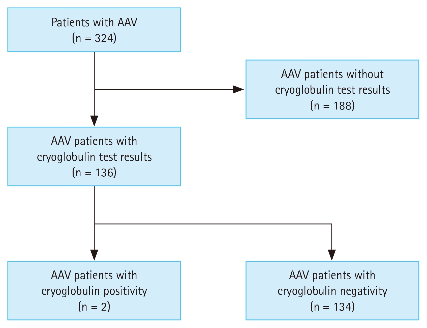
Patients’ selection. AAV, antineutrophil cytoplasmic antibody-associated vasculitis.
Characteristics of patients at diagnosis
Among the 136 patients with cryoglobulin test results, the median age of the patients was 61.0 years (50.0−69.0), and 41.2% were men. The median BMI was 22.6 kg/m2 (20.8−24.8), and only two patients had a history of smoking. Of these 136 patients, 75, 31, and 30 were diagnosed with microscopic polyangiitis (MPA), GPA, and EGPA, respectively. The detection rates of MPO-ANCA (or P-ANCA), and PR3-ANCA (or C-ANCA) were 74.3%, and 14.7%, respectively. The median BVAS was 11.0 (6.0−17.0), and the median FFS was 1.0 (0−2.0). The most frequently observed clinical manifestation was pulmonary manifestation (70.6%), followed by renal manifestation (55.1%). Additionally, the median ESR and CRP levels were measured as 58.0 (19.0−96.0) mm/h, and 8.5 (1.4−48.2) mg/L, respectively (Table 1).
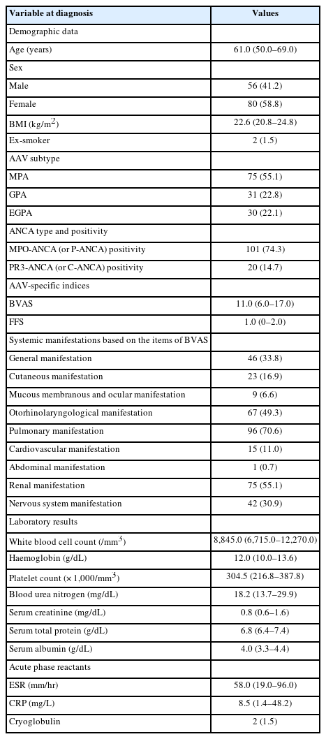
Characteristics of patients with AAV having the results of cryoglobulin tests at diagnosis (n = 136)
Cryoglobulin positivity
Cryoglobulin was detected in the sera of only two of the 136 patients, yielding a detection rate of 1.5% (Table 1).
Classification of CryoVas
The classification criteria for CryoVas were applied to AAV patients exhibiting repeated cryoglobulinaemia [6,7]. Patient #1, a female patient aged 65 years, demonstrated repeated cryoglobulinaemia. She was diagnosed with MPA based on MPO-ANCA positivity, histologically confirmed ANCA-associated glomerulonephritis (GN) and interstitial lung disease. She had red skin spots and experienced discoloration when they disappeared several times before or at diagnosis, which met the questionnaire item of CryoVas. She presented with fatigue, high fever, arthralgia, purpura in her lower extremities, and peripheral neuropathy at diagnosis, all of which met the clinical criteria for CryoVas. Additionally, the patient exhibited reduced C4 levels and RF positivity at diagnosis, meeting the laboratory criteria for CryoVas. Accordingly, patient #1 was classified as having CryoVas alongside MPA (Table 2). Patient #2, a female aged 75 years, also exhibited repeated cryoglobulinaemia. She was diagnosed with MPA based on MPO-ANCA positivity, ANCA-associated GN, and fibrotic lung alterations. She fulfilled the same questionnaire and clinical items as patient #1, except for the absence of peripheral neuropathy. No abnormal findings were observed in the laboratory sub-item. Consequently, patient #2 was also classified as having CryoVas alongside MPA (Table 2). Based on these findings, we conclude that the incidence rate of CryoVas in patients with AAV is 1.5%.
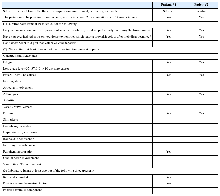
The classification criteria for cryoglobulinaemic vasculitis
DISCUSSION
Given that AAV is an uncommon aetiology of cryoglobulinaemia and that both CryoVas and AAV are classified as small vessel vasculitides despite differing histological features [1,2,5], CryoVas might reasonably be expected to develop in patients with AAV. Consequently, this study investigated the incidence of cryoglobulinaemia and the potential classification of CryoVas among patients with AAV without chronic viral hepatitis or haematologic malignancies. Several notable findings emerged. First, cryoglobulin was detected in the sera of two of the 136 patients with AAV, both exhibiting repeated cryoglobulinaemia, and these patients were classified as having CryoVas alongside AAV, yielding a detection rate of cryoglobulin and an incidence of CryoVas of 1.5%. Second, all patients with AAV exhibiting repeated cryoglobulinaemia met the criteria for CryoVas. Third, all patients classified as having concomitant CryoVas presented with purpuric skin lesions, constitutional (febrile) symptoms, and articular manifestations. Fourth, neurological manifestations and CryoVas-related laboratory findings varied between the two patients. We conclude that cryoglobulin can be detected in sera of patients with AAV and it may critically contribute to the classification of concomitant CryoVas.
The findings raise several critical considerations. The initial question concerns whether AAV or CryoVas primarily accounts for the observed clinical symptoms. Fever, arthralgia, and purpuric lesions in patients classified as having CryoVas alongside AAV play critical roles in the classification criteria for CryoVas [6]. At the same time, they also belong to the items of the BVAS used to assess AAV activity: general and cutaneous manifestations [19]. Nonetheless, these symptoms were deemed more likely associated with CryoVas than AAV, as general and cutaneous manifestations were observed in only 33.8% and 16.9% of patients with AAV, respectively, compared with 100.0% of those with CryoVas in this study. Peripheral neuropathy, observed in only one patient classified as having CryoVas alongside AAV, could not be definitely attributed to either condition, given its prevalence in both AAV and CryoVas [21]. Had a nerve biopsy been performed-similar to the kidney biopsy confirming ANCA-associated GN-it might have aided in distinguishing between the two diseases [22–24]. Additionally, regarding reduced C4 levels and RF positivity, given the relationship between serum C3 levels and ANCA-associated GN rather than serum C4 levels and the direct role of RF in cryoglobulinaemia, it was inferred that these laboratory subitems might be more likely to be associated with CryoVas than AAV [6,25].
The second consideration is whether the observed cases represent concurrence (e.g., overlap syndrome) or coincidental individual occurrences. Per the inclusion criteria, no patients with concomitant malignancies (e.g., monoclonal gammopathies), serious infectious diseases (e.g., chronic viral hepatitis), or autoantibody-related diseases other than AAV were included [1,2,6], thereby minimising confounding factors unrelated to AAV. However, as the mechanism by which CryoVas develops in patients with AAV remains unclear, the following hypotheses are proposed: patient #1 had RF and exhibited low C4 levels at diagnosis. RF may have played a central role in cryoglobulin formation, and ANCA itself, or antibodies other than ANCA generated by immune tolerance impairment due to AAV, may have bound to RF to promote cryoglobulin production. Therefore, patient #1 could be classified as having concurrent AAV and CryoVas because cryoglobulin production might be reasonably inferred. Conversely, patient #2 was RF-negative and had normal C4 levels. Therefore, patient #2 was diagnosed with a coincidental individual occurrence of AAV and CryoVas, since no relationship between the two diseases could be inferred.
The third consideration addresses the need for differential diagnosis between concurrent and coincidental occurrence of AAV and CryoVas in patients with both conditions. This distinction is critical due to differences in treatment strategies, particularly for severe cases. In terms of therapeutic strategies for severe AAV (MPA and GPA), a severe case of MPA and GPA is currently defined as the following cases: diffuse alveolar haemorrhage, rapidly progressive GN (RPGN), central nervous system involvement, peripheral neuropathy, myocarditis, mesenteric ischaemia, and digital ischaemic necrosis [26]. A combination of high-dose glucocorticoids with either rituximab or cyclophosphamide is recommended as the first line [26,27]. Conversely, no consensus exists for severe CryoVas; treatment typically focuses on underlying conditions (e.g., chronic viral hepatitis or haematologic malignancies) and general immunosuppression tailored to affected organs [28,29]. In this study, the two patients with CryoVas and AAV, lacking chronic viral hepatitis or haematologic malignancies, received high-dose glucocorticoids and rituximab for histologically confirmed RPGN associated with MPA rather than CryoVas. Their lung lesions were not severe enough to warrant rituximab as induction therapy. Excluding renal and pulmonary involvement, peripheral neuropathy might have guided treatment decisions: if attributed to AAV, rituximab would be prioritized; if linked to CryoVas, additional options like apheresis or plasma exchange might be considered [30,31]. Considering these circumstances, we believe that in patients classified as having CryoVas alongside AAV and in those with evident symptoms of major organ involvement, biopsy of the affected organ should be actively considered when clinically feasible.
The final consideration is the potential for false-positive or false-negative cryoglobulin test results due to multiple factors: i) if blood collection and sample processing are not maintained at 37°C, cryoprecipitate may form during clotting; ii) inadequate storage of serum at the proper temperature (4°C) after centrifugation may affect cryoprecipitate formation and concentration; iii) serum turbidity, caused by lipemia or fibrin formation during clotting, can interfere with result interpretation; and iv) the presence of immune complexes, which may form cryoglobulins due to inflammatory or infectious diseases, can also lead to inaccurate results [32]. Additionally, in clinical practice, because cryoglobulin testing relies on visual inspection rather than sensitive methods such as spectrophotometry [33], its sensitivity may be reduced when cryoglobulin concentrations are low, thereby increasing the risk of false negatives. However, as repeated cryoglobulinaemia was accepted as cryoglobulin-positive in this study, the likelihood of false results was likely minimised.
This study has several advantages: i) it is the first to quantify the rate of positive cryoglobulin test results and the concurrence of AAV and CryoVas in a cohort of patients with AAV; ii) it also provides a framework for considering concurrent versus coincidental occurrences of these conditions; and iii) it excludes the potential causes of cryoglobulinaemia, such as chronic viral hepatitis or haematologic malignancies, enhancing the focus on AAV-related findings, though not comprehensively. However, several limitations should be acknowledged. First, although the AAV cohort included in this study was the largest and most well-established cohort in Korea, the single-centre design limited patient numbers and institutional diversity. Second, although data collection was conducted prospectively, retrospective analyses introduced risks of missing or inconsistent data. Third, cryoglobulin retesting was performed only in patients with cryoglobulin positivity at diagnosis, potentially missing subsequent positivity in initially negative patients during follow-up. Fourth, the lack of standardised procedures in cryoglobulin testing might have contributed to the insufficient confirmation of the concurrence of AAV and CryoVas. Finally, a nerve biopsy was not performed in patient #1, who presented with symptoms of peripheral neuropathy, as a nerve biopsy would have provided useful information for differentiating between AAV and CryoVas.
In conclusion, this study demonstrated that the incidence rate of cryoglobulinaemia and CryoVas in patients with AAV without chronic viral hepatitis or haematologic malignancies is 1.5%. Although evidence is insufficient to strongly recommend routine cryoglobulin testing for all patients diagnosed with AAV, given the several differences in clinical features, disease progression, prognosis, and treatment modalities between AAV and CryoVas, we would recommend a cryoglobulin test at the time of AAV diagnosis and active biopsy of organs affected by vasculitis, if possible. This is particularly encouraged in patients presenting with purpuric skin lesions, constitutional (febrile) symptoms, and articular manifestations.
KEY MESSAGE
1. Cryoglobulin was detected in the sera of two of the 136 patients with AAV, both exhibiting repeated cryoglobulinaemia, and these patients were classified as having CryoVas alongside AAV, yielding a detection rate of cryoglobulin and an incidence of CryoVas of 1.5%.
2. All patients classified as having concomitant CryoVas presented with purpuric skin lesions, constitutional (febrile) symptoms, and articular manifestations.
3. Neurological manifestations and CryoVas-related laboratory findings varied between the two patients.
Notes
CRedit authorship contributions
J.Y. Whang: Conceptualization, Methodology, Resources, Investigation, Data curation, Formal analysis, Software, Writing - original draft, Writing - review & editing, Visualization, Project administration; J.W. Ha: Conceptualization, Methodology, Resources, Investigation, Data curation, Formal analysis, Software, Writing - original draft, Writing - review & editing, Visualization, Project administration; J. Chung: Methodology, Investigation, Data curation, Formal analysis, Software, Writing - review & editing; Y.B. Park: Conceptualization, Resources, Investigation, Validation, Writing - review & editing; J. Song: Conceptualization, Resources, Investigation, Validation, Writing - review & editing; Y. Park: Conceptualization, Methodology, Resources, Investigation, Data curation, Formal analysis, Software, Writing - original draft, Writing - review & editing, Visualization, Supervision, Project administration; S.W. Lee: Conceptualization, Methodology, Resources, Investigation, Data curation, Formal analysis, Software, Writing - original draft, Writing - review & editing, Visualization, Supervision, Project administration, Funding acquisition
Conflicts of interest
The authors disclose no conflicts.
Funding
This study was funded by Eisai Korea Inc. Seoul, Republic of Korea (4-2024-0700), and Yuhan Corporation, Seoul, Republic of Korea, as part of their “2024 Investigator Initiated Translation Research Program” (4-2025-0044). The funder was not involved in the study design, collection, analysis, interpretation of data, the writing of this article, or the decision to submit it for publication.