Acute gouty arthritis of the atlantoaxial joint
Article information

A 77-year-old man was admitted through emergency room complaining of 3-day history of non-traumatic neck pain. He had a 23-year history of diabetes mellitus. He was also diagnosed with gouty arthritis 7 year earlier; however, he did not take any urate lowering medication. He had a blood pressure 167/90 mmHg, pulse rate 102 beats/min, body temperature 38.1°C, and respiratory rate 19 breaths/ min. Physical examination revealed severe pain with neck stiffness on rotation in any direction. Laboratory results showed a white blood cell count of 12,200/mm3 (reference 4,000–10,000), C-reactive protein of 8.23 mg/dL (reference 0.0–0.5), serum creatinine of 1.63 mg/dL (reference 0.60–1.50) and serum uric acid of 9.1 mg/dL (reference 2.1–7.4). Blood culture showed negative results and cerebrospinal fluid tests (CSFs) were normal. Initial C-spine x-ray showed degenerative change of C-spine (Fig. 1A). Gadolinium-enhanced cervical spine magnetic resonance imaging (MRI) revealed heterogeneously enhancing lesion around odontoid process with bone marrow edema (Fig. 1B). Possible differential diagnosis at this stage included rheumatoid arthritis, pseudogout, infection and tumor. The patient showed no clinical evidence of rheumatoid arthritis; infection was excluded from his blood culture and CSF test. Moreover, no tumorous condition was identified in the MRI. Additional dual-energy computed tomography (DECT) showed erosion and tophi involving odontoid process (Fig. 2). Based on the clinical and imaging findings, a diagnosis of acute gouty arthritis of atlantoaxial joint was established. The patient was treated with steroid (prednisolone 15 mg twice a day). His symptoms and laboratory data dramatically improved.
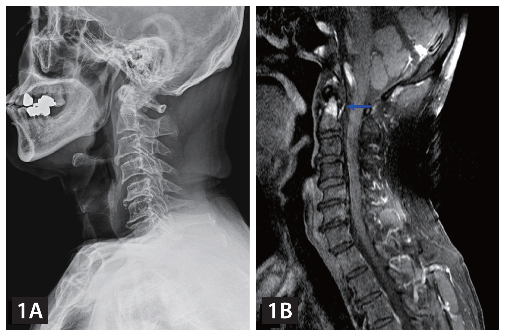
(A) X-ray of cervical spine. (B) Cervical spine magnetic resonance imaging. Heterogeneously enhancing lesion around odontoid process with bone marrow edema (blue arrow).
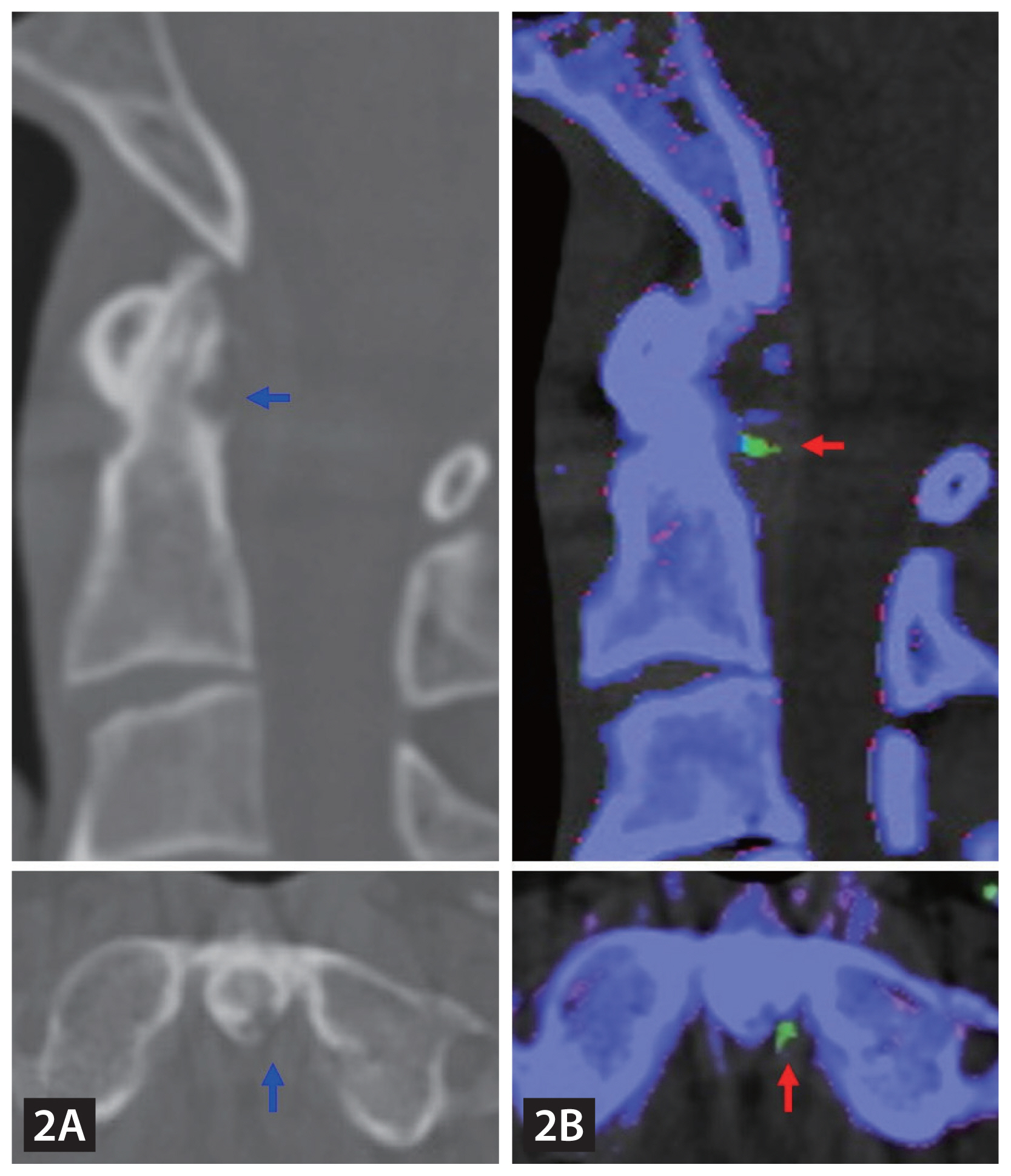
(A) Cervical spine computed tomography (CT). Erosion involving odontoid process (blue arrow). (B) Dual-energy CT. Tophi in posterior aspect of odontoid process (red arrow).
Gouty arthritis is one of the most prevalent form of inflammatory arthritis among men [1]. Cervical spine involvement of gout was rare, but can lead to serious complications such as myelopathy or atlantoaxial subluxation [2]. Gouty arthritis can be confirmed by identification of monosodium urate crystals. However, due to its nonspecific presentation and the deep complex structures, cervical spinal gout is difficult to confirm on traditional imaging modalities. In this respect, DECT has the potential to be a useful tool in identifying spinal gout and avoiding invasive interventions.
Notes
CRedit authorship contributions
Su Jin Choi: methodology, investigation, data curation, writing - original draft, writing - review & editing; Min Wook So: writing - review & editing; Sunggun Lee: writing - review & editing; Seung Won Choi: writing - review & editing; Doo-Ho Lim: conceptualization, resources, investigation, data curation, writing - review & editing, project administration
Conflicts of interest
The authors disclose no conflicts.
Funding
None