Clinical characteristics of male and female Korean patients with systemic lupus erythematosus: a comparative study
Article information
Abstract
Background/Aims
To compare the clinical characteristics and outcomes of systemic lupus erythematosus between male and female Korean patients.
Methods
A retrospective analysis was performed at a single tertiary hospital from August 1994 to May 2010. Male patients were matched with two to three female patients based on age and disease duration. Organ damage was assessed using the Systemic Lupus International Collaborating Clinics/American College of Rheumatology damage index (SLICC/ACR DI).
Results
Fifty-three male patients were compared with 150 female patients. Renal disorders were found more frequently in male patients at disease onset (p < 0.001); the adjusted odds ratio (OR) demonstrated a significant sex preponderance for renal manifestations (OR, 3.26; 95% confidence interval [CI], 1.62 to 6.57). Diffuse proliferative lupus nephritis and end-stage renal disease requiring dialysis during the disease course were more prevalent in male patients (p = 0.025 and p < 0.001, respectively). The risk for requiring long-term dialysis was significantly higher in male than in female patients (OR, 4.02; 95% CI, 1.07 to 15.06), as was the mean SLICC/ACR DI (1.55 ± 1.35 vs. 1.02 ± 1.57, respectively; p = 0.028).
Conclusions
Our data demonstrate that Korean patients with lupus have characteristics similar to those of cohorts reported previously. Male patients had significantly higher incidences of renal manifestations and organ damage.
INTRODUCTION
Systemic lupus erythematosus (SLE) is an autoimmune disease of unknown etiology and is characterized by a multifaceted clinical presentation and disease course. Female preponderance is a well-known characteristic of SLE, particularly during the reproductive years. Sex differences are another peculiarity of SLE with respect to clinical manifestations and outcomes. Previous studies have recognized male patients with lupus as a distinct minority; such patients often exhibit a poorer prognosis, especially involving the renal, cardiovascular, and neurologic systems [1,2,3,4]. Higher mortality rates in male than in female patients with SLE have also been reported [4,5].
The results of these previous studies may not be applicable to all ethnic groups. Koreans are a racially homogeneous population, and few studies have assessed the differences between male and female Korean patients with SLE. However, one study investigated patients with lupus nephritis [6]. Thus, we investigated the key manifestations and outcomes of SLE in male and female patients in Korea.
METHODS
Medical records were reviewed to identify patients with SLE treated from August 1994 to May 2010 at Samsung Medical Center, Seoul, Korea. The diagnosis of SLE was made by experienced rheumatologists based on clinical and laboratory data according to the classification criteria of the American College of Rheumatology (ACR) [7,8]. Each male patient was matched with two to three female patients with SLE according to their age at diagnosis and duration of follow-up. Fifty-three male patients and 150 female patients were included in the final analysis.
All relevant information was obtained, including the time of disease onset, disease duration, clinical features, and medications used. Presenting features were defined according to the ACR classification criteria [7,8]. Autoantibody profiles determined at baseline included antinuclear antibody (by indirect immunofluorescence), antidouble-strand DNA antibody (by radioimmunoassay), and antibodies to extractable nuclear antigens for Smith, ribonucleoprotein, Ro/SSA, and La/SSB (by enzyme immunoassay). Anticardiolipin antibodies and anti-β2 glycoprotein I antibodies (by enzyme-linked immunosorbent assay) as well as lupus anticoagulant (by dilute Russell viper venom test and platelet neutralization) were tested to determine the presence of antiphospholipid antibodies. Regular laboratory tests during follow-up included complete blood cell counts, chemistry profiles, complement levels (C3, normal, 90 to 180 mg/dL; C4, normal, 10 to 40 mg/dL by immunoturbidimetry), antidouble-strand DNA, and urinalysis. Cumulative organ damage was measured using the Systemic Lupus International Collaborating Clinics/ACR damage index (SLICC/ACR DI) [9]. This index was obtained at the last follow-up visit. A kidney biopsy was performed to confirm the presence of lupus nephritis in patients with a urinary protein level of > 0.5 g/day and/or hematuria characterized by ≥ 10 red blood cells per high-power field and/or the presence of leukocytes. The pathological findings were reported according to the 1995 revised World Health Organization (WHO) classification criteria.
Statistical analysis
Differences between groups were compared using Student t test for continuous variables or the chi-square test for categorical variables. Survival was estimated by the log-rank/Kaplan-Meier curve. Odds ratios (ORs) and associated 95% confidence intervals (CIs) were calculated to assess the impact of sex on the disease course. Variables showing p values of < 0.1 in the univariate analysis were selected for further stepwise multivariate analysis. A p value of < 0.05 was considered to indicate statistical significance. All analyses were performed using the PASW version 18.0 (SPSS Inc., Chicago, IL, USA).
RESULTS
In total, 632 patients (57 male, 575 female) with SLE were identified. The mean follow-up duration was 63.4 ± 52.2 months. Most patients were diagnosed before 40 years of age (75.6%) with a peak incidence in the third decade of life (33.8%); this finding did not differ between male and female patients. The survival rates were also comparable between the two sexes: the 5-year survival rate was 90.0% in male patients and 95.9% in female patients, and the 10-year survival rate was 90.0% in male patients and 94.2% in female patients (p = 0.11 for both).
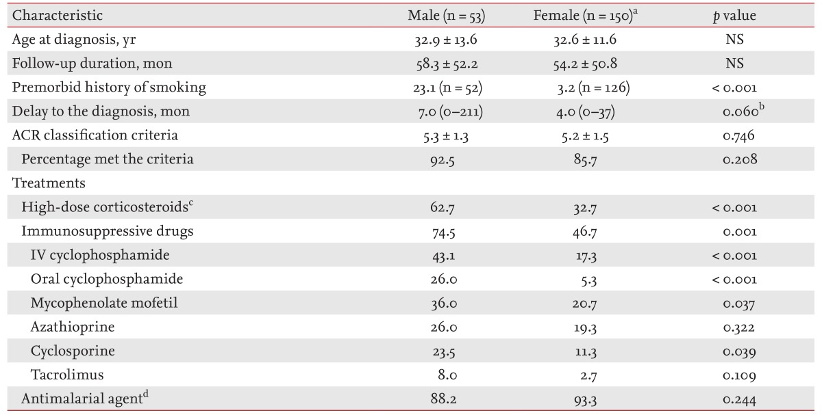
The general characteristics of the groups are shown in Table 1. A history of smoking was more prevalent in male than female patients before the diagnosis of SLE (p < 0.001). The median time between the onset of the first SLE-related symptom and diagnosis was delayed in male compared with female patients, but the difference was not significant (p = 0.06). High-dose corticosteroids, intravenous and oral cyclophosphamide, mycophenolate mofetil (MMF), and cyclosporine were used more frequently in male than in female patients.
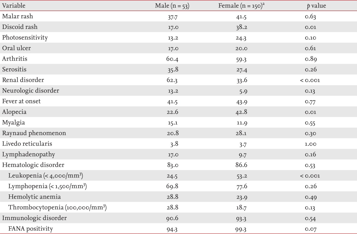
Renal disorder was the most common clinical feature at presentation. This was followed by arthritis and fever in male patients and by arthritis, fever, and alopecia in female patients; the hematologic and immunologic findings were similar in both groups (Table 2). Male patients displayed a lower incidence of discoid rash (17.0% vs. 38.2%, respectively; p = 0.01) and alopecia (22.6% vs. 42.8%, respectively; p = 0.01) while renal disorders were more common in male than in female patients (62.3% vs. 33.6%, respectively; p < 0.001). Male patients had fewer cases of leukopenia (24.5% vs. 53.2%, respectively; p < 0.001) and anti-Ro positivity (21.3% vs. 56.1%, respectively; p < 0.001). Hemolytic anemia and thrombocytopenia were more frequent in male patients, although the difference did not reach statistical significance. No differences were seen between the two groups in the prevalence of malar rash, photosensitivity, oral ulceration, arthritis, serositis, or neurologic disorders or in the other laboratory findings (data not shown). In terms of sex associations, SLE in male patients tended to cause less severe discoid rash (adjusted OR, 0.34; 95% CI, 0.14 to 0.81) and more renal disorders (adjusted OR, 3.26; 95% CI, 1.62 to 6.57) than in female patients. None of the other presenting features were found to be associated with sex (data not shown). Smoking had no association with the presenting features of male patients (data not shown).
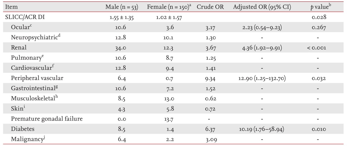
At the last follow-up visit, the mean SLICC/ACR DI was higher in the male than female patients (1.55 ± 1.35 vs. 1.02 ± 1.57, respectively; p = 0.028). Individual items of the SLICC/ACR DI were analyzed to assess the sex impact (Table 3). Male patients were more likely to have renal disorders (adjusted OR, 4.36; 95% CI, 1.92 to 9.91; p < 0.001), peripheral vascular disease (adjusted OR, 12.9; 95% CI, 1.25 to 132.75; p = 0.032), and diabetes (adjusted OR, 10.19; 95% CI, 1.76 to 58.94; p = 0.010). However, other items, including those associated with the cardiovascular and neuropsychiatric systems, did not differ between the two groups (data not shown). Biopsy-confirmed lupus nephritis, including WHO class IV diffuse proliferative lupus nephritis, was more prevalent in males at both presentation and during follow-up. Dialysis was performed more frequently in male than in female patients (18.0% vs. 3.6%, respectively; p = 0.002), and this outcome was associated with the male sex (adjusted OR, 4.02; 95% CI, 1.07 to 15.06).
DISCUSSION
Our data indicate that male patients are more prone than female patients to having renal disorders at presentation and during the SLE disease course; they also have a higher risk for requiring dialysis and a higher index of organ damage.
These data are consistent with those of previous reports showing poorer renal outcomes in male than in female patients (Table 4) [1,3,4,10,11,12,13,14]. Higher mortality rates associated with worse renal outcomes are thought to occur in male patients because renal involvement in SLE is a major determinant of the outcome [4,5]. However, in our male patients with SLE, the survival rates seemed to be lower but statistically comparable despite the fact that they had poorer renal outcomes than did the female patients. This may have been due to the relatively short follow-up period and because renal damage was not the sole determinant of survival. Meanwhile, smoking is a well-known risk factor for the progression of renal disease and the development of proteinuria in the general population, especially males [15,16]. Thus, the more frequent history of smoking in our male patients may have contributed to the worse renal outcomes. We also expected that in a long-term observation of male patients with SLE, survival, including cardiovascular mortality, would be influenced by smoking.

Major characteristics of male lupus in selected studies shown poorer renal outcomes or investigated in Asia
Our data are in accordance with those of previous reports of a lower incidence of alopecia and Raynaud phenomenon and a lower level of anti-Ro positivity in male than in female patients with lupus [1,11,13]. Compared with Asian male patients with lupus in other studies, the Korean male patients with lupus in the present study displayed several notable findings: less frequent discoid rash and leukopenia, a higher incidence of WHO class IV lupus nephritis and dialysis during the follow-up period, and more frequent use of immunosuppressive agents. Discoid lesions are characterized by discrete, erythematous, slightly infiltrated plaques covered by adherent scales on the scalp, face, and neck. Because they are not usually associated with pain or pruritus, patients may under-report their presence; this may have led to the low rate of presentation of such lesions in our male patients. Less sun exposure among Korean male than female patients may also contribute because discoid lesions are highly photosensitive. On the other hand, with respect to renal disorders as the most common presenting manifestations of lupus in male patients and discoid rash as the most common chronic lesion, the diagnosis of SLE in male patients might be achieved by kidney biopsy before the development of discoid skin lesions. Less frequent leukopenia in male than in female patients with lupus was reported in a previous study from Singapore, but the authors did not comment on the potential cause of this finding [11]. The genes and their allelic variants of Fc gamma receptors (FcγR) are regarded as regulators of immune responses, and they may predispose to the development of SLE [17,18]. Moreover, FcγRIIa and FcγRIIIa polymorphisms show some clinically different associations between the sexes, and these associations are also present with respect to ethnicity [19,20,21]. Therefore, a genetic predisposition involving FcγR polymorphisms might be associated with the hematologic findings and renal diseases in our male patients, particularly with respect to the less frequent leukopenia and higher incidence of WHO class IV lupus nephritis. Moreover, dialysis was performed more frequently during follow-up in our male patients despite the fact that their mean age at diagnosis was similar to that of other Asian male patients with lupus. This discrepancy could have been caused by differences in disease durations among studies; in terms of risk factors for the progression of renal disease; however, other contributory factors may include smoking, hypertension, and various factors that were not assessed in this retrospective medical record review. The more frequent use of immunosuppressive agents in our male patients was mostly related to the higher prevalence of renal disease at presentation and during follow-up. Nonclassic immunosuppressive agents such as MMF, cyclosporine, and tacrolimus are now more widely available than in previous eras in which traditional cyclophosphamide was implicitly used. This variability in treatment options might be one of the reasons for the differences among studies.
Surprisingly, cardiovascular and neuropsychiatric manifestations were not commonly observed in male patients. Only three cases of myocardial infarction (one female and two male patients) and five cases of stroke (two female and three male patients) were identified, which limited further statistical analysis. Peripheral vascular disease, including claudication and venous thrombosis, was more common among male patients; however, the correlation with antiphospholipid antibodies could not be assessed because few patients were screened for these antibodies. The sex association with the development of diabetes may have been a consequence of frequent corticosteroid use for more severe disease, including nephritis, in male patients. Moreover, MMF was recently suggested to be an independent risk factor for glucocorticoid-induced diabetes in patients with SLE [22].
The present study had several strengths: homogenous ethnicity, controls matched for both age and follow-up duration, higher incidence of renal disease but comparable survival between groups, and a sex association with the incidence of diabetes.
This study also had limitations. Its retrospective nature limited access to clinical information, preventing comprehensive analysis of data, including disease activity. Neither socioeconomic status nor education level, important prognostic factors for SLE, was included in the analysis. Finally, the SLICC/ACR DI was assessed only once. Thus, it was difficult to assess the change in scores between two time points during the disease course.
In conclusion, SLE in male Korean patients appears to be associated with more severe renal impairment and organ damage than in female Korean patients with SLE.
KEY MESSAGE
This study provides evidence in support of a male association with poor renal manifestation in Korean patients with systemic lupus erythematosus (SLE).
The incidence of organ damage was higher in male than in female patients with SLE, but the survival rate was comparable between male and female patients.
Notes
No potential conflict of interest relevant to this article was reported.


