Bilateral Emphysematous Pyelonephritis
Article information
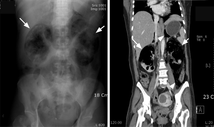
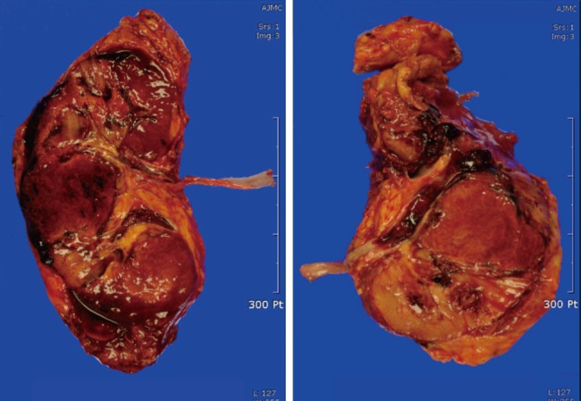
A 46-year-old woman presented to the emergency department with a 1-day history of lethargy and a 7-day history of general weakness. The patient had diabetes mellitus with poor glucose control and degenerative arthritis that had been treated intermittently with steroid medication over the past several years. A plain radiograph revealed diffuse gas formation throughout both kidneys (Fig. 1). Abdominal computed tomography showed air densities in the kidney parenchyma bilaterally, as well as in the collecting system, subcapsular and perinephric spaces, and retroperitoneum (Fig. 1). The patient was treated with empirical antibiotics and underwent a bilateral nephrectomy. Grossly, both kidneys showed diffuse suppurative nephritis with abscess formation, and gas in the necrotic areas, all of which were consistent with emphysematous pyelonephritis (Fig. 2). Escherichia coli was cultured from the patient's blood and urine. The patient was started on hemodialysis and was well at the 1-year follow-up.

Simple X-ray and abdominal computed tomography show large amounts of air throughout both kidneys (arrows) and gas dissecting in the retroperitoneal space (arrowheads).

Examination of the removed kidneys showed the characteristics consistent with emphysematous pyelonephritis.
Notes
No potential conflict of interest relevant to this article is reported.