A case of scar sarcoidosis
Article information
Abstract
Infiltration of sarcoid granuloma in old cutaneous scars is one of the uncommon cutaneous manifestations of sarcoidosis. Here, we report the case of a 47-year-old female who presented with swelling and irritation in 5 old scars. She had acquired these scars 9 years ago in a traffic accident. An incisional scar biopsy revealed noncaseating granulomas consistent with sarcoidosis. High-resolution CT (HRCT) revealed right paratracheal, both hilar, paraaortic, and subcarinal lymphadenopathy without any nodular densities in both lung fields. Successful regression of cutaneous inflammation was achieved using a short course of oral steroids.
INTRODUCTION
Sarcoidosis is a systemic noncaseating granulomatous disorder of unknown origin that involves mainly the lungs, mediastinal and peripheral lymph nodes, eyes, and skin. Cutaneous involvement occurs in 25% of cases1).
Skin manifestations include erythema nodosum, lupus pernio papules, nodules, and plaques. Infiltration of old cutaneous scars with sarcoid granuloma in the active phase of disease, which is known as scar sarcoidosis, is the most clinically characteristic and an uncommon cutaneous manifestation of sarcoidosis2). Some cases of scar sarcoidosis have been reported at the dermatology department of various hospitals in Korea3, 4).
Here, we present a case of a patient who developed scar sarcoidosis in old wounds that were acquired 9 years ago in a traffic accident.
CASE REPORT
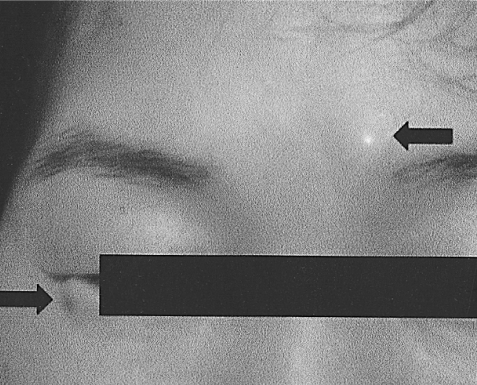
A 47-year-old female presented with swelling and irritation for 10 months in 5 old scars that she had acquired 9 years ago in a traffic accident: 3 scars on the face, 1 on the left knee, and 1 scar on the right leg. Dyspnea, night sweats, weight loss, or any other constitutional symptoms were absent. After incisional skin biopsy of the right leg scar performed at a private clinic, she was referred to our hospital for further consultation. Physical examination findings were normal, except for the presence of nodules at the sites of 5 scars on her lip, eyebrow, eyelid (Figure 1), knee, and leg.
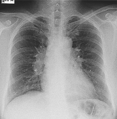
Routine laboratory tests, including white blood cell count and differential cell count, and liver and renal function tests, were normal. Erythrocyte sedimentation rate was 57 mm/h, and serum calcium was 10.8 mg/dL (normal: 8.5-10.8 mg/dL). A chest radiograph showed enlarged hilar lymph node that was suggestive of sarcoidosis (Figure 2). Pulmonary function tests revealed a normal ventilatory pattern with FVC, 2.99 L (99% of predicted); FEV1, 2.34 L (102% of predicted); FEV1/FVC, 78%; and diffusing capacity of the lung for carbon monoxide, 96% of predicted.
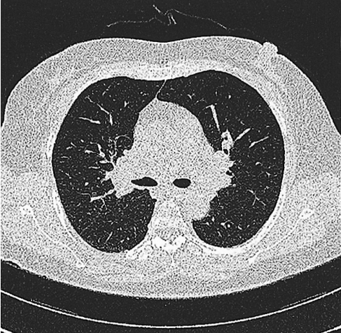
High-resolution CT (HRCT) revealed right paratracheal, both hilar, paraaortic, and subcarinal lymphadenopathy without any nodular densities in both lung fields (Figure 3). Serologic tests showed that both C-reactive protein and RA (rheumatoid arthritis) tests were negative. Antinuclear antibody was weakly positive, and serum angiotensin converting enzyme (ACE) level was 40 IU/L (normal: 18-55 IU/L). Sputum for acid-fast bacilli and tuberculin skin test were negative. Ophthalmoscopic examination, including a slit-lamp study, revealed normal findings.
Biopsy specimens from the eyebrow and knee scars showed a noncaseating epitheloid granuloma (Figure 4); this finding was similar to the result of biopsy performed at the private clinic. Detection of acid-fast bacilli was negative in the biopsy sample. The patient refused to undergo bronchoscopy. Scar sarcoidosis was diagnosed accompanying the involvement of hilar and mediastinal lymph nodes. The patient was initially treated with topical steroid, which did not cause any changes in the cutaneous lesion. After a short course of oral methylprednisolone (24 mg per day for 4 weeks and gradually reduced over the next month), the visible inflammation in the scars regressed. She remained stable without recurrence for a follow-up period of 12 months. However, hilar lymphadenopathy on chest radiography persisted.
DISCUSSION
Sarcoidosis is a multisystem disease of unknown etiology characterized by the formation of noncaseating granulomas in the affected organs. Skin sarcoidosis occurs in 25% of cases1), and the majority of patients present with lupus pernio, maculopapular eruption, subcutaneous nodules, and erythema nodosum2).
Scar sarcoidosis is itself rare but highly specific for cutaneous sarcoidosis in which old scars are infiltrated with noncaseating granulomas5, 6). In addition to reactivation of the scars obtained from previous wounds, scar sarcoidosis has been reported to occur at the sites of previous intramuscular injections, tattoo scars, blood donation sites, and herpes zoster sites7) after a varying period of 6 months to 59 years8). Previous contamination of these scars with foreign material has been suggested to be a possible underlying cause6).
A previous study reported that 30% of patients with isolated cutaneous lesions developed systemic involvement after a period of 1 month to 1 year9). Every patient with cutaneous sarcoidosis requires an initial work-up for systemic involvement, followed by periodic screening. Scar sarcoidosis is often associated with erythema nodosum and hilar as well as with generalized lymphadenopathy8, 9). Our patient had skin manifestation of scar sarcoidosis along with hilar and mediastinal lymphadenopathy.
Cutaneous sarcoidosis is often not suspected, and clinical diagnosis is made on subsequent biopsy after excluding other causes, particularly mycobacterial infection that is more prevalent in Korea. Recognition of cutaneous lesions is important because they provide a visible clue to diagnosis and serve as an easily accessible source of tissue for histologic examination. The treatment and prognosis of cutaneous sarcoidosis is primarily dependent on the degree of systemic involvement. Topical steroid therapy may be sometimes effective for purely cutaneous sarcoidosis1). For disfiguring lesions unresponsive to initial topical therapy or in the case of systemic involvement, oral therapy with prednisolone, hydroxychloroquine, and methotrexate may be instituted1, 10).
Based on the above findings, it is essential that patients with inflammation at the sites of preexisting scars should be examined and investigated for sarcoidosis.


