Retinal Vein Occlusion in Two Patients with Primary Antiphospholipid Syndrome
Article information
Abstract
Primary antiphopholipid syndrome (APS) is a disease producing vascular thrombus with antiphospholipid antibody without association with autoimmune diseases as systemic lupus erythematosus. Retinal vein occlusion is a rare vascular manifestation in primary APS.
We describe 2 cases of primary APS presenting with developing blurred vision. Each had central retinal vein occlusion and high titer of IgG anticardiolipin antibody.
INTRODUCTION
Antiphospholipid syndrome (APS) is a disorder of recurrent arterial or venous thrombosis, pregnancy losses and/or thrombocytopenia associated with persistently positive results of anticardiolipin (aCL) antibody or lupus anticoagulant tests1). This disorder may occur in association with systemic lupus erythematosus or other autoimmune diseases, or it may occur alone (Primary APS). The deep and superficial veins of the lower extremity are the most common sites of the venous thrombosis2). But central retinal vein occlusion is rare in primary APS. The highest level of IgG aCL antibody is more often associated with retinal vein occlusion3). We want to report two cases of retinal vein occlusion with IgG aCL antibody as the first manifestation of the primary APS in two male patients.
CASE REPORTS
Case 1
A 21-year-old male patient was referred for evaluation of thrombocytopenia and blurred vision in his left eye for one month. He did not have any ocular or systemic diseases before. He was neither a smoker nor an alcoholic, nor did he have a history of drug abuse. Family history was also unremarkable. His height was 175 centimeters and his body weight was 68 kilograms. His blood pressure was 130/80 mmHg. The corrected visual acuity on his first visit was 20/20 in his right eye and 20/20 in his left eye.
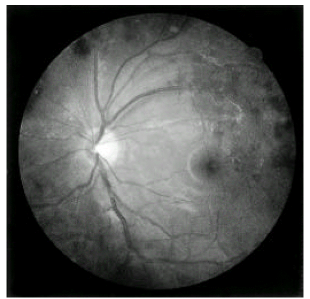
On ophthalmic examination, the left eye disclosed branch retinal vein occlusion and superficial flame-shaped retinal hemorrhages on superotemporal arcade (Figure 1). The right eye showed normal sizes of the retinal vessels and retina.
On laboratory studies, platelets were 37,700/mm3, 7,520/mm3 leukocytes (67% neutrophils, 21.6% lymphocytes), 15.6 g/dL hemoglobulin. Erythrocyte sedimenstration rate (ESR) was 2 mm/hr, 0.2 mg/dL CRP. Prothrombin (PT) and partial thromboplastin time (aPTT) were in normal range. Serum creatinine, aspartate aminotransferase, alanine aminotrasferase, alkaline phosphatase and urine analysis were all normal range. An immunologic study revealed high titer (>120 U/mL) IgG aCL antibody (normal, <10 U/mL) but the antinuclear antibody, anti-dsDNA antibody, lupus anticoagulant, IgM antiphospholipid antibody and antiplatelet antibody were negative. The serologic test for syphilis was negative; protein C and S antigen were also normal range.
He was started on warfarin to maintain an international normalization unit (INR) of 2.0 to 3.0 and high dose prednisolone (1 mg/kg/day) for a month at first. One month later, the patient’s visual acuity deteriorated to 20/400 in his left eye during anticoagulant therapy. Warfarin was switched to a low dose of aspirn (100 mg/day) after developing viterus hemorrhage. On the second month, prednisolone was tapered to 10 mg daily because platelet kept above 50,000/mm3. His visual acuity did not change during follow-up a year’s.
Case 2
A 60-year-old male patient presented with blurred vision in his right eye. There was no history of diabetes mellitus or hypertension. He also did not have any ocular or systemic diseases before. He is neither a smoker nor did he have a history of drug abuse. Family history was also unremarkable. His height was 173 centimeters and his body weight was 73 kilograms. His blood pressure was 130/80 mmHg. The uncorrected visual acuity on his first visit was 18/20 in his right eye and 18/20 in his left eye.
On ophthalmic examination, the right eye disclosed mild to moderate tortuous and dilated branch retinal vein and superficial flame-shaped retinal hemorrhages on superior and inferomedial arcade. The left eye showed normal sizes of the retinal vessels and retina.
On laboratory studies, platelets were 154,000/mm3, 4,500/mm3 leukocytes (63.6% neutrophils, 30.0% lymphocytes), 12.8 g/dL hemoglobulin. ESR was 7 mm/hr, 0.4 mg/dL CRP. PT and aPTT were in normal range. An immunologic study revealed 80 U/mL IgG aCL antibody (normal <10 U/mL), 5.1 PL IgM antiphospholipid antibody (normal <5.0 PL) and positive lupus anticoagulant antibody. But the antinuclear antibody, anti-dsDNA antibody and antiplatelet antibody were negative. The serologic test for syphilis was negative and protein C and S antigen were also normal range.
He has been maintained with low dose of aspirn (100 mg) daily. His visual acuity kept well during antiplatelet therapy but the blurred vision persisted.
DISCUSSION
Proposed diagnostic criteria for primary APS was a positive test of aCL antibody or lupus anticoagulant (LA) antibody, measured twice with a minimum interval of three months and one major clinical manifestation of APS, such as venous thrombosis, arterial thrombosis or thrombocytopenia in an individual without any underlying predisposing disorder3). Anticardiolipne antibodies, isotype IgG and Ig M were measured by means of an enzyme linked immunosorbent assay and the results were classified as negative (0 to 5 U/mL), low positive (5 to 15 U/mL), moderately positive (15 to 60 U/mL) or high positive (greater than 60 U/mL)4). The two cases were compatible with primary APS as they had retinal vein thrombosis with high titer of IgG aCL antibody without association to other immunologic diseases.
According to Asherson et al.5) the prevalence of ocular vaso-occlusive disease patients with lupus without antiphospholipid antibodies was less than 2%. When these antibodies were present, its prevalence increased fourfold. The recurrence of cerebral vascular thrombosis in APS was also more frequent in patients with high titers (over 100 U/mL) of aCL6). IgG isotype was significantly more frequent than IgM isotype and seemed to correlate best with clinical disease7). A previous study showed that all ocular vaso-occlusive patients with primary APS had high titer IgG aCL antibody, but the prevalence of lupus anticoagulant was as low as 29 percent3). In our 2 cases, IgG aCL antibodies were high titer (over 80 U/mL) and persisted for more than 4 months but IgM antiphopholipid antibody was normal or low positive.
Ocular symptoms associated with primary APS were transient blurring of vision, decreased vision, transient diplopia and transient field loss associated with headache and photophobia. Fundoscopic abnormalities were vein tortuosity, swelling optic disc, vitreous and preretinal hemorrhage, microaneurysms8). The two patients initially complained of blurred vision without decreased visual acuity, but the vision of one patient rapidly deteriorated because of vitreous hemorrhage during anticoagulant therapy.
Coexistent thrombosis and thrombocytopenia presented a major therapeutic dilemma in the anticoagulated patient. Lower levels of anticoagulation have been utilized (INR 2.0–3.0) for platelet count of 50,000–100,0009). In case 1, who had retinal vein thrombosis and thrombocytopenia, viterus hemorrhage occurred during anticoagulation therapy with warfarin.
In conclusion, it is necessary to evaluate the APS in patients with retinal vein thrombosis without considerable causes, as well as to further study whatever APS is related with retinal vein thrombosis.
