Systemic Lupus Erythematosus Presenting Earlier as Retinal Vaso-Occlusion
Article information
Abstract
Retinal vascular lesions are the most common ophthalmologic manifestation of systemic lupus erythematosus (SLE), occurring in 3% to 29% of cases, generally late in the disease. More rare is the severe vaso-occlusive disease, often termed “retinal vasculitis”, which includes central retinal artery occlusion, multifocal arteriolar occlusions, extensive capillary nonperfusion and central venous occlusion. Patients with SLE and raised serum concentrations of anticardiolipin antibodies (ACA) have a higher risk of developing occlusive ocular vascular disease. We report a case in which retinal involvement was an earlier manifestation of SLE in a patient without ACA.
INTRODUCTION
Systemic lupus erythematosus is a multisystem disease of unknown etiology characterized by numerous autoimmune phenomena with lesions in multiple organ systems. Retinal vascular lesion is the most common ophthalmologic manifestation, and the most common ocular findings are retinal hemorrhages and cotton-wool spots. These lesions occur in 3% to 29% of cases and generally are found late in the disease. We report a patient with SLE who had severe vaso-occlusive disease without ACA in early stage.
Case Report
An 18-year-old woman was admitted because of sudden decrease of visual acuity in both eyes. She had suffered general aching and malar rashes on her face 4 months before, at which time leukocyte count was 3.20×10⁹/L on routine examination at a private clinic. The visual loss began suddenly, and the skin lesions were aggravated several days before admission. She first visited the Division of Ophthalmology and was referred to the Rheumatology Department to exclude connective tissue diseases.
At admission, she looked acutely ill, but her vital signs were normal. On ophthalmologic examination, the visual acuity of both eyes was considerably decreased, the visual acuity of the right eye being 0.1×4/5 and that of the left eye FC20 (finger count 20cm). On physical examination, erythematous elevated eruptions were noted on the fingertips, knuckle areas, arms, and malar rash was developed on areas of the face, and 2nd, 3rd and 4th proximal interphalangeal joints of both hands were swollen slightly (Figure 1). These skin lesions were tender on pressure.
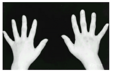
Cutaneous lesions of SLE.
Initial laboratory findings revealed mild anemia, with a hemoglobin value of 11.7 gm/dL. The white blood cell count was 5.10×10⁹/L, with 45.5% neutrophils, 43.8% lymphocytes and 8.2% monocytes, and the platelet count was 149×109/L. Blood urea nitrogen and creatinine concentrations were normal. C-reactive protein was 0.3 mg/dL (normal <0.8 mg/dL) and the erythrocyte sedimentation rate was 32 mm/h (normal <20 mm/h) by the Wintrobe method. Urinalysis revealed normal findings without blood or casts. The results of immunologic tests were as follows: antinuclear antibody 1:1280 with a speckled pattern; anti-Sm antibody, VDRL, and anti-Ro/La negative, anticardiolipin antibody IgM 6.2 MPL U/mL (normal <7 U/mL); IgG 8.3 GPL U/mL (normal <10 U/mL); anti-ds DNA antibody 5.15 IU/ml (normal <5.3 I U/mL); C3 69.5 mg/dL (normal range 55120 mg/dL); and C4 10.5 mg/dL (normal range 2050 mg/dL).
On admission, the ophthalmoscopic examination with mydriasis showed multiple retinal hemorrhages and cotton-wool spots in the right eye. The retinal arteries were constricted, and tortuous veins were surrounded by exudates. In the left eye, there was profuse vitreous and disc hemorrhage and ghost vessels (Figure 2). Retinal fluorescein angiography showed ischemic changes secondary to venous nonperfusion and leakage of dye from the retinal vessels. These features strongly suggested the clinical diagnosis of vaso-occlusive disease secondary to ocular SLE.
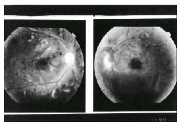
Fundoscopic examination showed multiple hemorrhages and cotton-wool spots in both eyes.
Panretinal argon laser photocoagulation therapy was done on the first, second and fourth day after admission but, on 6th day, the signs and symptoms of both eyes were unchanged. She was treated with pulsed methylprednisolone, 1 gm/day for 3 consecutive days and subsequently received oral prednisone (1 mg/kg per day) and hydroxychloroquine 400 mg/day. On the 22nd day of treatment, the retinal status was stable and her visual acuity was considerably recovered. Prednisone was gradually tapered to 15 mg/day. We added oral warfarin 5 mg daily to prevent recurrence of retinal vaso-occlusion.
Five months later, the vitreous and disc hemorrhage in the left eye had disappeared and the vessels had a more normal appearance (Figure 3). At present, the vision of both eyes is much improved (right 0.3 and left 0.1×4/5), and she is being maintained on prednisone 15 mg/day, hydroxychloroquine 400 mg/day and warfarin 5 mg/day in the outpatient department.

View of both eyes after 5 months of treatment.
Discussion
Systemic lupus erythematosus is a multisystem disease of unknown etiology in which the basic pathogenetic mechanism is a deposition of immune complexes, activation of complement and resulting inflammation1). Multifocal retinal vascular occlusive disease can be seen in a number of systemic disorders including SLE, Behcet’s disease, polyarteritis nodosa, and Takayasu’s disease. The ocular manifestations of lupus are most commonly recognized as a nonprogressive retinopathy with hemorrhages, exudates and cotton-wool spots caused by occlusive changes in the small retinal arterioles2). These lesions occur in 3% to 29% of cases and are generally found late in the disease2, 3). Other findings include arteriovenous crossing changes, arteriolar narrowing, retinal edema, papilledema, hard exudates and microaneurysm2). While these changes are seldom associated with abnormalities of visual function, extensive obliteration of larger retinal vessels, which occurs less frequently, can have potentially devastating effects on the visual function1).
Central nervous system involvement has been present in the majority of patients with significant retinal vascular lesions according to some reports4, 5), whereas others believe there is no association between SLE retinopathy and CNS lupus3). However, simultaneous involvement of the CNS occurred more frequently in patients with than in those without such retinal disease (73%)6). Also, in these patients with circulating lupus anticoagulant, an immunoglobulin, usually of the IgG class, is commonly associated with a tendency to venous and arterial thrombosis7).
The clinical significance of the lupus anticoagulant and related antiphospholipid antibodies in patients with SLE is their association with thrombotic events, recurrent fetal wastage and thrombocytopenia. The frequency of the lupus anticoagulant or related antiphospholipid antibodies in patient with SLE has been estimated to be 6.7% to 25%, according to the method used8). The differences in incidence of lupus anticoagulants and anticardiolipin antibodies in SLE is not merely a matter of test sensitivities. Love and Santoro analyzed series in which patients with SLE had both tests performed; only 45% of anticardiolipin antibodies-positive patients were also lupus anticoagulants positive, while only 59% of lupus anticoagulant-positive patients were also anticardiolipin antibodies-positive9). These findings are consistent with the concept that the two tests identify different populations of antibodies and that these two populations of antibodies can be physically separated. Almost all cases of severe vaso-occlusive retinal diseases should be found in patients who have the lupus anticoagulant. However, there are no data on the frequency of lupus anticoagulant in patients with retinopathy. We suspect a correlation between the vaso-occlusive retinal disease and the lupus anticoagulant. The patients with SLE and raised levels of ACA have a high risk of developing occlusive ocular vascular disease, and these lesions generally occur late in the disease. All the patients with lupus retinopathy were seriously ill with active systemic disease and high ds-DNA antibody levels. Also, these patients manifested antiphospholipid antibodies and CNS involvement.
Complete agreement has not been reached concerning the medical treatment of ocular thrombotic disease in SLE. Anticoagulant therapy is generally recommended. The second choice is an antiplatelet drug. The role of corticosteroid and immunosuppressive agents in preventing the thrombotic complications remains unclear. These drugs should probably be given, to patients with severe and progressive lesions that compromise the visual prognosis.
Interestingly, in our patient, although ds-DNA antibodies and ACA were not detected, retinal vaso-occlusive disease manifested earlier. We hope to see more reports of similar cases with vaso-occlusion or CNS lupus in the early stage.