Pleural Empyema Due To Salmonella: A Case Report
Article information
Abstract
Pleuropulmonary involvement of salmonella infection is very rare and only two cases of salmonella empyema have been reported in Korea. We report the case of a 70-year-old female diabetic patient who presented with right flank pain and right lower chest pain. The chest radiographs revealed fibrostreaky and hazy density at right lower lung field and blunting of right costophrenic angle. Thoracentesis revealed turbid yellowish fluid. Salmonella group B was identified from the cultures of blood and pleural fluid. After antimicrobial therapy and repeated therapeutic thoracentesis, the patient was improved.
INTRODUCTION
Salmonella are non-spore-forming gram-negative rods of the family Enterobacteriaceae. The relative prominence of certain clinical manifestations in persons infected with salmonella forms the basis for the designation of several clinical syndromes: enterocolitis (gastroenteritis), enteric fever, bacteremia, localized infection and chronic enteric or urinary carrier state. Localization of infection may occur at any site after salmonella bacteremia, irrespective of the associated clinical syndrome. But salmonella infection outside of the gastrointestinal tract remains uncommon and the pleural effusion or empyema infected with salmonella species is extremely rare.
Saphra et al. reported 85 cases of respiratory tract involvement with salomella1). Thereafter, pleural empyema due to salmonella has rarely been reported in immunocompromised patients. Only two cases of salmonella empyema have been reported in Korea2). Pneumonia or empyema, the predominant types of serious respiratory disease, occur usually in elderly patients or in patients with underlying diseases such as diabetes mellitus, malignancy or pulmonary disease.
We present a case of salmonella empyema in a 70-year-old female diabetic patient with a review of the literature.
CASE
A 70-year-old female was admitted to the hospital because of right flank pain. She had been relatively well until 15 days earlier when she began to have discomfort during urination. One week later, she had fever, chills and right flank pain.
There was a long history of non-insulin dependent diabetes mellitus and essential hypertension. She was treated with oral hypoglycemic and antihypertensive agents.
On admission, blood pressure was 120/80 mmHg, pulse rate 72/min, respiration rate 26/min and temperature 37.2 °C.
The results of physical examination were normal except for tenderness over the right costovertebral-angle.
The laboratory findings on admission were as follows: hemoglobin, 12.0 g/dL; hematocrit, 36.1%; WBC count, 14500/mm3 with 91.6% neutrophils and 6.0% lymphocytes; platelet, 188000/mm3; Na, 139 mEq/L; K, 3.2 mEq/L; Cl, 104 mEq/L; BUN, 9.9 mg/dL, creatinine, 0.6 mg/dL, total serum bilirubin, 0.7 mg/dL; ALP, 132 IU/L, ALT, 34 IU/L, AST, 16 IU/L; total protein, 6.2 g/dL; albumin, 2.8 g/dL; blood sugar, 145 mg/dL; PT, 84%; APTT, 30 sec. Urinalysis revealed glucosuria (300mg/dL) and the sediment contained more than 100 white cells per high-Power field. Stool examination revealed no abnormal findings.
Radiographs of the abdomen showed slightly distended loops of the small bowel and mottled density of feces in the large bowel. Radiographs of the chest were normal. An ultrasonographic examination of the abdomen showed slightly enlarged right kidney.
On admission day, the temperature rose to 38.6°C. Specimens of blood and urine were obtained for routine culture. We believed that she had urinary tract infection. So, ciprofloxacin was empirically administered intravenously.
On the third hospital day, urine culture yielded E. coli (>105 CFU) and blood culture yielded salmonella group B. We were perplexed by that result. At that time, there were not any noticeable alterations in bowel habits. Widal test, stool microscopic examination and culture were performed. Widal test showed O agglutinin titer positive at 1:160, H agglutinin titer positive at 1:320, para A agglutinin titer positive at 1:40 and para B agglutinin titer positive at 1:80. Stool culture yielded no pathogens and microscopic examination also showed no abnormal findings. So, we were not concerned about the possibility of salmonella bacteremia. However, ciprofloxacin was active in both organisms. Antimicrobial treatment with ciprofloxacin was continued. On the fifth hospital day, the fever went down some and the patient’s condition began to improve.
On the 15th hospital day, she complanied of right pleuritic chest pain and low-grade fever. Physical examination at that time revealed decreased breath sounds and fine crackles over the lateral aspect of right lower chest. The white blood cell count was 19900/mm3 with 88.6% neutrophils and 7.1% lymphocytes. The sputum cultures did not reveal an identifiable pathogen. Radiographs of the chest revealed fibrostreaky and hazy density at right lower lung field and blunting of right costophrenic angle (Figure 1).

Chest radiographs
Diagnostic thoracentesis was performed. Thoracentesis revealed turbid yellowish fluid (Figure 2). Laboratory data of pleural fluid revealed the following: pH, 7.168; RBC, 10000/mm3; WBC, 8300/mm3 with 70% neutrophils and 30% lymphocytes; LDH, 4470 IU/L; Protein, 3.9 g/dL; Glucose, 76 mg/dL; ADA, 81 U/L. Pleural fluid culture yielded salmonella group B.
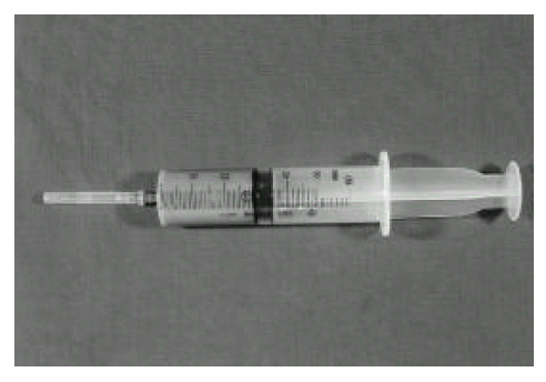
Gross appearance of pleural fluid
An indwelling catheter was inserted, which drained turbid yellowish pleural fluid. Ciprofloxacin was given for 9 weeks. After antibiotic therapy and repeated therapeutic thoracentesis, the patient was improved.
DISCUSSION
Acute enterocolitis is the most common clinical expression of salmonella infection. But salmonella can produce illness characterized by fever and sustained bacteremia without manifestations of enterocolitis or enteric fever. Salmonella bacteremia may be caused by any salmonella serotype. Threlfall et al. reported that the highest numbers of blood stream isolates were from infections caused by S. enteritidis and S. typhimurium, but the highest incidence of septicemia was attributable to infections with S. choleraesuis, S. dublin, and S. virchow in England and Wales3). S. choleraesuis seems to be especially invasive. Mortality in S. choleraesuis infections is higher than in other salmonella infections.
The clinical syndrome of salmonella bacteremia is characterized by a hectic febrile course lasting for days or weeks. The organism is isolated from blood, but stool cultures are often negative. Localized suppurative infections develop in about 10 percent of the patients and may become apparent days, months or even years after the initial bacteremia.
Localization of infection may occur at any site after salmonella bacteremia, irrespective of the associated clinical syndrome. As might be anticipated, localization at distant sites occurs relatively frequently in patients with the salmonella bacteremia syndrome but rarely in patients with enterocolitis. Localized infection has been reported in the thyroid, meninges, bone, heart, lungs, adrenals, pancreas, spleen, liver, testes, pericardium, soft tissues, areas of necrosis or infarction, benign or malignant tumors and cysts. Huang et al. reported 78 cases of nontyphoid salmolella bacteremia4). In that report, the concomitant focal infections with bacteremia included septic arthritis (5.1%), urinary tract infection (3.8%), peritonitis (2.6%) and empyema (1.3%). The site determines to a large extent the clinical manifestations, although most patients have spiking fever and polymorphonuclear leukocytosis.
Saphra et al. reported 85 cases of respiratory tract involvement with salomella1). Thereafter, pleural empyema due to salmonella has rarely been reported in immunocompromised patients. Pneumonia or empyema, the predominant types of serious respiratory disease, occurs usually in elderly patients or in patients with underlying diseases such as diabetes mellitus, malignancy, cardiovascular disease or pulmonary disease.
Salmonella syndrome is common in patients with AIDS. Salmonella bacteremia may be the clinical manifestation of AIDS. Wolday et al. reported a case of pleural empyema due to S. paratyphi in a patient with AIDS5). In patients with AIDS, organisms are difficult to eradicate from tissue even with prolonged therapy with bactericidal agents, and repeated relapses of infection are common.
For diagnosis of localized salmonella infection, culture of specimens that are normally sterile, such, as blood, joint fluid, CSF and pleural fluid, can be done on ordinary media such as blood agar. Salmonella may colonize the upper respiratory tract. So, the presence of these organisms in sputum does not necessarily imply lower tract infections.
Ampicillin, amoxicillin, chloramphenicol, trlmethoprim-sulfamethoxazole or third generation cephalosporins, such as cefotaxime or cefoperazone, can be used in the treatment of salmonella bacteremia. However, chloramphenicol should not be used when there is localization of infection at intravascular sites (endocarditis or aneurysmal infection). Ampicillin, amoxacillin or third generation cephalosporin is preferred under these circumstances. Bacteremia patients with impaired systemic resistance, for example, patients with AIDS, should also be treated with ampicillin, amoxicillin or a third generation cephalosporin. Ciprofloxacin is also reported to be effective in the therapy of recurrent salmonella sepsis. Gill et al. reported a case of malignant pleural effusion infected with S. enteritidis6). The infection was eventually eradicated with ciprofloxacin.
Localized infection with abscess formation usually requires surgical drainage in addition to antimicrobial therapy. Yang et al. reported that a patient with salmonella pericarditis and empyema was completely recovered by pericardiocentesis and repeated thoracentesis in addition to antibiotics therapy7). Burney et al. reported that cure was achieved by decortication and obliteration of pleural empyema spaces8). Carol et al. reported that intrapleural administration of antibiotics resulted in a rapid rise of the antibacterial activity of pleural fluid, leading to rapid clinical improvement and eradication of the infection in malignant pleural effusions9).
The duration of therapy is influenced by the site of infection and by the antimicrobial agent. Bacteremia without symptoms of localization should be treated for 10–14 days, whereas localized, infections, such as osteomyelitis or endocarditis, can require therapy for 4–6 weeks or longer. In patients with AIDS, organisms are difficult to eradicate from tissue, even with prolonged therapy with bactericidal agents, and repeated relapses of infection are common. So, patients with AIDS should be treated for 3–4 weeks in an effort to prevent relapse and long term therapy with oral antimicrobials may be required.
Mortality of salmonella pleuropulmonary disease is high. Aguado et al. reported 11 patients with pleuropulmonary infections due to nontyphoid strains of salmonella10). The overall mortality in that report was 63%. It was higher than that in others, perhaps due to the high number of immunosuppressed patients in that study.
In summary, we report a case of salmonella empyema in a 70-year-old female diabetic patient. Salmonella can produce illness characterized by bacteremia without manifestations of enterocolitis. When a blood culture yields salmonella in elderly patients or in patients with an underlying disease, such as diabetes mallitus or malignancy, it must be considered as salmonella bacteremia and subsequent localized salmonella infection.