Epidermoid splenic cyst with elevated serum level of CA19-9
Article information
The 25-year old patient, was hospitalized due to the abdomen pain in the left upper quadrant, accompanied by watery diarrhoea, nausea, vomits, periodically occurring fever up to 39°C degrees with shivers and the body weight loss (15 kg during 2 months). There was conducted the extensive laboratory diagnostics and there was noted the high level of tumour markers in the plasma: cancer antigen 19.9 (CA19.9) 2,878 U/mL (N < 37 U/mL) and CA-125 95 U/mL (N < 35 U/mL). The results of several tests performed on fecal for parasite eggs and immunological tests of blood infection with hydit (Elis’s and Western blot’s methods) were negative. Within the diagnostic imaging was performed the ultrasound test of the abdomen, a static scintigraphy of the spleen and the liver with sulphidicid colloid—99mTc + single-photon emission computed tomography/computed tomography (CT), magnetic resonance of the abdomen and the lesser pelvic with giving the contrast agent (Fig. 1). The ultrasonography (US) revealed typical splenic cysts’ features: the complex of homogeneous, anechoic, well separated cysts, with the largest of 90 × 80 mm, divided with a septum into two chambers. One was filled with homogenous hyperechoic content. In the magnetic resonance imaging (MRI) the cysts were: hypointense on T1-weighted images and hyperintense on T2-weighted images and intensity of the signal was similar to water and with no contrast uptake.
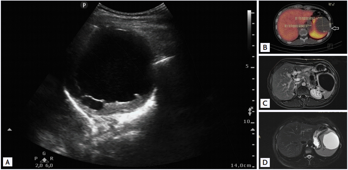
Imaging diagnostic tests. (A) Ultrasound examination of abdomen shows: anechoic, well demarcated, and capsulated spleen cyst. (B) Static scintigraphy of the liver and spleen in the lesion with sulphidic colloid—99mTc + single-photon emission computed tomography/computed tomography: there is no uptake of the marked sulphidic colloid. An arrow shows spleen cyst. (C, D) Magnetic resonance imaging of abdomen shows: hypointense cyst T1-weighted image and hyperintense T2-weighted image of the spleen.
Complaints reported by the patient, weight loss and large sizes and unclear character of the splenic cysts qualified for the laparoscopic splenectomy. The liquid culture from the cysts was negative. A histopathology indicated an epidermoid cyst. A cyst wall composed of a thick layer of fibrous tissue was partly lined by thin multilayered nonkeratinizing squamous epithelium; squamous differentiation was confirmed by strong diffuse cytoplasmic expression of CK5/6 (Fig. 2). The postoperative course was not complicated. During the laboratory control (3 weeks after the surgery) was stated the normalization if the CA-125 level and significant (×20) lowering of the CA19.9 level in plasma.
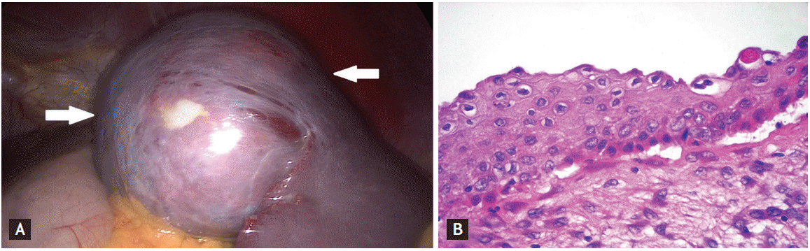
(A) Intraoperative image of the spleen cyst (arrows). (B) Histopathology: a cyst wall composed lined by thin multilayered nonkeratinizing squamous epithelium. Epidermoid splenic cyst (×40).
Coexistence of the increased CA19-9 and CA-125 in plasma with the epidermoid splenic cyst seems to be substantial in the differential diagnostics of the spleen lesions. In the diagnostics are also useful imaging tests like: the US and next CT or MRI. However the essential meaning has the histopathological test’s result. In the case of the symptomatic cysts and of the large size (> 5 cm), the chosen treatment is the partial or complete splenectomy.
Notes
No potential conflict of interest relevant to this article was reported.