Large venous malformation of right colonic flexure
Article information
Colonoscopy of a 38-year-old woman with persistent diarrhea despite drug therapy revealed no inflammatory changes in the colonic mucosa, but a dark blue, elevated lesion with a diameter of 3 cm was incidentally discovered in the right colonic flexure (Fig. 1A). Computed tomography showed vascular lesions with a clear boundary, 3 cm in diameter, with a patchy contrast effect in the right colonic flexure. The late arterial phase of angiography revealed a scattered light cotton-like stain that was consistent with the location of the lesion and vein flow into the lesion was noticeable during the venous phase (Fig. 1B). According to the International Society for the Study of Vascular Anomalies (ISSVA) classification, this lesion was diagnosed as a venous malformation (formerly known as a cavernous hemangioma). Intervention therapy is not considered suitable for this type of vascular anomaly, but therapy such as endoscopic mucosal resection has been described for venous malformations. However, the size of the lesion and the shape of the base precluded endoscopic therapy. The patient was therefore treated by laparoscopic ascending colon resection. Histologically, vascular endothelial cells had proliferated from the submucosa through the muscle layer and subserosa, which was consistent with a venous malformation (Fig. 2).
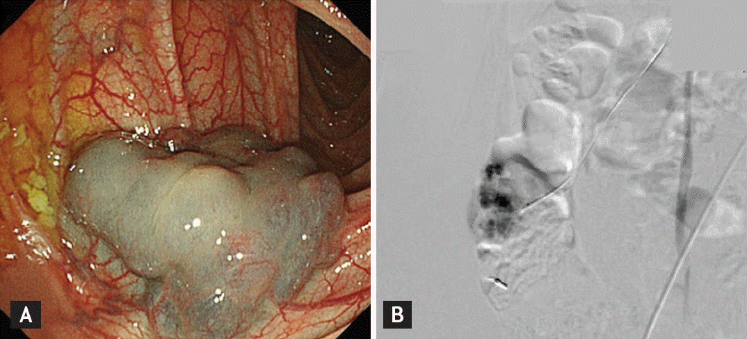
(A) Colonoscopy showing a dark blue elevated lesion in the right colonic flexure. (B) Abdominal angiography showing scattered light cotton-like stain of the lesion in the late arterial phase.

Microphotograph showing vascular endothelial cells proliferated from the submucosa through the muscle layer and subserosa (H&E, ×100).
Notes
No potential conflict of interest relevant to this article was reported.