Hepatocellular carcinoma hidden by multiple infarcted regenerative nodules
Article information
To the Editor,
Infarcted regenerative nodules in a patient with liver cirrhosis result from a sudden decrease in portal and arterial blood flow followed by ischemic necrosis. These lesions are rare in stable patients but typically occur in patients with a recent episode of gastrointestinal hemorrhage, usually due to variceal bleeding [1]. Distinguishing these lesions from other focal lesions, including hypovascular hepatocellular carcinomas (HCCs) and metastases, is challenging [2]. We describe a case of HCC that was indistinguishable from multiple infarcted regenerative nodules in a patient with cirrhosis and a previous episode of variceal bleeding. A 70-year-old male with alcoholic cirrhosis visited the emergency room because of epigastric discomfort and dizziness. He was diagnosed with a 1.3-cm single nodular HCC 4 years ago and had achieved a complete response after three sessions of transarterial chemoembolization (TACE) with doxorubicin. On arrival, his blood pressure was 63/45 mmHg, and pulse rate was 72 beats per minute (baseline resting heart rate, 45 to 55). Laboratory tests showed the following results: hemoglobin, 10.4 g/dL; hematocrit, 31.8%; white blood cell count, 4,230/μL; and platelet count, 69,000/mm3. The blood chemistry profile was as follows: total protein, 5.5 g/dL; albumin, 3.1 g/dL; total bilirubin, 0.7 mg/dL; aspartate aminotransferase (AST), 45 U/L; and alanine aminotransferase (ALT) 31 U/L. His prothrombin time (14.4 seconds) and international normalized ratio (1.31) were prolonged. Endoscopy revealed recent bleeding stigmata on the esophageal varices; thus, an endoscopic variceal ligation was performed.
Serum liver enzyme levels increased abruptly the next day: AST, 2,604 U/L and ALT, 1,684 U/L. Tests for serum tumor markers revealed normal levels of α-fetoprotein (11.4 ng/mL) and protein induced by the absence of vitamin K or antagonist II (14 mAU/mL). Serological tests for hepatitis B and C were negative. A dynamic liver computed tomography (CT) scan was performed and arterial-phase CT scans showed multiple, clustered, irregularly marginated, hypoattenuating lesions scattered in the left hepatic lobe, the caudate lobe, and the periphery of the right lobe (Fig. 1A). The peripheral portions of these lesions were enhanced subtly on portal-venous (Fig. 1B) and delayed-phase images. Infarcted regenerative nodules were firstly suspected. However, we subsequently performed gadoxetate disodium-enhanced magnetic resonance imaging (MRI) because diffuse infiltrative HCC had not been completely excluded. MRI revealed multiple, clustered, nodular lesions with peripheral high signal intensities on T2-weighted images (Fig. 2A) and low signal intensities on T1-weighted images (Fig. 2B). The center portion of these lesions remained at low signal intensity; however, the peripheral portion was rim-enhanced during the arterial, portal, and delayed phases. These lesions showed low signal intensities with fuzzy margins during the hepatobiliary phase (Fig. 2C-2F). Furthermore, a 1-cm nodule with arterial enhancement and rapid washout was newly noted. This lesion was missed on the initial dynamic CT examination. Hepatic angiography showed nodular hypervascular tumor staining, corresponding to the MRI. TACE followed by radiofrequency ablation resulted in a complete response. Most of the lesions other than the newly detected HCC were found to be resolved on a 2-month follow-up CT scan (Fig. 1C and 1D).
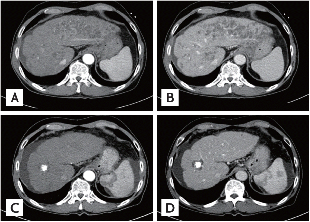
(A) An arterial-phase computed tomography (CT) image showing multiple, clustered, hypoattenuating nodules with peripheral rim enhancement in the left hepatic lobe, the caudate lobe, and the periphery of the right lobe. (B) A portal venous-phase CT image also showing multiple, clustered, hypoattenuating nodules with peripheral rim enhancement. The follow-up CT (C, arterial phase; D, portal phase) scan acquired 2 months later showing that the multiple hypoattenuating lesions have resolved. A lipiodol-laden hepatocellular carcinoma surrounded by non-enhancing ablation zone is also noted.
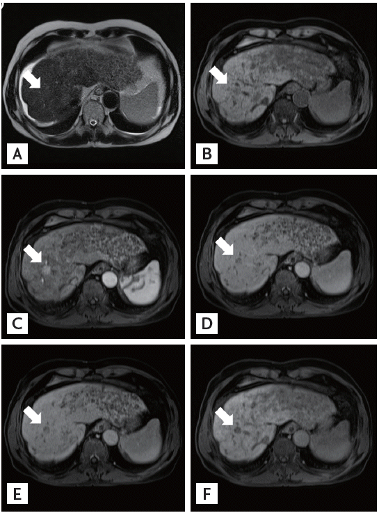
Magnetic resonance imaging (MRI) findings of the liver reveal multiple, clustered, nodular lesions with peripheral high signal intensities on (A) T2-weighted image and low signal intensities on (B) unenhanced T1-weighted image. The peripheral portion of these lesions was rim-enhanced during the (C) arterial-, (D) portal-, (E) delayed-, and (F) hepatobiliary-phase MRI images. MRI also show a 1 cm-nodule with high signal intensity on T2-weighted image and low signal intensity on unenhanced T1-weighted image (arrows). This nodule shows arterial enhancement and portal venous washout and appears as a defect in the hepatobiliary phase.
Infarcted regenerative nodules are rare and observed primarily in patients with a recent episode of variceal bleeding or various types of shock. These nodules are characterized by ischemic coagulative necrosis resulting from a sudden reduction in portal and arterial blood circulation [1]. A rapid rise in serum AST and ALT levels, which reflect hepatic ischemia, is often observed.
The infarcted regenerative nodules were depicted as low-attenuating nodular lesions on an unenhanced CT scan and as hypoattenuating zones with heterogeneous enhancement on contrast-enhanced CT scans. The infarcted regenerative nodules were of high signal intensity on T2-weighted images but showed isointensity on unenhanced T1-weighted images [3]. However, it is difficult to distinguish infarcted regenerative nodules from hepatic malignancies based on imaging findings alone, due to their overlapping radiological appearance. Some HCCs are hypovascular and show iso- or hypoattenuation on an arterial-phase CT scan, similar to infarcted regenerative nodules. Furthermore, infarcted regenerative nodules often show peripheral rim enhancement that mimics hepatic metastases [2].
Although liver biopsy is the gold standard for distinguishing infarcted regenerative nodules from hepatic malignancies, patients with cirrhosis and suspected infarcted regenerative nodules often are at risk of serious bleeding. Therefore, an acute episode of variceal bleeding or shock, a rapid elevation in serum AST and ALT levels, newly appearing lesions, and interval resolution of nodules on follow-up imaging can be helpful for a radiological diagnosis of these lesions [1,2,4]. In our case, the clinical history, laboratory findings, and new low-attenuation lesions suggested multiple infarcted regenerative nodules. However, among the suspected multiple infarcted regenerative nodules, an HCC that was not seen on the initial dynamic CT scan was detected by gadoxetate disodium-enhanced MRI. In retrospective review of initial dynamic CT scan, a faint arterial enhancing and washout lesion was suspected in the same location.
As multiple infarcted regenerative nodules manifest as hypoattenuating lesions, venous or delayed phase wash-out of HCCs may be indistinguishable from these lesions. The diagnostic ability of gadoxetate disodium-enhanced MRI is better for small hypervascular and hypovascular HCCs than is dynamic CT [5]. Therefore, gadoxetate disodium-enhanced MRI may assist in detecting hidden HCC among multiple infarcted regenerative nodules.
In summary, infarcted regenerative nodules should be included in the differential diagnosis of focal hepatic lesions in patients with cirrhosis who have a history of substantial variceal bleeding or shock. These lesions can hide HCC portal venous washout; thus, gadoxetate disodium-enhanced MRI or a follow-up CT scan must be performed for differential diagnosis and to detect hidden nodules.
Notes
No potential conflict of interest relevant to this article was reported.