Extranodal natural killer/T-cell lymphoma of the tenosynovium of the hand
Article information
To the Editor,
Extranodal natural killer/T-cell lymphoma(ENKL) usually occurs in the nasal area [1]. In cases involving extranasal regions, the skin, gastrointestinaltract, lung, and liver were reported as involvement sites [2], and soft tissue involvement of ENKL is extremely rare.Recently, our patient was diagnosed-with extranasal ENKL arising in the tenosynovium of the hand, and was successfully treated with dexamethasone, methotrexate, ifosfamide, L-asparginase, etoposide (SMILE) chemotherapy followed by autologous hematopoietic stem cell transplantation (HSCT). To the best of our knowledge, this is the first report of ENKL of the tenosynovium of the hand.
A 51-year-old male was admitted to our hospital for incomplete extension and tingling sensations in his left fourth and fifth fingers for 2 months.He was a left handed-carpenter with no prior history of illness and had experienced no fever, night sweats, or weight loss. His Eastern Cooperative Oncology Group performance state score was 0. Physical examination revealed that the fourth finger of his left hand was bent towards the palm and his left fourth and fifth fingers could not be fully extended. A subcutaneous mass on the left palm along the fifth finger without erythema or tenderness was noted.
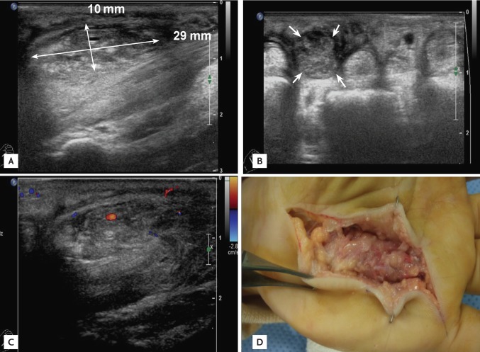
Nerve conduction velocity of the left median and ulnar nerves, and electromyogram of the left upper extremity, were normal. Ultrasonography revealed a 29 × 10 mm hypoechoic mass at the tendon sheath of the left fourth and fifth flexor digitorum profundus (FDP) (Fig. 1A-C). Tenosynovectomy of the left fifth finger was planned under the preoperative diagnosis of Dupuytren's contracture.

Ultrasonographic (A-C) and operative (D) findings. A 29 × 10-mm heterogeneous isoechoic to hypoechoic mass (A) was noted at the 4th to 5th flexor tendon of the left hand (B, white arrows). There was no increased blood flow in this mass on the color Doppler image (C). Intraoperative findings showed hypertrophied tenosynovium enveloping the flexor tendon and discolored lumbrical muscle involvement (D).
Upon surgery, inflammatory hypertrophied tenosynovial tissue enveloping the flexor tendon sheaths of the third to fifth fingers, and necrotized lumbrical muscle of the third to fifth fingers were observed (Fig. 1D). As infective tenosynovitis was suspected, tenosynovectomy of the third to fifth FDP tendon and excision of necrotized lumbrical muscle of the respective fingers were performed. Bacterial and tuberculosis cultures were negative. Histopathologic examination showed diffuse infiltrative atypical cells with geographic necrosis (Fig. 2A and 2B). Immunohistochemical staining for CD20 was negative (Fig. 2C), and CD56, CD3, and Epstein-Barr virus encoded RNA were positive (Fig. 2D-F), confirming the diagnosis of ENKL. A positron emission tomography-computed tomography scan showed increased fluorodeoxyglucose uptake at the left fourth and fifth fingers, and left axillary lymph node. Lactate dehydrogenase was increased up to 280 IU/L. Polymerase chain reaction for Epstein-Barr virus was negative. During the staging workup the patient's left palm began to swell and he developed fever. The fever was thought to be B symptom-related because it occurred almost a month after surgery and did not appear to be infection related. The Ann Arbor stage was IIBE, the international prognostic index (IPI) score was 1, and the Korean Prognostic Index (KPI) was 3 [1]. The patient was treated with three cycles of SMILE chemotherapy followed by autologous HSCT. The subject remained disease-free at the 2-year follow-up.

(A) Diffuse infiltration of atypical lymphoid cells with geographic necrosis was noted (H&E, ×12). (B) Atypical lymphocytes infiltrated the soft tissue (H&E, ×200). Immunohistochemical staining showing cells in the mass were CD-20 negative (C, ×200), CD-3 positive (D, ×200), and CD-5 positive (E, ×200). The lesion was positive for Epstein-Barr virus encoded RNA based on in situ hybridization (F, ×200).
Natural killer/T-cell lymphomas usually occur in middle-aged patients. They present as localized disease in about two thirds of patients; however, there is a high frequency of B symptoms despite apparently limited disease and rare bone marrow involvement [1]. Our patient was middle-aged and had B symptoms, despite localized disease without bone marrow involvement. The prognosis for ENKL is poor, even though more than half of ENKL patients have IPI scores of less than 2 [1]. Because IPI is not efficient at discriminating between low- and low-intermediate risk groups, a new prognostic model, KPI, was proposed [1]. Our patient was allocated to group 4 based on the KPI model, indicating poor expected survival.
There is no established treatment protocol for extranasal ENKL, and outcomes remain poor when it is treated with cyclophosphamide, doxorubicin, vincristine, prednisolone (CHOP), radiotherapy, or a combination of the two [3]. A recent retrospective study by Jo et al. [2] reported that SMILE was relatively effective as a first-line therapy for extranasal ENKL, and Kwong et al. [4] showed that extranasal ENKL patients treated with SMILE had good response rates. Autologous HSCT has also been shown to confer a survival benefit in patients who attained complete response on post-remission consolidation therapy, especially those patients with a high KPI score at diagnosis [5]. Thus, our patient was treated with SMILE followed by autologous HSCT, and has remained disease-free for 2 years.
In conclusion, this is the first case report of extranasal ENKL of the tenosynovium of the hand. It was successfully treated with SMILE followed by autologous HSCT. Extranasal ENKL could be considered as a differential diagnosis when Dupuytren's contracture is suspected.
Notes
No potential conflict of interest relevant to this article was reported.